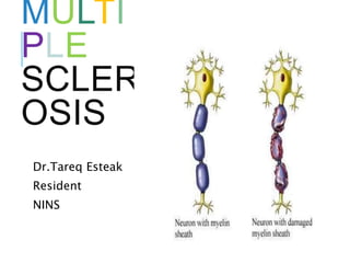
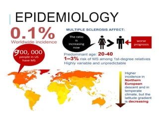
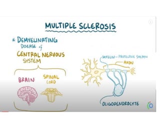
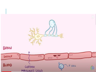
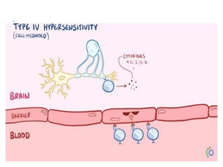
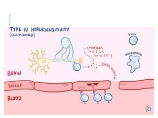
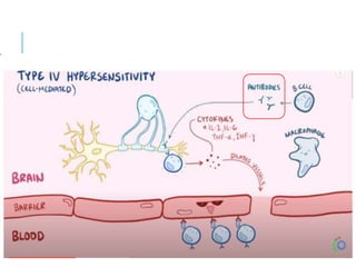
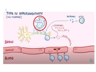
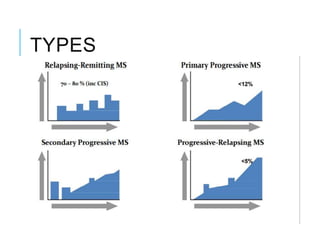
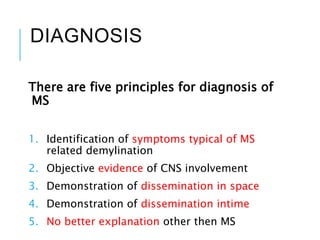
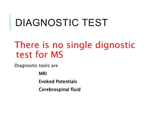
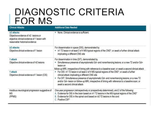
MULTI
PLE
SCLER
OSIS
Dr.Tareq Esteak
Resident
NINS
MULTIPLE SCLEROSIS
Multiple Sclerosis is an autoimmune
disease of the central nervous system
characterized by
Chronic inflammation,
Demyelination,
Gliosis
Neuronal loss.
EPIDEMIOLOGY
More common in female
Age onset 20 to 40
Temperate zone(Scotland*,North America,
Australia and New zealand)
EPIDEMIOLOGY
RISK FACTORS
 Genetic predisposion
 Vitamin D deficiency
 Epstein-Bar virus exposure after early
childhood
 Cigarette smoking
N.B: Vitamin D deficiency also increase disease
activity in MS patients.
PATHOGENESIS
PHYSIOLOGY
PATHOLOGY
CLINICAL FEATURE
Sensory loss(38%)
Optic neuritis(37%)
Weakness(36%)
Transverse Myelities (Clinically isolated syndrome)
Cerebellar ataxia
Brain stem syndrome
Vertigo
Facial pain
Dysarthria
Diplopia
ANCILLARY SYMPTOMS
Lhermitte’s symptoms
Heat sensitivity(Uhthoff’s symptom)
Tonic contraction of a limb, face or trunk(tonic
seizure)
Paroxysmal sensory disturbances
Trigeminal neuralgia
Facial myokymia
TYPES
DIAGNOSIS
There are five principles for diagnosis of
MS
1. Identification of symptoms typical of MS
related demylination
2. Objective evidence of CNS involvement
3. Demonstration of dissemination in space
4. Demonstration of dissemination intime
5. No better explanation other then MS
DIAGNOSTIC TEST
There is no single dignostic
test for MS
Diagnostic tools are
MRI
Evoked Potentials
Cerebrospinal fluid
DIAGNOSTIC CRITERIA
FOR MS
MRI
Revolutionized the diagnosis and
management of MS
Characteristic abnormalities are
found >95% of patients
MRI: TYPICAL SITES OF
MS
MRI:DIT & DIS
Dissemination in Time
MRI can demonestrate
dissemination of time by
the presence of gadolinium
–enhancing and
nonenhancing lesions in
same film or by the
appearance of a new T2-
hyperintence or
gadolinium –enhancing
lesions on a follow up MRI.
Dissemination of space
MRI demonestrate
dissemination of space if
at least one T2 –
hyperintense lesion found
two of the following region
1. Periventricular
2. Cortical or
juxtracortical
3. Infratentorial
4. The spinal cord
EVOKED POTENTIALS
Afferent: Visual, Auditory, Somatosensory
Efferent: Motor
Not specifice for MS but Suggestive
Abnormalities on one or more EP
modalities occur in 80-90% of MS
patients.
CEREBROSPINAL FLUID
CSF monoeuclear pleocytosis (more then 5
cells/uL in 25%)
CSF protein usually normal or mildly elevated
Protein concentration more then 100mg/dl
and pleocytosis more then 75 cells/uL raise the
concern that patient may not have MS.
CEREBROSPINAL
FLUID:IGG INDEX
The CSF IgG index is often
used to help diagnose
multiple sclerosis (MS), but
it is not specifically an MS
test.
Normal value is <0.7
Increased value means
local production of IgG
rather than infusion from
blood
Increased in : MS ,SSPE
CEREBROSPINAL
FLUID:OCB
Oligoclonalbands are bands of
immunoglobulins that are seen when
serum/csf is analyzed.
CSF OCBs (may be absent at onset and
number of OCBs may increase with time)
>2 discrete OCBs, not present in a
paired serum sample , taken
simultaneously found around 75% of
cases.
VARIANTS OF MULTIPLE
SCLEROSIS
T1WI C+Large
hypointense area with
incomple ring
enhancement, a
classical Tumefective
MS
MRS: shows large choline
pick with low NAA.(tumor
like)
Prefusion scan: rCBV
reduced in
demyelinationbut
increased in tumor.
MARBURG
DISEASE
(ACUTE
FULMINANT
MS)
T1WI C+ :Large
Enhancing
demyelinating lesion
in deep and
periventricular white
matter lesion
BALO’S
CONCENTRIC
MS
T1WI C+ :
showsConcentriclaminat
ed “ONION BULB”
appearance.
This is a rare aggresive
variant MS where
patients usually
presents with rapid
onset worsening
symptoms.
BLACK
HOLES
T1WI shows multiple
hypointense lesion
in the periventricular
area related to
Axonal
destruction.Note,
moderate ventricular
and sulcal
enlargement.
MS MIMICS
Many Neurological disease may mimic
MS clinically and/ radiologically.
Most common MS mimics are of
vascular origin.
The possible differentials for white-
matter lesion are long.
SMALL
VESSEL
DISEASE
(LEUKOARAI
OSIS)
Multiple white
matter lesion near
the ventricles and
sub-cortical space.
May give history of
HTN,DM or
Homocystinurea
MS VS
VASCULAR(LEUKOARAO
SIS)
MS: FLAIR
• Juxtra-cortical/u fibre
involvement.
• Corpus callosal
involvement.
• Tempor.
• Peri-ventricular
distribution.
• Dawson finger present
• Tempora
lobe,infratentoreal,s.cord
involvement.
Vascular:T2 WI
• Sub-cortical distribution
• Lesion doesn’t touch the
cortex.
• Asysmtrical distribution.
• Dawson finger absent
• No temporal or
infratentorealor s.cord
involvement.
MS VS NMO
• A very important
differential to keep in
mind, especially in
patients with a bilateral
optic neuritis
• We should think of NMO
when there are extensive
spinal cord lesions (more
than 3 vertebral
segments) with low T1-
signalintensity and
swelling of the cord.
• Often there are few/no
T2-lesions in the brain.
• The clue to the diagnosis
is the high AQP4-AB titer
ADEM VS MS
ADEM VS MS
MS VS METS
PERILESIONAL EDEMA
GOES IN FAVOR OF
METS
SINGLE,LARGE, RING
ENHANCEMENT GOES
INFAVOR OF METS THAN
MS
SARCOIDOSI
S VS MS
• Multi-system disorder.
• Associated Respiratory,
cardiovascular and skin
manifestation may be
present.
• The distribution of lesions is
quite similar to MS.
Besides lesions in the deep
WM, there are some
juxtaiventricular lesions and
even Dawson finger-like
lesions.
• Typical for sarcoid in this
case is the leptomeningeal
SARCOIDOSIS VS MS
T1WI C+:
Leptomeningeal
Enhancement
T1WI C+: Linear
enhancement due to
inflammation of vircow
Robbin’s space.
PML VS MS
•Caused by JC virus in
immunosuppressed
patients.
•Usually have history of
HIV infection or
treatment with
monoclonalantibodylik
e Natalizumab.
•Lesion usually are
subcortical, diffuse, ill
defined, usually size
>5mm (MS-<5mm)
MS VS
CADASIL
• Cerebral Autosomal Dominant
Arteriopathy with Subcortical
Infarcts and
Leukencephalopathy.
• Clinical clues: family history
migraine, dementia.
• MRI:fLAIR shows Multiple sub-
cortical infracts
WMLs in the anterior temporal pole
and external capsule
TREATMENT
ACUTE ATTACK
Intravenous Methylprednesolone 500 to
1000mg/day for 5 days without an oral tapper.
Alternatively, Oral steroid is effective as the IV
infusion.
Patients who cant tolerate oral steroid with
poor venous access IM/SubQ corticotropin gel
may be used.
In patients with severe neurological deficit
refractory to high dose steroid, plasma
exchange is another option.
DISEASE MODIFYING
THERAPIES
GOAL:
A decrease in relapse rate.
A slower accumulation of brain lesion
on MRI
Lower the risk of disease progression.
WHAT DMT TO CHOOSE?
High
diseas
e
activity
WHAT DMT TO CHOOSE?
PPMS(PrimaryProgressive
MS)
Ocrelizumab
SPMS
(Secodary Progressive MS)
IFN-B(if Active)
Mitoxantrone
(lack in evidence)
MECHNISM OF ACTION:
DMTS
MECHNISM OF ACTION
DMF
MONITORING
RESPONSE
Tools:
Clinical followup
MRI brain:
o Before starting or changing DMT
oAfter 6months
o After that,annually
oPCR-Jcvirus -6monthly-for patients on
Natalezumab
Expanded disability status scale – 3monthly
Patient refractory to initial DMT change into another 1st
line drug.
(single mild relapse or a single lesion within 6month is not refractory
MS)
Recommendations:
If initially treated with glatiramer or inf-beta switch to-
•Di-methylfumerate, Fingolimod or Teriflunamide.
Or
•Natalezumab or Ocrelezumab.
 Initially treated with Natalezumab who have inadequate
response or developed PML-
•Stop the drug and switch to Ocrelezumab or Alemtuzumab
REFRACTORY DISEASE
PREGNANCY
Data shows activity of MS isreduced during
pregnancy.
Baby child may beborn with IUGR or congenital
fetal malformations.
Drug should be avoided are –Fingolimod and
Mitoxantrone.
Druds that are safe are- Glatiramer,Inf,
Alamtuzumab, Natalizumab etc
OFF LABEL TREATMENT
OPTIONS
Options Preferred situation Drawback
Azathioprine rrMS Benefit on disability progression yet
to be demonstrated.
Methotrexate SPMS Irreversible liver damage,
Cyclophosphamide Treatment-refractory MS-
Abmulatory, <40y, apperntly in
good health
Bladder carcinoma, Hemorrhagic
cystitis
Intravenous
immunoglobulin
monthly pulses (up to 1 g/kg) for
up to 2y, appears to reduce
annual exacerbation rates
High cost
Methylprednisolone in one study, administered as
monthly iv pulses, reduced
disability progression
Steroid related Side effects
Hematopoietic stem cell
transplantation
rrMS: highly effective in reducing
the occurrence of relapses and
may improve disability in
procedure carries a significant
mortality risk
COVID-19 RELATED
UPDATE
Available data shows that DMT does not
increase the risk of COVID-19
DMT was not associated with increase severity
of Covid-19.
THANK YOU….

Multiple Sclerosis

  • 1.
  • 2.
    MULTIPLE SCLEROSIS Multiple Sclerosisis an autoimmune disease of the central nervous system characterized by Chronic inflammation, Demyelination, Gliosis Neuronal loss.
  • 3.
    EPIDEMIOLOGY More common infemale Age onset 20 to 40 Temperate zone(Scotland*,North America, Australia and New zealand)
  • 4.
  • 5.
    RISK FACTORS  Geneticpredisposion  Vitamin D deficiency  Epstein-Bar virus exposure after early childhood  Cigarette smoking N.B: Vitamin D deficiency also increase disease activity in MS patients.
  • 6.
  • 7.
  • 8.
  • 19.
    CLINICAL FEATURE Sensory loss(38%) Opticneuritis(37%) Weakness(36%) Transverse Myelities (Clinically isolated syndrome) Cerebellar ataxia Brain stem syndrome Vertigo Facial pain Dysarthria Diplopia
  • 21.
    ANCILLARY SYMPTOMS Lhermitte’s symptoms Heatsensitivity(Uhthoff’s symptom) Tonic contraction of a limb, face or trunk(tonic seizure) Paroxysmal sensory disturbances Trigeminal neuralgia Facial myokymia
  • 23.
  • 24.
    DIAGNOSIS There are fiveprinciples for diagnosis of MS 1. Identification of symptoms typical of MS related demylination 2. Objective evidence of CNS involvement 3. Demonstration of dissemination in space 4. Demonstration of dissemination intime 5. No better explanation other then MS
  • 25.
    DIAGNOSTIC TEST There isno single dignostic test for MS Diagnostic tools are MRI Evoked Potentials Cerebrospinal fluid
  • 26.
  • 27.
    MRI Revolutionized the diagnosisand management of MS Characteristic abnormalities are found >95% of patients
  • 28.
  • 29.
    MRI:DIT & DIS Disseminationin Time MRI can demonestrate dissemination of time by the presence of gadolinium –enhancing and nonenhancing lesions in same film or by the appearance of a new T2- hyperintence or gadolinium –enhancing lesions on a follow up MRI. Dissemination of space MRI demonestrate dissemination of space if at least one T2 – hyperintense lesion found two of the following region 1. Periventricular 2. Cortical or juxtracortical 3. Infratentorial 4. The spinal cord
  • 30.
    EVOKED POTENTIALS Afferent: Visual,Auditory, Somatosensory Efferent: Motor Not specifice for MS but Suggestive Abnormalities on one or more EP modalities occur in 80-90% of MS patients.
  • 31.
    CEREBROSPINAL FLUID CSF monoeuclearpleocytosis (more then 5 cells/uL in 25%) CSF protein usually normal or mildly elevated Protein concentration more then 100mg/dl and pleocytosis more then 75 cells/uL raise the concern that patient may not have MS.
  • 32.
    CEREBROSPINAL FLUID:IGG INDEX The CSFIgG index is often used to help diagnose multiple sclerosis (MS), but it is not specifically an MS test. Normal value is <0.7 Increased value means local production of IgG rather than infusion from blood Increased in : MS ,SSPE
  • 33.
    CEREBROSPINAL FLUID:OCB Oligoclonalbands are bandsof immunoglobulins that are seen when serum/csf is analyzed. CSF OCBs (may be absent at onset and number of OCBs may increase with time) >2 discrete OCBs, not present in a paired serum sample , taken simultaneously found around 75% of cases.
  • 34.
  • 35.
    T1WI C+Large hypointense areawith incomple ring enhancement, a classical Tumefective MS MRS: shows large choline pick with low NAA.(tumor like) Prefusion scan: rCBV reduced in demyelinationbut increased in tumor.
  • 36.
    MARBURG DISEASE (ACUTE FULMINANT MS) T1WI C+ :Large Enhancing demyelinatinglesion in deep and periventricular white matter lesion
  • 37.
    BALO’S CONCENTRIC MS T1WI C+ : showsConcentriclaminat ed“ONION BULB” appearance. This is a rare aggresive variant MS where patients usually presents with rapid onset worsening symptoms.
  • 38.
    BLACK HOLES T1WI shows multiple hypointenselesion in the periventricular area related to Axonal destruction.Note, moderate ventricular and sulcal enlargement.
  • 39.
    MS MIMICS Many Neurologicaldisease may mimic MS clinically and/ radiologically. Most common MS mimics are of vascular origin. The possible differentials for white- matter lesion are long.
  • 40.
    SMALL VESSEL DISEASE (LEUKOARAI OSIS) Multiple white matter lesionnear the ventricles and sub-cortical space. May give history of HTN,DM or Homocystinurea
  • 41.
  • 42.
    MS: FLAIR • Juxtra-cortical/ufibre involvement. • Corpus callosal involvement. • Tempor. • Peri-ventricular distribution. • Dawson finger present • Tempora lobe,infratentoreal,s.cord involvement. Vascular:T2 WI • Sub-cortical distribution • Lesion doesn’t touch the cortex. • Asysmtrical distribution. • Dawson finger absent • No temporal or infratentorealor s.cord involvement.
  • 43.
    MS VS NMO •A very important differential to keep in mind, especially in patients with a bilateral optic neuritis • We should think of NMO when there are extensive spinal cord lesions (more than 3 vertebral segments) with low T1- signalintensity and swelling of the cord. • Often there are few/no T2-lesions in the brain. • The clue to the diagnosis is the high AQP4-AB titer
  • 44.
  • 45.
  • 46.
    MS VS METS PERILESIONALEDEMA GOES IN FAVOR OF METS SINGLE,LARGE, RING ENHANCEMENT GOES INFAVOR OF METS THAN MS
  • 47.
    SARCOIDOSI S VS MS •Multi-system disorder. • Associated Respiratory, cardiovascular and skin manifestation may be present. • The distribution of lesions is quite similar to MS. Besides lesions in the deep WM, there are some juxtaiventricular lesions and even Dawson finger-like lesions. • Typical for sarcoid in this case is the leptomeningeal
  • 48.
    SARCOIDOSIS VS MS T1WIC+: Leptomeningeal Enhancement T1WI C+: Linear enhancement due to inflammation of vircow Robbin’s space.
  • 49.
    PML VS MS •Causedby JC virus in immunosuppressed patients. •Usually have history of HIV infection or treatment with monoclonalantibodylik e Natalizumab. •Lesion usually are subcortical, diffuse, ill defined, usually size >5mm (MS-<5mm)
  • 50.
    MS VS CADASIL • CerebralAutosomal Dominant Arteriopathy with Subcortical Infarcts and Leukencephalopathy. • Clinical clues: family history migraine, dementia. • MRI:fLAIR shows Multiple sub- cortical infracts WMLs in the anterior temporal pole and external capsule
  • 51.
  • 52.
    ACUTE ATTACK Intravenous Methylprednesolone500 to 1000mg/day for 5 days without an oral tapper. Alternatively, Oral steroid is effective as the IV infusion. Patients who cant tolerate oral steroid with poor venous access IM/SubQ corticotropin gel may be used. In patients with severe neurological deficit refractory to high dose steroid, plasma exchange is another option.
  • 53.
    DISEASE MODIFYING THERAPIES GOAL: A decreasein relapse rate. A slower accumulation of brain lesion on MRI Lower the risk of disease progression.
  • 54.
    WHAT DMT TOCHOOSE? High diseas e activity
  • 55.
    WHAT DMT TOCHOOSE? PPMS(PrimaryProgressive MS) Ocrelizumab
  • 56.
    SPMS (Secodary Progressive MS) IFN-B(ifActive) Mitoxantrone (lack in evidence)
  • 57.
  • 58.
  • 59.
    MONITORING RESPONSE Tools: Clinical followup MRI brain: oBefore starting or changing DMT oAfter 6months o After that,annually oPCR-Jcvirus -6monthly-for patients on Natalezumab Expanded disability status scale – 3monthly
  • 60.
    Patient refractory toinitial DMT change into another 1st line drug. (single mild relapse or a single lesion within 6month is not refractory MS) Recommendations: If initially treated with glatiramer or inf-beta switch to- •Di-methylfumerate, Fingolimod or Teriflunamide. Or •Natalezumab or Ocrelezumab.  Initially treated with Natalezumab who have inadequate response or developed PML- •Stop the drug and switch to Ocrelezumab or Alemtuzumab REFRACTORY DISEASE
  • 61.
    PREGNANCY Data shows activityof MS isreduced during pregnancy. Baby child may beborn with IUGR or congenital fetal malformations. Drug should be avoided are –Fingolimod and Mitoxantrone. Druds that are safe are- Glatiramer,Inf, Alamtuzumab, Natalizumab etc
  • 62.
    OFF LABEL TREATMENT OPTIONS OptionsPreferred situation Drawback Azathioprine rrMS Benefit on disability progression yet to be demonstrated. Methotrexate SPMS Irreversible liver damage, Cyclophosphamide Treatment-refractory MS- Abmulatory, <40y, apperntly in good health Bladder carcinoma, Hemorrhagic cystitis Intravenous immunoglobulin monthly pulses (up to 1 g/kg) for up to 2y, appears to reduce annual exacerbation rates High cost Methylprednisolone in one study, administered as monthly iv pulses, reduced disability progression Steroid related Side effects Hematopoietic stem cell transplantation rrMS: highly effective in reducing the occurrence of relapses and may improve disability in procedure carries a significant mortality risk
  • 63.
    COVID-19 RELATED UPDATE Available datashows that DMT does not increase the risk of COVID-19 DMT was not associated with increase severity of Covid-19.
  • 64.