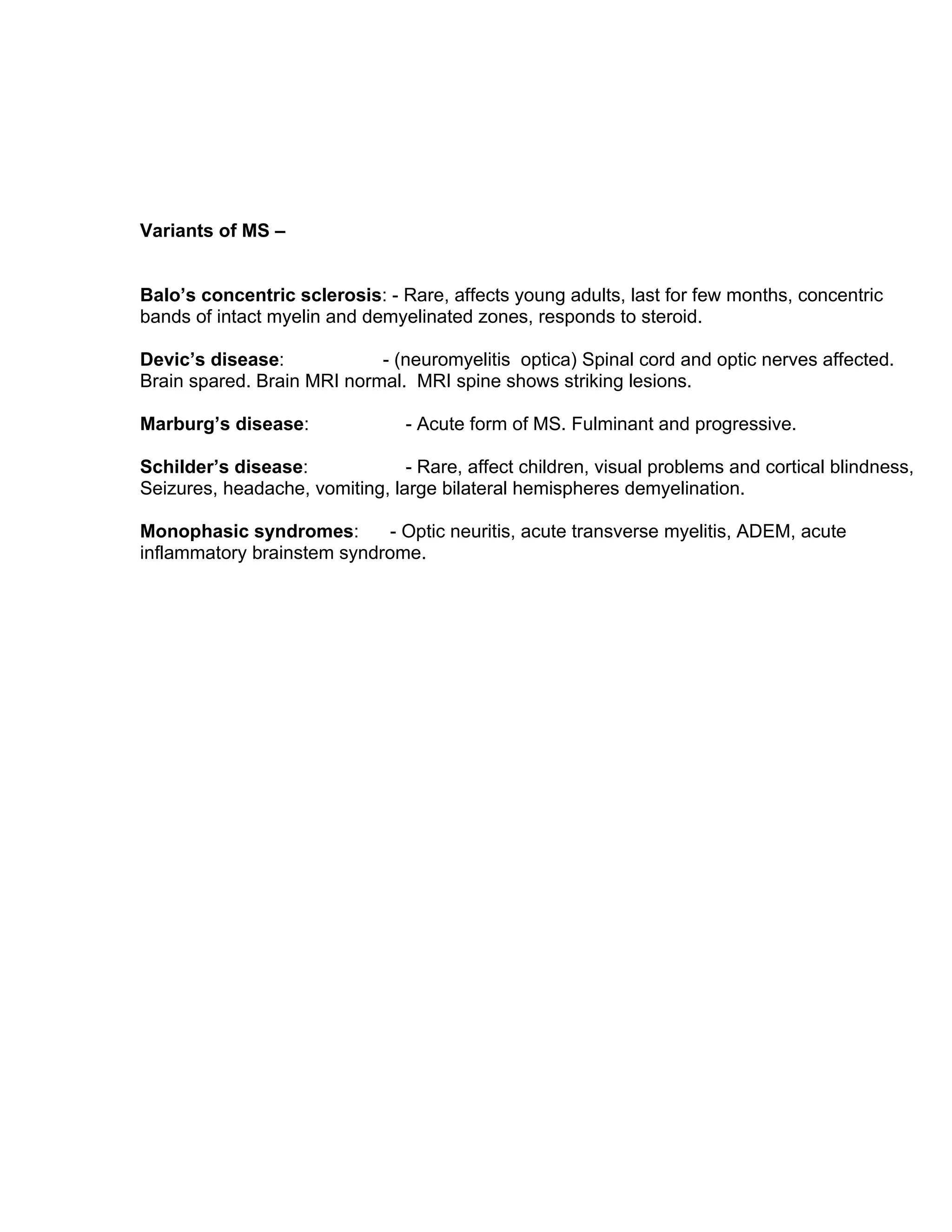
This document provides an overview of multiple sclerosis (MS), including its causes, pathophysiology, clinical features, diagnosis, course, classifications, and the role of MR imaging. MS is a demyelinating disease of the central nervous system that typically affects people aged 20-40. It has an unknown cause but is thought to involve genetic, viral, autoimmune, and environmental factors. Clinically, it presents with sensory issues, optic neuritis, spasticity, and other symptoms. Diagnosis involves identifying neurological abnormalities via history, exam, and MRI findings. The disease course is highly variable but can be classified as relapsing-remitting, secondary-progressive, primary-progressive, or progressive-
Neuroradiology in multiple sclerosis
MRI in diagnosis of MS
MRI in D.D. of MS
MRI in monitoring disease progression and response to DMT
New imaging techniques
Neuroradiology in multiple sclerosis
MRI in diagnosis of MS
MRI in D.D. of MS
MRI in monitoring disease progression and response to DMT
New imaging techniques
A case of Neuromyelitis optica as a presenting manifestation of Systemic Lupu...Apollo Hospitals
Neuromyelitis optica (NMO) is a well characterised, autoimmune, clinicopathological syndrome, which is uncommon and occurs as an isolated entity. Unlike multiple sclerosis, in NMO, the autoimmunity is humorally mediated and the recent availability of Antiaquaporin antibody testing has increased the positive diagnosis of this condition. NMO can also occur in patients with established Systemic Lupus Erythematosis (SLE) who have multiple autoantibodies. The presence of Antiaquaporin antibody is specific for NMO and is seen in patients with SLE who develop inflammatory CNS disease. However, Neuromyelitis optica occurring as a presenting manifestation of SLE is extremely rare and we report one such case.
Multiple sclerosis (MS) is a demyelinating disease in which the insulating covers of nerve cells in the brain and spinal cord are damaged.This damage disrupts the ability of parts of the nervous system to communicate, resulting in a range of signs and symptoms, including physical, mental, and sometimes psychiatric problems
Brain MRI biomarkers for improved follow up of people with Multiple Sclerosis...Wim Van Hecke
MRI is increasingly used for the diagnosis and follow-up of people with Multiple Sclerosis (MS). However, there is a need for objective MRI biomarkers that can be used in clinical practice. This is now possible. By sending MRI data to a icometrix, reliable and objective reports of brain atrophy and lesion load can be obtained.
If you really want to get a chance in radiology fellowship programs, You need an amazing statement that may attract reader's eyes or you may select for the program. So you can use our radiology fellowship statement. For more information, visit http://www.radiologyfellowship.net/
The aim of the 3DOR Workshop series is to stimulate researchers from different fields to present state-of-the-art work in the field. 3DOR 2013 took place as the 6th workshop in this series on May 11, 2013 in Girona (Spain), in conjunction with Eurographics 2013. Prof. Henning Muller presented the keynote talk about Medical 3D data retrieval.
Market Research Report : Medical diagnostics market in india 2014 - SampleNetscribes, Inc.
For the complete report, get in touch with us at: info@netscribes.com
Abstract :
Netscribes’ latest market research report titled Medical Diagnostics Market in India 2014 analyses the growth of the market due to cutting edge technology which provides better disease diagnosis. The Indian diagnostics market can be divided into equipment, reagents and services. The service sector is characterized by a large number of laboratories in the unorganized sector, which are clustered in the suburban areas and metros. In order to have better regulations and proper definition for the market a clear and structured format is being established. The laboratories are rapidly expanding using various business models. Large numbers of laboratories are registered only with the state health departments and not recognized by NABL (National Accreditation Board for Testing and Calibration Laboratories under Department of Science and Technology, Govt. of India).
The market is driven by increasing number of lifestyle diseases, rise in life expectancy due to preventive healthcare practices and increasing medical tourism. Currently there is trend of diagnostic medical imaging due to innovation in the technology sector which is helping the market to grow. Few numbers of accredited labs and high dependence on imported medical diagnostics products are the challenges this market is facing. Even though a range of diagnostic tests for various diseases are available in the market, there is a need for more reliable and better diagnostics. The market is dominated by private diagnostics companies in India who are expanding in Tier I and Tier II cities. Providing after-sales services for medical instruments and competitive pricing of the tests will catalyze the growth of the diagnostic sector.
Table of Contents :
Slide 1: Executive Summary
Macroeconomic Indicators
Slide 2: GDP at Factor Cost: Quarterly (2010-11, 2011-12, 2012-13, 2013-14), Inflation Rate: Monthly (Jul 2013 – Dec 2013)
Slide 3: Gross Fiscal Deficit: Monthly (Feb 2013 – Jul 2013), Exchange Rate: Half Yearly (Aug 2013 – Jan 2014)
Slide 4: Lending Rate: Annual (2008-09, 2009-10, 2010-11, 2011-12), Trade Balance: Annual (2009-10, 2010-11, 2011-12, 2012-13), FDI: Annual (2009-10, 2010-11, 2011-12, 2012-13)
Introduction
Slide 5: Medical Diagnostics Sector – Overview
Market Overview
Slide 6: Medical Diagnostics– Market Overview Forecasted Market Size and Growth (Value-Wise; 2013 – 2018e)
Slide 7: Medical Diagnostics Market – Overview of Franchise Business Model
Slide 8: Medical Diagnostics Market – Overview of Other Business Models
Slide 9: Accreditation of Laboratories
Slide 10: Technological Developments Used for the Diagnosis of a Wide Spectrum of Infectious Diseases
Drivers & Challenges
Slide 11: Medical Diagnostics Market – Drivers and Challenges summary
Slide 12-16: Drivers
Slide 17-18: Challenges
Trends
Slide 19: Trends – Summary
Slide 20-22: Trends
Go
Magnetic resonance imaging (MRI) is an essential tool for multiple sclerosis (MS) diagnosis and treatment, understanding MS natural history and pathophysiology, and as an outcome measure in clinical trials. This review will provide descriptions of the features, Pathophysiological substrates, and clinical utility of MRI measures of MS including T2-weighted, proton density (PD), and fluid-attenuated inversion recovery (FLAIR) hyperintense lesions, T1-weighted hypointense lesions, gadolinium-enhancing lesions, and measures of brain atrophy. Lesion presence and atrophy within both the brain and spinal cord will be described. This review will also provide a description of non-conventional MRI markers including diffusion tensor imaging (DTI), functional MRI (fMRI), magnetization transfer ratio(MTR) imaging, relaxometry/quantitative magnetic susceptibility (QS) mapping, and magnetic resonance spectroscopy (MRS). Basic descriptions of how these measures are obtained, the pathological substrates, clinical correlates (e.g. with physical disability, cognition, fatigue, etc.) and advantages/ drawbacks of each technique will be reviewed. Conclusions will be drawn on the overall clinical utility and future directions for use of MRI in MS.
This project was developed for a competitive intelligence company by mining data from the various information sources e.g. Company (News, Investor Section, SEC filings, Annual Reports, Presentations etc), Universities/Medical Schools/Organizations, Medical Affairs Companies, Non- Profit Medical Agency, Government Agencies, Drug Delivery Companies, Contract Manufacturing Organizations, Contract Research Organizations, Consultancies and Financial Institutions. The complete information available there complied into a single MS word document, listed in MS Excel and then by using MS publisher it was converted into the report which finally converted into PDF.
Explore natural remedies for syphilis treatment in Singapore. Discover alternative therapies, herbal remedies, and lifestyle changes that may complement conventional treatments. Learn about holistic approaches to managing syphilis symptoms and supporting overall health.
micro teaching on communication m.sc nursing.pdfAnurag Sharma
Microteaching is a unique model of practice teaching. It is a viable instrument for the. desired change in the teaching behavior or the behavior potential which, in specified types of real. classroom situations, tends to facilitate the achievement of specified types of objectives.
HOT NEW PRODUCT! BIG SALES FAST SHIPPING NOW FROM CHINA!! EU KU DB BK substit...GL Anaacs
Contact us if you are interested:
Email / Skype : kefaya1771@gmail.com
Threema: PXHY5PDH
New BATCH Ku !!! MUCH IN DEMAND FAST SALE EVERY BATCH HAPPY GOOD EFFECT BIG BATCH !
Contact me on Threema or skype to start big business!!
Hot-sale products:
NEW HOT EUTYLONE WHITE CRYSTAL!!
5cl-adba precursor (semi finished )
5cl-adba raw materials
ADBB precursor (semi finished )
ADBB raw materials
APVP powder
5fadb/4f-adb
Jwh018 / Jwh210
Eutylone crystal
Protonitazene (hydrochloride) CAS: 119276-01-6
Flubrotizolam CAS: 57801-95-3
Metonitazene CAS: 14680-51-4
Payment terms: Western Union,MoneyGram,Bitcoin or USDT.
Deliver Time: Usually 7-15days
Shipping method: FedEx, TNT, DHL,UPS etc.Our deliveries are 100% safe, fast, reliable and discreet.
Samples will be sent for your evaluation!If you are interested in, please contact me, let's talk details.
We specializes in exporting high quality Research chemical, medical intermediate, Pharmaceutical chemicals and so on. Products are exported to USA, Canada, France, Korea, Japan,Russia, Southeast Asia and other countries.
Recomendações da OMS sobre cuidados maternos e neonatais para uma experiência pós-natal positiva.
Em consonância com os ODS – Objetivos do Desenvolvimento Sustentável e a Estratégia Global para a Saúde das Mulheres, Crianças e Adolescentes, e aplicando uma abordagem baseada nos direitos humanos, os esforços de cuidados pós-natais devem expandir-se para além da cobertura e da simples sobrevivência, de modo a incluir cuidados de qualidade.
Estas diretrizes visam melhorar a qualidade dos cuidados pós-natais essenciais e de rotina prestados às mulheres e aos recém-nascidos, com o objetivo final de melhorar a saúde e o bem-estar materno e neonatal.
Uma “experiência pós-natal positiva” é um resultado importante para todas as mulheres que dão à luz e para os seus recém-nascidos, estabelecendo as bases para a melhoria da saúde e do bem-estar a curto e longo prazo. Uma experiência pós-natal positiva é definida como aquela em que as mulheres, pessoas que gestam, os recém-nascidos, os casais, os pais, os cuidadores e as famílias recebem informação consistente, garantia e apoio de profissionais de saúde motivados; e onde um sistema de saúde flexível e com recursos reconheça as necessidades das mulheres e dos bebês e respeite o seu contexto cultural.
Estas diretrizes consolidadas apresentam algumas recomendações novas e já bem fundamentadas sobre cuidados pós-natais de rotina para mulheres e neonatos que recebem cuidados no pós-parto em unidades de saúde ou na comunidade, independentemente dos recursos disponíveis.
É fornecido um conjunto abrangente de recomendações para cuidados durante o período puerperal, com ênfase nos cuidados essenciais que todas as mulheres e recém-nascidos devem receber, e com a devida atenção à qualidade dos cuidados; isto é, a entrega e a experiência do cuidado recebido. Estas diretrizes atualizam e ampliam as recomendações da OMS de 2014 sobre cuidados pós-natais da mãe e do recém-nascido e complementam as atuais diretrizes da OMS sobre a gestão de complicações pós-natais.
O estabelecimento da amamentação e o manejo das principais intercorrências é contemplada.
Recomendamos muito.
Vamos discutir essas recomendações no nosso curso de pós-graduação em Aleitamento no Instituto Ciclos.
Esta publicação só está disponível em inglês até o momento.
Prof. Marcus Renato de Carvalho
www.agostodourado.com
Lung Cancer: Artificial Intelligence, Synergetics, Complex System Analysis, S...Oleg Kshivets
RESULTS: Overall life span (LS) was 2252.1±1742.5 days and cumulative 5-year survival (5YS) reached 73.2%, 10 years – 64.8%, 20 years – 42.5%. 513 LCP lived more than 5 years (LS=3124.6±1525.6 days), 148 LCP – more than 10 years (LS=5054.4±1504.1 days).199 LCP died because of LC (LS=562.7±374.5 days). 5YS of LCP after bi/lobectomies was significantly superior in comparison with LCP after pneumonectomies (78.1% vs.63.7%, P=0.00001 by log-rank test). AT significantly improved 5YS (66.3% vs. 34.8%) (P=0.00000 by log-rank test) only for LCP with N1-2. Cox modeling displayed that 5YS of LCP significantly depended on: phase transition (PT) early-invasive LC in terms of synergetics, PT N0—N12, cell ratio factors (ratio between cancer cells- CC and blood cells subpopulations), G1-3, histology, glucose, AT, blood cell circuit, prothrombin index, heparin tolerance, recalcification time (P=0.000-0.038). Neural networks, genetic algorithm selection and bootstrap simulation revealed relationships between 5YS and PT early-invasive LC (rank=1), PT N0—N12 (rank=2), thrombocytes/CC (3), erythrocytes/CC (4), eosinophils/CC (5), healthy cells/CC (6), lymphocytes/CC (7), segmented neutrophils/CC (8), stick neutrophils/CC (9), monocytes/CC (10); leucocytes/CC (11). Correct prediction of 5YS was 100% by neural networks computing (area under ROC curve=1.0; error=0.0).
CONCLUSIONS: 5YS of LCP after radical procedures significantly depended on: 1) PT early-invasive cancer; 2) PT N0--N12; 3) cell ratio factors; 4) blood cell circuit; 5) biochemical factors; 6) hemostasis system; 7) AT; 8) LC characteristics; 9) LC cell dynamics; 10) surgery type: lobectomy/pneumonectomy; 11) anthropometric data. Optimal diagnosis and treatment strategies for LC are: 1) screening and early detection of LC; 2) availability of experienced thoracic surgeons because of complexity of radical procedures; 3) aggressive en block surgery and adequate lymph node dissection for completeness; 4) precise prediction; 5) adjuvant chemoimmunoradiotherapy for LCP with unfavorable prognosis.
ARTIFICIAL INTELLIGENCE IN HEALTHCARE.pdfAnujkumaranit
Artificial intelligence (AI) refers to the simulation of human intelligence processes by machines, especially computer systems. It encompasses tasks such as learning, reasoning, problem-solving, perception, and language understanding. AI technologies are revolutionizing various fields, from healthcare to finance, by enabling machines to perform tasks that typically require human intelligence.
TEST BANK for Operations Management, 14th Edition by William J. Stevenson, Ve...kevinkariuki227
TEST BANK for Operations Management, 14th Edition by William J. Stevenson, Verified Chapters 1 - 19, Complete Newest Version.pdf
Tom Selleck Health: A Comprehensive Look at the Iconic Actor’s Wellness Journeygreendigital
Tom Selleck, an enduring figure in Hollywood. has captivated audiences for decades with his rugged charm, iconic moustache. and memorable roles in television and film. From his breakout role as Thomas Magnum in Magnum P.I. to his current portrayal of Frank Reagan in Blue Bloods. Selleck's career has spanned over 50 years. But beyond his professional achievements. fans have often been curious about Tom Selleck Health. especially as he has aged in the public eye.
Follow us on: Pinterest
Introduction
Many have been interested in Tom Selleck health. not only because of his enduring presence on screen but also because of the challenges. and lifestyle choices he has faced and made over the years. This article delves into the various aspects of Tom Selleck health. exploring his fitness regimen, diet, mental health. and the challenges he has encountered as he ages. We'll look at how he maintains his well-being. the health issues he has faced, and his approach to ageing .
Early Life and Career
Childhood and Athletic Beginnings
Tom Selleck was born on January 29, 1945, in Detroit, Michigan, and grew up in Sherman Oaks, California. From an early age, he was involved in sports, particularly basketball. which played a significant role in his physical development. His athletic pursuits continued into college. where he attended the University of Southern California (USC) on a basketball scholarship. This early involvement in sports laid a strong foundation for his physical health and disciplined lifestyle.
Transition to Acting
Selleck's transition from an athlete to an actor came with its physical demands. His first significant role in "Magnum P.I." required him to perform various stunts and maintain a fit appearance. This role, which he played from 1980 to 1988. necessitated a rigorous fitness routine to meet the show's demands. setting the stage for his long-term commitment to health and wellness.
Fitness Regimen
Workout Routine
Tom Selleck health and fitness regimen has evolved. adapting to his changing roles and age. During his "Magnum, P.I." days. Selleck's workouts were intense and focused on building and maintaining muscle mass. His routine included weightlifting, cardiovascular exercises. and specific training for the stunts he performed on the show.
Selleck adjusted his fitness routine as he aged to suit his body's needs. Today, his workouts focus on maintaining flexibility, strength, and cardiovascular health. He incorporates low-impact exercises such as swimming, walking, and light weightlifting. This balanced approach helps him stay fit without putting undue strain on his joints and muscles.
Importance of Flexibility and Mobility
In recent years, Selleck has emphasized the importance of flexibility and mobility in his fitness regimen. Understanding the natural decline in muscle mass and joint flexibility with age. he includes stretching and yoga in his routine. These practices help prevent injuries, improve posture, and maintain mobilit
Tom Selleck Health: A Comprehensive Look at the Iconic Actor’s Wellness Journey
Overview & role of imaging of ms
1. MULTIPLE SCLEROSIS: Overview and role of MR imaging
Himadri Sikhor Das,MD
Multiple sclerosis (MS) is an idiopathic inflammatory and most common demyelinating
disease of the CNS. Most people with this disease are affected in their prime of their lives,
usually between 20 and 40 years of age though exceptions have been documented. Cause of
this disease remains unknown. Genetic, viral, autoimmune and environmental factors have
been implicated in the disease.
Pathologic hallmark of MS is multicentric and multiphasic CNS inflammation and remyelination
scattered over space and time. In MS, cells of the immune system invade the CNS and
destroys the myelin cover leading to demyelination of the axon and damage to the axon itself.
In response, other cells of the CNS produce a hard sclerotic lesion (“ the MS plaque”)
around the multiple demyelinated sites. Areas of axonal damage can be measured by
magnetic resonance spectroscopy (MRS) and is found to correlate with clinical disability. Few
lesions in non-eloquent areas do not produce clinical symptoms or neurological dysfunction.
Such lesions are referred to as “silent lesions”. Approximately 1 per 1000,000 people acquire
MS internationally. Throughout adulthood, the female to male ratio is 2:1.
Clinical features:
Sensory problems occur in 20%-50% of patients and are often the earliest symptoms. These
manifest as tingling, tight band feeling, crawling sensations etc are found in the extremities and
in the trunk and are referred to as paresthesias. Few patients may experience an electric like
sensation that goes down the back and legs with head or neck motion (Lhermitte’s sign).
Optic neuritis is the presenting symptom in 15%-20% of patients with MS and usually starts
with blurring of vision followed by loss of vision. May appear on one side followed by a later
appearance in the other. It rarely involves both eyes simultaneously.
Spasticity occurs due to cortico-spinal tract involvement . Occurs with the initial attack of MS
in 30%-40% of patients. It is present in 60% of patients with progressive disease. Usually legs
are involved more than the arms.
Other clinical features of MS includes gait and balance incoordination, bladder & bowel
dysfunction, fatigue (the single most complaint of people with MS), heat sensitivity, cognitive
and emotional dysfunctions etc.
Diagnosis of MS is based on a classic presentation (optic neuritis, transverse myelitis,
paresthesias etc) and on the identification of other neurological abnormalities, which is
indicated by the patients history and clinical examination. Typical findings in MRI greatly help
to establish diagnosis of MS. Patients with atypical presentations and /or a normal or atypical
MRI may require evoked potential studies to know about subclinical neurological abnormality.
CSF analysis is done to exclude treatable conditions and to document immunological activity in
the CNS. Oligoclonal bands are present in over 90% of definite MS, though these can be seen
in other inflammatory diseases and in 7% of
normal controls. An IgG index of >0.7 is seen in 86%-94% of MS patients and is usually the
first CSF abnormality in early MS. 25% patients show elevated protein levels. Presence of
myelin basic protein in CSF indicates demyelination though these also can be seen in other
neurological conditions like infections, infarct etc. However this protein can be found in the first
2 weeks after a substantial exacerbation in 50%-90% of patients.
2. Course of disease: The natural course of MS is highly variable and it is impossible to predict
the nature, severity or timing of progression in a given patient. Patients with sensory problems
tends to have a better prognosis than those with spasticity or paralysis. Another factor that
influences prognosis is age of onset. Disease progression tends to be more rapid in patients
who experience their first symptoms after age 40. Other factors predictive of rapid progression
include male gender, frequent attacks and burden of disease as detected by MRI scans.
Classifications of MS :
Clinically definite MS is further categorized according to disease course. Relapsing-remitting
MS (RR-MS) is characterized by symptoms that develop over a period of a few hours to a few
days, followed by recovery and a stable course between relapses. Approximately 80% of
patients are initially dignosed with relapsing-remitting MS. Almost 50% of patients with
relapsing- remitting MS eventually develop secondary-progressive MS (SP-MS)
characterized by gradual neurological deterioration with or without superimposed acute
relapses. If there is continual disease progression from onset with only minor fluctuation the
classification becomes primary-progressive MS (PP-MS). PP-MS occurs in approximately 10
% of patients and mostly who are > 40 years of age. Progressive-relapsing MS (PR-MS), a
rare from of the disease, is characterized by gradual neurological deterioration from the onset
of symptoms to subsequent relapses.
MR IMAGING IN MS:
MR imaging is the modality of choice in patients with MS. Use of MRI in MS was first
described by Young et al in 1981. Previously spin echo (SE) sequences like T1, T2 and PD
weighted images are commonly used to screen patients with MS. Recently fast or turbo spin
echo (FSE & TSE) techniques with similar PD and T2 weighted lesion contrast has become
popular because this sequences utilize ¼ to 1/3rd
of acquisition time. Very small lesions can be
missed on FSE sequences because of edge blurring, but taking thinner slices compensates it.
Recent MR developments in imaging of white matter disease :
Nowadays FLAIR (fluid attenuated inversion recovery) sequences are widely used because
heavily T2 weighted images can be obtained with CSF suppression and enables greater lesion
conspicuity in the gray white interface areas. Another technique is EPI (echo planar imaging).
Use of EPI FLAIR is very useful in detecting early lesions that do not enhance such as
Demyelinating disease, acute infarcts and infection.
Diffusion weighted imaging (DWI) :
Normal white matter exhibit anisotropic diffusion with increased diffusion parallel to white
matter fibers and restricted diffusion present perpendicular to these fibers. Demylination results
in increase in extracellular space which in turn results in increase in water diffusion and
diffusion coefficient as compared to normal white matter. Hence in MS, both in acute and
chronic plaques there will be increase in diffusion coefficient. Acute plaques has higher
diffusion coefficient than chronic plaques probably due to gliosis in chronic plaques. Currently
3. modalities like DTI (diffusion tensor imaging) and FA (fractional anisotropy) are being utilized
for more research in MS.
Quantative magnetisation transfer (MT) technique :
Useful in MS patients on drug therapies to know the disease activity. In active plaques there is
little demyelination and their MT ratio is slightly reduced which indicates that lesions are most
likely to respond to methylprednisolone and more likely to disappear. In contrast chronic
plaques have more demyelination and very low MT ratio. These are unlikely to respond to any
drug therapy. This technique is now applied to other white matter disease also.
Magnetic resonance spectroscopy (MRS):
This technique does not produce images but graphs that display levels of metabolites as zones
of different colors or shades of gray known as spectroscopic images. In MS tissue metabolite
like NAA ( N - acetyl aspartate ) is decreased in chronic plaques and remains normal in active
plaques.
MR appearance of MS lesions:
Lesions are typically nodular or ovoid in appearance. Size varies from few mm to more than
1cm. Lesions have propensity to involve the large white matter tracts particularly corpus
callosum, medial longitudinal fasciculus and middle cerebellar peduncle. Lesions can also be
found in juxtacortical location involving the “U” fibres, along the perimedullary veins at the
calloso-septal interface and also in periventricular location giving rise to the classical
“Dawson’s fingers appearance”. MS lesions however can involve any portion of the white
matter. Recently presence of “ Subcallosal Striations”has been described using sagittal
FLAIR sequence. These are thin white lines radiating from the calloso-septal interface and
represents the earliest manifestation of MS in this location. Occasionally, MS lesions present
as large lesions with mass effect and vasogenic edema indistinguishable from brain tumor by
MR imaging (tumefactive MS plaque). Other nonspecific findings include thinning of the
corpus callosum, dirty white matter on T2 weighted images and deposition of non haem iron in
the basal ganglia with progression of the disease
Spinal MS : Spinal MS has a predilection for the cervical spinal cord ( 67 % of cases), with
preferential eccentric involvement of the dorsal and lateral areas of the spinal cord abutting the
subarachnoid space around the cord. About 55 to 75 % of patients with MS have spinal lesions
at some point of time during the course of the disease. As many as 20% of spinal MS lesions
are isolated. Spinal lesions enhance after contrast administration. Enhancement may last for 2
to 8 weeks. Steroids do not suppress enhancement of active plaques. Chronic plaques do not
enhance and often demonstrate focal cord atrophy. Lesions of other etiologies ( eg, viral
myelitis, ADEM ) may resemble MS plaques and must be considered along with the clinical
history and the patients sign and symptoms.
Typical MR morphology of MS lesion:
Initially MS lesions are isointense to mildly hypointense (black) on T1 weighted images. With
time, the hypointensity progresses to develop the so-called “T1 black hole”. Some lesions
show slight peripheral hyperintensity surrounding the lesion due to presence of free radicles in
the surrounding inflammatory tissues. On T2 and PD weighted images the lesions are usually
hyperintense (bright).
4. Role of contrast administration in MS :
In MS contrast enhanced MRI plays an important role depending on the clinical context.
Contrast enhancement in general indicates the presence of active inflammatory process. Non
enhancing lesions are thought to be chronic lesions. Presence of enhancing and non-
enhancing lesions is strong evidence to indicate that these multiple lesions are separated in
time supporting diagnosis of MS. Presence of ring enhancement suggest reactivation of an old
lesion, the central nonenhancing portion representing the “burnt out” portion of the lesion. An
incomplete or open ring enhancement is more indicative of an MS lesion.
MR imaging criteria for clinical progression to MS in patients with clinically isolated
syndromes (CIS). MS typically presents as an acute reversible episode of neurologic
dysfunction.
Paty et al ( 1988 ) : 4 lesions ( Paty A)
: 3 or more lesions, including 1 periventricular lesion (Paty B)
Sensitivity 86 %, Specificity 54 %
Fazekas et al (1988): 3 lesions with 2 of the following properties.
: 5 or > 5 mm diameter of lesion.
: Infratentorial or periventricular location.
: Sensitivity 86 %, Specificity 54 %
Barkhof et al ( 19 97 ) : 4 lesions criteria
: 1 or > 1 Juxtacortical lesion.
: 1 or > 1 enhancing lesion or > 9 nonenhancing lesion.
: 1 or > 1 infratentorial lesion.
: 3 or > 3 periventricular lesions. (Sensitivity and specificity 73%)
Proposed new diagnostic category: MR imaging supported definite multiple sclerosis
(MRISDMS)
1. Age – Not older than 45 yrs.
2. At least one MS –like clinical episode with appropriate clinical findings, remission not
necessary.
3. Abnormal MR image findings (strongly suggestive of MS).
a. Four or more white matter lesions ( > 3 mm diameter ).
b. 3 lesions with at least one located in periventricular location.
4. One or more of the following specific features.
a. Involvement of corpus callosum.
b. Infratentorial location.
c. Oval shape.
d. > 6 mm in diameter.
e. Some but not all enhancing.
5. Variants of MS –
Balo’s concentric sclerosis: - Rare, affects young adults, last for few months, concentric
bands of intact myelin and demyelinated zones, responds to steroid.
Devic’s disease: - (neuromyelitis optica) Spinal cord and optic nerves affected.
Brain spared. Brain MRI normal. MRI spine shows striking lesions.
Marburg’s disease: - Acute form of MS. Fulminant and progressive.
Schilder’s disease: - Rare, affect children, visual problems and cortical blindness,
Seizures, headache, vomiting, large bilateral hemispheres demyelination.
Monophasic syndromes: - Optic neuritis, acute transverse myelitis, ADEM, acute
inflammatory brainstem syndrome.