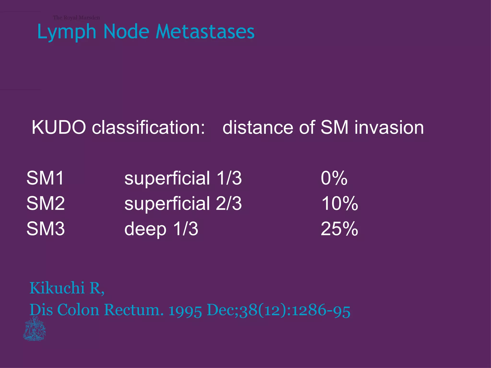
The Royal Marsden


MRI staging of
early tumours

Gina Brown
The Royal Marsden


Lymph Node Metastases


KUDO classification: distance of SM invasion

SM1                    superficial 1/3   0%
SM2                    superficial 2/3   10%
SM3                    deep 1/3          25%


Kikuchi R,
Dis Colon Rectum. 1995 Dec;38(12):1286-95
The Royal Marsden
EUS with high fequency probe detects sm1 tumours
 The Royal Marsden


suitable for local excision


                     1. Akasu T, Kondo H, Moriya Y, Sugihara K, Gotoda
                     T, Fujita S, et al. Endorectal ultrasonography and
                     treatment of early stage rectal cancer. World J Surg
                     2000;24(9):1061-8.
The Royal Marsden


Konishi, K., Y. Akita, et al. (2003). "Evaluation of endoscopic ultrasonography
in colorectal villous lesions." Int J Colorectal Dis

– Large (>/=20 mm wide, >/=5 mm high) or
  rectal villous lesions were more likely than
  nonvillous lesions to be misjudged with
  regard to the differentiation between M/SM-s
  and non-M/SM-s.
– It is difficult to determine the depth of
  invasion in villous lesions, especially large or
  rectal lesions, using only EUS.
– EUS-based evaluation alone cannot
  determine the appropriate treatment for
  colorectal villous lesions.
Early stage disease
The Royal Marsden
The Royal Marsden


UK TEMS trial (NCRI)

1. Biopsy proven adenocarcinoma
2. MRI defined stage I rectal cancer (less than
   or equal to pT2 N0)
3. Endorectal ultrasound defined rectal cancer
   less than or equal to uT2
4. Patients who have undergone submucosal
   excision for villous adenoma that on
   histopathological examination contains
   discrete invasion less than 3 cm diameter
The Royal Marsden


Staging of Early tumours MRI vs
Histopathology
The Royal Marsden
The Royal Marsden
The Royal Marsden
The Royal Marsden
The Royal Marsden
The Royal Marsden
The Royal Marsden
The Royal Marsden
The Royal Marsden
The Royal Marsden


MRI staging of early polyps disease


– MRI T1/T2
  – If local excision, should be deep
    submucosal excision and standard of care =
    Histological assessment, If deep T1 for
    primary completion TME surgery
  – May consider randomisation TME vs local
    excision + short course RT (NCRI TREC
    trial)
The Royal Marsden


Conclusions

– Early stage tumours can be usefully evaluated using
  MRI
  – Technique important
  – Options to consider especially for low lying early
    stage tumours
  – Follow up after less radical therapy: MRI is
    important

MRI Staging of Early Rectal Tumours- Gina Brown

  • 1.
    The Royal Marsden MRIstaging of early tumours Gina Brown
  • 2.
    The Royal Marsden LymphNode Metastases KUDO classification: distance of SM invasion SM1 superficial 1/3 0% SM2 superficial 2/3 10% SM3 deep 1/3 25% Kikuchi R, Dis Colon Rectum. 1995 Dec;38(12):1286-95
  • 3.
  • 4.
    EUS with highfequency probe detects sm1 tumours The Royal Marsden suitable for local excision 1. Akasu T, Kondo H, Moriya Y, Sugihara K, Gotoda T, Fujita S, et al. Endorectal ultrasonography and treatment of early stage rectal cancer. World J Surg 2000;24(9):1061-8.
  • 5.
    The Royal Marsden Konishi,K., Y. Akita, et al. (2003). "Evaluation of endoscopic ultrasonography in colorectal villous lesions." Int J Colorectal Dis – Large (>/=20 mm wide, >/=5 mm high) or rectal villous lesions were more likely than nonvillous lesions to be misjudged with regard to the differentiation between M/SM-s and non-M/SM-s. – It is difficult to determine the depth of invasion in villous lesions, especially large or rectal lesions, using only EUS. – EUS-based evaluation alone cannot determine the appropriate treatment for colorectal villous lesions.
  • 6.
  • 7.
    The Royal Marsden UKTEMS trial (NCRI) 1. Biopsy proven adenocarcinoma 2. MRI defined stage I rectal cancer (less than or equal to pT2 N0) 3. Endorectal ultrasound defined rectal cancer less than or equal to uT2 4. Patients who have undergone submucosal excision for villous adenoma that on histopathological examination contains discrete invasion less than 3 cm diameter
  • 8.
    The Royal Marsden Stagingof Early tumours MRI vs Histopathology
  • 9.
  • 10.
  • 11.
  • 12.
  • 13.
  • 14.
  • 15.
  • 16.
  • 17.
  • 18.
    The Royal Marsden MRIstaging of early polyps disease – MRI T1/T2 – If local excision, should be deep submucosal excision and standard of care = Histological assessment, If deep T1 for primary completion TME surgery – May consider randomisation TME vs local excision + short course RT (NCRI TREC trial)
  • 19.
    The Royal Marsden Conclusions –Early stage tumours can be usefully evaluated using MRI – Technique important – Options to consider especially for low lying early stage tumours – Follow up after less radical therapy: MRI is important