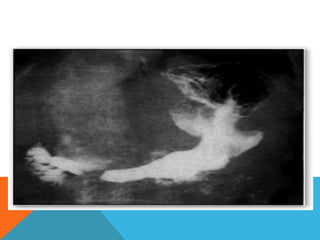
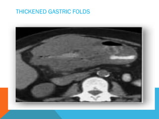
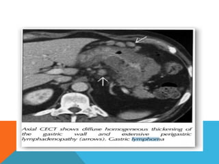
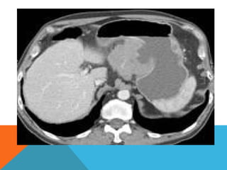
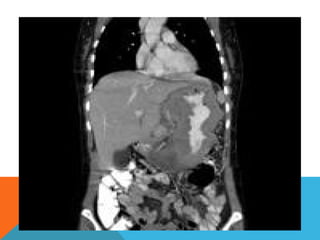
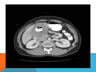
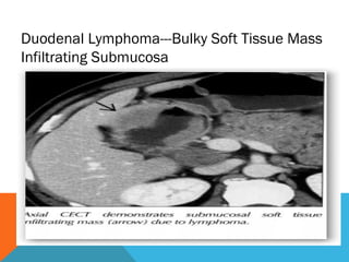
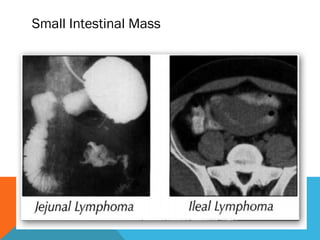
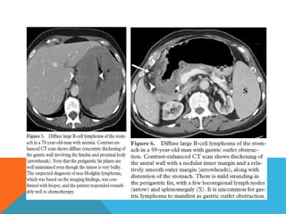
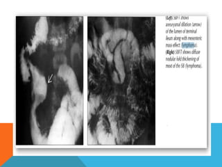
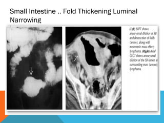
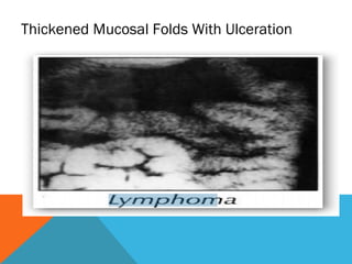
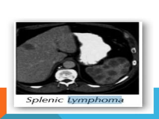
GIT LYM
PHOM
AS
Role Of Radiology in
Gastrointestinal Lymphoma
DEFINITION
 Lymphoma Is A Type Of Cancer That Arises
In the Immune Cells Called As Lymphocytes.
 Extra nodal Lymphoma Is Term Used To
Describe Lymphomas Occuring Outside The
Lymphatic Tissues.
INTRODUCTION
 Distinct group of lymphoma that primarily arises in lymphoid
tissue of the bowel rather than in lymph nodes
 G I lymphoma may either represent secondary involvement by
systemic disease or primary malignancy confined to the GI tract
 GIT-lymphoid Elements Seen In The Lamina Propria and Sub
Mucosa
 Secondary GI Involvement Is Common
 Primary Lymphomas Involve Only One Site
 
Five Criteria Put Forth By Dawson et al
To Diagnose Primary GI Lymphomas
 No Palpable Superficial Lymph Nodes
 Normal CXR
 WBC Count are Normal
 At Laparotomy Alimentary Tract Is Involved With Lymph node
Involvement if Any confined to the drainage area of gut
involved
 No Spleen Or Liver Involvement
 Advanced Stages Mimic Secondary GI Lymphoma
Gastrointestinal Lymphoma
Most Commonly Non-hodgkin Lymphomas That Are High Grade
Large Cell Or Immunoblastic Cell Types
 Burkitt More Common In Pediatric Patients
 Most Common Symptoms: Abdominal Pain, Weight Loss, fever,
Anemia
T-cell Lymphoma If In GIT Occurs In Small Bowel
(64%)esp.Duodenum+jejunum.
Oesophagus
 Esophagus: Least common site within GI tract
 Accounts for only about 1% of all cases
 Usually non-Hodgkin & less commonly Hodgkin
 Patients almost always have generalized lymphoma
 Primary esophageal lymphoma seen in AIDS cases
Imaging
 Usually contiguous spread from gastric
 cardia/fundus to distal esophagus
 Polypoid or ulcerated mass or infiltrating stricture
 Submucosal infiltration (less common)
 Enlarged, tortuous longitudinal folds mimicking varices
Diagnosis: Endoscopy with deep esophageal biopsy
MULTIPLE ESOPHAGEAL NODULES
 gastric lymphoma represents the most common
site of extra nodal lymphoma, accounting for 25%
of all such lymphomas, 50% of all gastrointestinal
lymphomas, but comprise only 1-5% of all gastric
malignancies .
 Typically primary gastric lymphoma occurs in
adults in the 6th decade of life, without gender
predilection. Secondary gastric lymphoma matches
the demographics of the underlying lymphoma.
Gastric lymphoma
Pathology
Three distinct types of gastric lymphoma are recognized :
low-grade MALT lymphoma : 60% of all primary gastric
lymphomas
primary sporadic lymphoma : vast majority are B-cell non-
Hodgkins lymphoma
secondary involvement of the stomach by systemic lymphoma
(usually high grade)
Mucosa-associated lymphoid tissue (MALT) lymphoma and
are strongly associated with Helicobacter pylori  (85 - 98% of
cases). These are low-grade lymphomas and may regress following
treatment of Helicobacter infection .
Radiographic features Gastric
lymphoma
Fluoroscopy : Barium meal
Appearances vary from normal, to grossly abnormal.
Possible appearances include:
 bull's eye appearance due to central ulceration
 filling defects
 thickened gastric rugae
 linitis plastica
Computed tomography
Typically gastric lymphomas demonstrate marked
thickening of the stomach wall (2-4cm) with extensive
lateral extension of the tumour (i.e. along the wall of
the stomach) representing submucosal spread.
 Submucosal spread may
 Encompasses the majority of the stomach giving a
linitis plastica appearance.
 Can extend across the pylorus into the duodenum
and superiorly into the oesophagus.
 uncommon for lymphoma to result in
gastric outlet obstruction
UPPER GI SERIES SHOWS MASSIVE NODULAR THICKENINGS
OF GASTRIC WALL/FOLDS; LYMPHOMA
PERSISTENT COLLECTION OF CONTRAST WITH
MUCOSAL ULCERATION
LINITIS PLASTICA—SMALL NON DISTENSIBLE
STOMACH
BULL’S EYE LESION
THICKENED GASTRIC FOLDS
GI lymphoma staging
 I: Tumor confined to bowel wall
 II: Limited nodal spread to local nodes
 III: Widespread nodal mets
 IV: Spread to bone marrow, solid viscera,
liver
Small bowl
 1/5 of all small bowel malignancies.
 Most common malignant small bowel tumor.

 Multiple sites involvmentin1/5.
Most common cause of INTUSSUSEPTION in
children>6yrs
 Location
 ileum(51%)
 jejunum(47%)
 duodenum(2%)
 Site
payer patches
 Radiographic findings;
 Nodular pattern
 Single mass
 Infiltrating pattern
 Exophytic
 Mesenteric/retroperitoneal Adenopathy
Duodenal Lymphoma---Bulky Soft Tissue Mass
Infiltrating Submucosa
Small Intestinal Mass
Colon
 Less Comonly Involved Than Stomach/Small Bowel
1.5% Of All Abdominal Lymphomas
 Location
Cecum Mostly
 Presentation
Single mass>diffuse infiltrating>polypoid
Paradoxical dilatation
Gross mural circumferential thickening
Massive regional+distantmesenteric+retroperitoneum
adenopathy.
APPENDICULAR LYMPHOMA--- SOFT TISSUE
MASS NEAR TIP OF CAECUM
Mesentry And Omentum
 Infiltration and thickenining of mesentry
 Omental caking
 Nodular or strand like soft tissue density
 Calcified foci
 Rounded mass
Peritoneal And Omental Mass
Small Intestine .. Fold Thickening Luminal
Narrowing
Thickened Mucosal Folds With Ulceration
THANK YOU

Git lymphomas

  • 1.
  • 2.
    Role Of Radiologyin Gastrointestinal Lymphoma
  • 3.
    DEFINITION  Lymphoma IsA Type Of Cancer That Arises In the Immune Cells Called As Lymphocytes.  Extra nodal Lymphoma Is Term Used To Describe Lymphomas Occuring Outside The Lymphatic Tissues.
  • 4.
    INTRODUCTION  Distinct groupof lymphoma that primarily arises in lymphoid tissue of the bowel rather than in lymph nodes  G I lymphoma may either represent secondary involvement by systemic disease or primary malignancy confined to the GI tract  GIT-lymphoid Elements Seen In The Lamina Propria and Sub Mucosa  Secondary GI Involvement Is Common  Primary Lymphomas Involve Only One Site  
  • 5.
    Five Criteria PutForth By Dawson et al To Diagnose Primary GI Lymphomas  No Palpable Superficial Lymph Nodes  Normal CXR  WBC Count are Normal  At Laparotomy Alimentary Tract Is Involved With Lymph node Involvement if Any confined to the drainage area of gut involved  No Spleen Or Liver Involvement  Advanced Stages Mimic Secondary GI Lymphoma
  • 6.
    Gastrointestinal Lymphoma Most CommonlyNon-hodgkin Lymphomas That Are High Grade Large Cell Or Immunoblastic Cell Types  Burkitt More Common In Pediatric Patients  Most Common Symptoms: Abdominal Pain, Weight Loss, fever, Anemia T-cell Lymphoma If In GIT Occurs In Small Bowel (64%)esp.Duodenum+jejunum.
  • 7.
    Oesophagus  Esophagus: Leastcommon site within GI tract  Accounts for only about 1% of all cases  Usually non-Hodgkin & less commonly Hodgkin  Patients almost always have generalized lymphoma  Primary esophageal lymphoma seen in AIDS cases
  • 8.
    Imaging  Usually contiguousspread from gastric  cardia/fundus to distal esophagus  Polypoid or ulcerated mass or infiltrating stricture  Submucosal infiltration (less common)  Enlarged, tortuous longitudinal folds mimicking varices Diagnosis: Endoscopy with deep esophageal biopsy
  • 9.
  • 10.
     gastric lymphomarepresents the most common site of extra nodal lymphoma, accounting for 25% of all such lymphomas, 50% of all gastrointestinal lymphomas, but comprise only 1-5% of all gastric malignancies .  Typically primary gastric lymphoma occurs in adults in the 6th decade of life, without gender predilection. Secondary gastric lymphoma matches the demographics of the underlying lymphoma. Gastric lymphoma
  • 11.
    Pathology Three distinct typesof gastric lymphoma are recognized : low-grade MALT lymphoma : 60% of all primary gastric lymphomas primary sporadic lymphoma : vast majority are B-cell non- Hodgkins lymphoma secondary involvement of the stomach by systemic lymphoma (usually high grade) Mucosa-associated lymphoid tissue (MALT) lymphoma and are strongly associated with Helicobacter pylori  (85 - 98% of cases). These are low-grade lymphomas and may regress following treatment of Helicobacter infection .
  • 12.
    Radiographic features Gastric lymphoma Fluoroscopy: Barium meal Appearances vary from normal, to grossly abnormal. Possible appearances include:  bull's eye appearance due to central ulceration  filling defects  thickened gastric rugae  linitis plastica
  • 13.
    Computed tomography Typically gastriclymphomas demonstrate marked thickening of the stomach wall (2-4cm) with extensive lateral extension of the tumour (i.e. along the wall of the stomach) representing submucosal spread.
  • 14.
     Submucosal spreadmay  Encompasses the majority of the stomach giving a linitis plastica appearance.  Can extend across the pylorus into the duodenum and superiorly into the oesophagus.  uncommon for lymphoma to result in gastric outlet obstruction
  • 15.
    UPPER GI SERIESSHOWS MASSIVE NODULAR THICKENINGS OF GASTRIC WALL/FOLDS; LYMPHOMA
  • 16.
    PERSISTENT COLLECTION OFCONTRAST WITH MUCOSAL ULCERATION
  • 18.
    LINITIS PLASTICA—SMALL NONDISTENSIBLE STOMACH
  • 20.
  • 21.
  • 26.
    GI lymphoma staging I: Tumor confined to bowel wall  II: Limited nodal spread to local nodes  III: Widespread nodal mets  IV: Spread to bone marrow, solid viscera, liver
  • 27.
    Small bowl  1/5of all small bowel malignancies.  Most common malignant small bowel tumor.   Multiple sites involvmentin1/5. Most common cause of INTUSSUSEPTION in children>6yrs
  • 28.
     Location  ileum(51%) jejunum(47%)  duodenum(2%)  Site payer patches
  • 29.
     Radiographic findings; Nodular pattern  Single mass  Infiltrating pattern  Exophytic  Mesenteric/retroperitoneal Adenopathy
  • 30.
    Duodenal Lymphoma---Bulky SoftTissue Mass Infiltrating Submucosa
  • 32.
  • 35.
    Colon  Less ComonlyInvolved Than Stomach/Small Bowel 1.5% Of All Abdominal Lymphomas  Location Cecum Mostly  Presentation Single mass>diffuse infiltrating>polypoid Paradoxical dilatation Gross mural circumferential thickening Massive regional+distantmesenteric+retroperitoneum adenopathy.
  • 36.
    APPENDICULAR LYMPHOMA--- SOFTTISSUE MASS NEAR TIP OF CAECUM
  • 37.
    Mesentry And Omentum Infiltration and thickenining of mesentry  Omental caking  Nodular or strand like soft tissue density  Calcified foci  Rounded mass
  • 39.
  • 40.
    Small Intestine ..Fold Thickening Luminal Narrowing
  • 41.
    Thickened Mucosal FoldsWith Ulceration
  • 43.