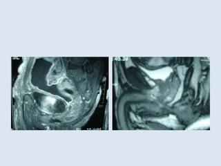
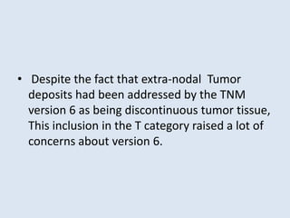
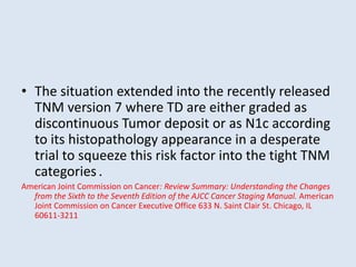
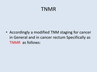
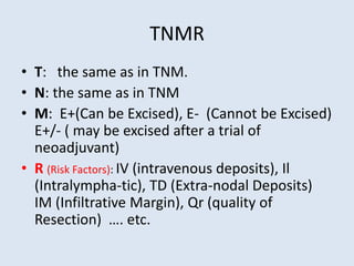
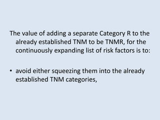
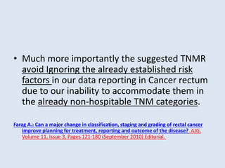
TNMR Vs.TNM and MRI Vs.Rigid
Endoscopy for CA rectum:
A New Tuxedo for a Growing young
Athlete.
Prof. Dr. Ahmed Farag.
Dept. Of G. Surgery – Cairo University
Hypothesis
Can a Major change In classification, Staging and
Grading of Rectal Cancer improve planning
for treatment , Reporting and outcome
of the disease?
• The value of all grading and staging systems of
cancers is to inform on the outcome be it with
or without treatment.
• Classification, TNM staging and Histologic
Grading of the cancer rectum had undergone
minimal changes during the last 20 years
despite the major impact of them on
planning, reporting and outcome of the
disease.
Classification of cancer rectum.
• The classification of the rectal cancer into the
upper, middle and lower thirds based on the
distance from the anal verge using Rigid
sigmoidospy is recommended by different
international Guidelines (AJCC, NCCN and
others)
Classification of cancer rectum.
• Rigid Sigmoidoscopy is not ideal for this task
due to the discomfort with the use of rigid
endoscopy, the variations in measurements
due to inter-observer errors, intra-observer
errors as well as the different stature and body
mass index of the patients.
Rigid sigmoidoscopy makes measures as 0-5cm, 6-10 cm and 11-
15 cm. This mean different locations in different patients .
Preoperative MRI Vs. Rigid
Sigmoidoscopy
• On the other hand, the use of MRI for
preoperative staging of rectal cancer and the
circumferential resection margin had been
reported as compared to postoperative
Histopathology and recommended by AJCC,
NCCN and others as a routine preoperative
investigation of Ca rectum.
• Can we use MRI as an alternative tool to
classify rectal cancer?
• In other words can we have another added
value from the recommended preoperative
MRI?
• In the era of routine preoperative staging
using MRI where the Peritoneal reflection can
be accurately located in each individual
patient irrespective to his stature and BMI
using T2 weighted image, Ca rectum can be
reclassified preoperatively as:
• CA of Intra-peritoneal rectum where the
lower edge of the tumor ends 2 cm above the
peritoneal reflection of the Douglas pouch
irrespective to the distance from the anal
verge. Those patients should not receive
neoadjuvant treatment except may be in T4
cases.
• CA of extra-peritoneal rectum where the
lower edge of the tumor is < 2cm from the
peritoneal reflection on preoperative MRI
where neoadjuvant treatment can be given.
Those patients. Those cases can be
subdivided into :
• Posterior Extra-peritoneal: where neoadjuvant a
can be given to T3 lesions due to the mesorectal
buffer.
• Anterior Extra-peritoneal: where the
neoadjuvant can be given to T2 lesions due to the
lack of mesorectal buffer and marked proximity
to the dennonvier fascia ( usually < 1cm) which by
definition is a close circumferential margin.
TNM 6th and 7th editions
• The TNM classification in its present status
does not address the other risk factors Proved
by high quality evidence based research in the
field such as:
TNM 6th and 7th editions
• Intravascular and Intra-lymphatic deposits,
where lympho-vascular invasion-positive tumors
metastasized to systemic lymph nodes more
often (P < .001).
• These tumors also recurred at systemic lymph
nodes after curative intent surgery more often (P
= .007
Lim, Seok-Byung; Yu, Chang Sik; Jang, Se Jin; Kim, Tae Won; Kim, Jong Hoon; Kim, Jin Cheon: Prognostic Significance of
Lymphovascular Invasion in Sporadic Colorectal Cancer. Diseases of the Colon & Rectum. 53(4):377-384, April
2010. doi: 10.1007/DCR.0b013e3181cf8ae5
Other risk factors
• Intraoperative blood transfusion.
• Intra-operative tumor perforation.
• Preoperative High CEA.
• Quality of TME.
• Number of retrieved (examined) LNs.
• Type of advancing margin of the tumor (infiltrative vs.
Pushing)
• Tumor Ploidy.
• The decreased monocyte chemoattractant protein-1 ratio.
• A Ki-67 labeling index of 5 percent.
• A positive cytoplasmic p53 expression …..etc.
• Despite the fact that extra-nodal Tumor
deposits had been addressed by the TNM
version 6 as being discontinuous tumor tissue,
This inclusion in the T category raised a lot of
concerns about version 6.
• The situation extended into the recently released
TNM version 7 where TD are either graded as
discontinuous Tumor deposit or as N1c according
to its histopathology appearance in a desperate
trial to squeeze this risk factor into the tight TNM
categories.
American Joint Commission on Cancer: Review Summary: Understanding the Changes
from the Sixth to the Seventh Edition of the AJCC Cancer Staging Manual. American
Joint Commission on Cancer Executive Office 633 N. Saint Clair St. Chicago, IL
60611-3211
TNMR
• Accordingly a modified TNM staging for cancer
in General and in cancer rectum Specifically as
TNMR as follows:
TNMR
• T: the same as in TNM.
• N: the same as in TNM
• M: E+(Can be Excised), E- (Cannot be Excised)
E+/- ( may be excised after a trial of
neoadjuvant)
• R (Risk Factors): IV (intravenous deposits), Il
(Intralympha-tic), TD (Extra-nodal Deposits)
IM (Infiltrative Margin), Qr (quality of
Resection) …. etc.
• Similarly Grading of cancer with special
reference to Cancer rectum should include the
R Sign beside the G, in order to indicate the
same risk factors in the suggested
modification in TNM above as well as any
other added risk factor in the Future.
• Alternatively: Grading can be added to the
new R category.
The value of adding a separate Category R to the
already established TNM to be TNMR, for the
continuously expanding list of risk factors is to:
• avoid either squeezing them into the already
established TNM categories,
• Warn the treating team about the need to
have a more aggressive treatment strategies
in those patients with early TNM staging with
one or more risk factors.
• Much more importantly the suggested TNMR
avoid Ignoring the already established risk
factors in our data reporting in Cancer rectum
due to our inability to accommodate them in
the already non-hospitable TNM categories.
Farag A.: Can a major change in classification, staging and grading of rectal cancer
improve planning for treatment, reporting and outcome of the disease? AJG.
Volume 11, Issue 3, Pages 121-180 (September 2010) Editorial.
www.drfarag.org
Thank You
www.drfarag.org

TNMR Vs TNM and new Classification of Rectal Cancer

  • 1.
    TNMR Vs.TNM andMRI Vs.Rigid Endoscopy for CA rectum: A New Tuxedo for a Growing young Athlete. Prof. Dr. Ahmed Farag. Dept. Of G. Surgery – Cairo University
  • 2.
    Hypothesis Can a Majorchange In classification, Staging and Grading of Rectal Cancer improve planning for treatment , Reporting and outcome of the disease?
  • 3.
    • The valueof all grading and staging systems of cancers is to inform on the outcome be it with or without treatment.
  • 4.
    • Classification, TNMstaging and Histologic Grading of the cancer rectum had undergone minimal changes during the last 20 years despite the major impact of them on planning, reporting and outcome of the disease.
  • 5.
    Classification of cancerrectum. • The classification of the rectal cancer into the upper, middle and lower thirds based on the distance from the anal verge using Rigid sigmoidospy is recommended by different international Guidelines (AJCC, NCCN and others)
  • 6.
    Classification of cancerrectum. • Rigid Sigmoidoscopy is not ideal for this task due to the discomfort with the use of rigid endoscopy, the variations in measurements due to inter-observer errors, intra-observer errors as well as the different stature and body mass index of the patients.
  • 7.
    Rigid sigmoidoscopy makesmeasures as 0-5cm, 6-10 cm and 11- 15 cm. This mean different locations in different patients .
  • 8.
    Preoperative MRI Vs.Rigid Sigmoidoscopy • On the other hand, the use of MRI for preoperative staging of rectal cancer and the circumferential resection margin had been reported as compared to postoperative Histopathology and recommended by AJCC, NCCN and others as a routine preoperative investigation of Ca rectum.
  • 9.
    • Can weuse MRI as an alternative tool to classify rectal cancer? • In other words can we have another added value from the recommended preoperative MRI?
  • 11.
    • In theera of routine preoperative staging using MRI where the Peritoneal reflection can be accurately located in each individual patient irrespective to his stature and BMI using T2 weighted image, Ca rectum can be reclassified preoperatively as:
  • 12.
    • CA ofIntra-peritoneal rectum where the lower edge of the tumor ends 2 cm above the peritoneal reflection of the Douglas pouch irrespective to the distance from the anal verge. Those patients should not receive neoadjuvant treatment except may be in T4 cases.
  • 13.
    • CA ofextra-peritoneal rectum where the lower edge of the tumor is < 2cm from the peritoneal reflection on preoperative MRI where neoadjuvant treatment can be given. Those patients. Those cases can be subdivided into :
  • 14.
    • Posterior Extra-peritoneal:where neoadjuvant a can be given to T3 lesions due to the mesorectal buffer. • Anterior Extra-peritoneal: where the neoadjuvant can be given to T2 lesions due to the lack of mesorectal buffer and marked proximity to the dennonvier fascia ( usually < 1cm) which by definition is a close circumferential margin.
  • 15.
    TNM 6th and7th editions • The TNM classification in its present status does not address the other risk factors Proved by high quality evidence based research in the field such as:
  • 16.
    TNM 6th and7th editions • Intravascular and Intra-lymphatic deposits, where lympho-vascular invasion-positive tumors metastasized to systemic lymph nodes more often (P < .001). • These tumors also recurred at systemic lymph nodes after curative intent surgery more often (P = .007 Lim, Seok-Byung; Yu, Chang Sik; Jang, Se Jin; Kim, Tae Won; Kim, Jong Hoon; Kim, Jin Cheon: Prognostic Significance of Lymphovascular Invasion in Sporadic Colorectal Cancer. Diseases of the Colon & Rectum. 53(4):377-384, April 2010. doi: 10.1007/DCR.0b013e3181cf8ae5
  • 17.
    Other risk factors •Intraoperative blood transfusion. • Intra-operative tumor perforation. • Preoperative High CEA. • Quality of TME. • Number of retrieved (examined) LNs. • Type of advancing margin of the tumor (infiltrative vs. Pushing) • Tumor Ploidy. • The decreased monocyte chemoattractant protein-1 ratio. • A Ki-67 labeling index of 5 percent. • A positive cytoplasmic p53 expression …..etc.
  • 18.
    • Despite thefact that extra-nodal Tumor deposits had been addressed by the TNM version 6 as being discontinuous tumor tissue, This inclusion in the T category raised a lot of concerns about version 6.
  • 19.
    • The situationextended into the recently released TNM version 7 where TD are either graded as discontinuous Tumor deposit or as N1c according to its histopathology appearance in a desperate trial to squeeze this risk factor into the tight TNM categories. American Joint Commission on Cancer: Review Summary: Understanding the Changes from the Sixth to the Seventh Edition of the AJCC Cancer Staging Manual. American Joint Commission on Cancer Executive Office 633 N. Saint Clair St. Chicago, IL 60611-3211
  • 20.
    TNMR • Accordingly amodified TNM staging for cancer in General and in cancer rectum Specifically as TNMR as follows:
  • 21.
    TNMR • T: thesame as in TNM. • N: the same as in TNM • M: E+(Can be Excised), E- (Cannot be Excised) E+/- ( may be excised after a trial of neoadjuvant) • R (Risk Factors): IV (intravenous deposits), Il (Intralympha-tic), TD (Extra-nodal Deposits) IM (Infiltrative Margin), Qr (quality of Resection) …. etc.
  • 22.
    • Similarly Gradingof cancer with special reference to Cancer rectum should include the R Sign beside the G, in order to indicate the same risk factors in the suggested modification in TNM above as well as any other added risk factor in the Future. • Alternatively: Grading can be added to the new R category.
  • 23.
    The value ofadding a separate Category R to the already established TNM to be TNMR, for the continuously expanding list of risk factors is to: • avoid either squeezing them into the already established TNM categories,
  • 24.
    • Warn thetreating team about the need to have a more aggressive treatment strategies in those patients with early TNM staging with one or more risk factors.
  • 25.
    • Much moreimportantly the suggested TNMR avoid Ignoring the already established risk factors in our data reporting in Cancer rectum due to our inability to accommodate them in the already non-hospitable TNM categories. Farag A.: Can a major change in classification, staging and grading of rectal cancer improve planning for treatment, reporting and outcome of the disease? AJG. Volume 11, Issue 3, Pages 121-180 (September 2010) Editorial.
  • 26.
  • 27.