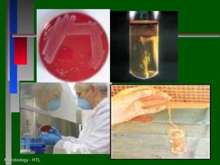
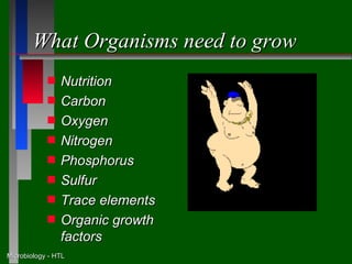
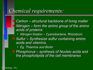
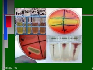
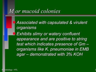
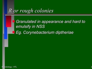
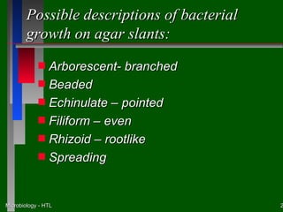
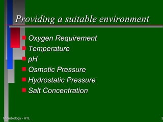
This document discusses the nutritional and environmental requirements for bacterial growth, including the need for carbon, nitrogen, phosphorus, and other elements. It describes different types of culture media like liquid broth, semi-solid agar, and solid agar plates and how they are used. The document also covers bacterial colony morphology, describing smooth, mucoid, and rough colony types. It discusses factors that provide a suitable growth environment for bacteria, such as oxygen levels, temperature, pH, and osmotic pressure requirements.
Oral exfoliative cytology is a technique that examines cells collected from the oral mucosa. It can help detect undiagnosed diseases through a simple procedure and confirm suspected diseases without trauma. The document discusses the definition, role, indications, advantages and preparation methods of oral cytology. It describes how to make smears and various staining and fixation techniques used. Cytological classification systems and what normal and abnormal oral cells look like are presented. The uses of oral cytology in detecting oral cancer and newer diagnostic methods like cytomorphometry are also summarized.
This document discusses various artifacts that can occur during biopsy and tissue processing. It defines an artifact as a defect or distortion resulting from how the tissue was handled from biopsy to fixation. Artifacts can occur during tissue manipulation, transport, fixation, processing, embedding and sectioning. Specific artifacts discussed include squeeze artifacts from improper forceps use, heat artifacts from electrosurgery, autolysis artifacts from delayed fixation, curling artifacts from thin specimens, and orientation artifacts from unlabeled specimens. Proper techniques such as gentle tissue handling, rapid fixation in adequate formalin volume, and specimen orientation labeling can prevent many artifacts.
This document discusses biosafety and biosecurity when working with infectious agents in a laboratory setting. It defines biosafety as safety precautions that reduce risk of exposure and contamination. There are four biosafety levels depending on the agent, with level 4 being the most restrictive for dangerous pathogens with no treatment. Key equipment for containment includes biosafety cabinets, which use HEPA filters to circulate air and prevent exposure, with various types for different risk levels. Proper use and certification of this equipment is important for protecting laboratory workers and the public.
The document discusses haemopoiesis (blood cell formation) and the histology of bone marrow. It describes how in fetal life, blood cells are formed in the yolk sac, liver, and later the bone marrow and liver. In newborns, children and adults, red bone marrow is found in all bones up to age 20, and later in flat bones and ends of long bones. Bone marrow contains hematopoietic stem cells that differentiate into the various blood cell lineages through progenitor and precursor cells in the bone marrow microenvironment.
Candida albicans is a yeast that can cause infections in humans. It exists in different morphological forms, including yeast, hyphae, and pseudohyphae. Its cell wall contains beta-glucan, chitin, lipids, and mannans. Morphological shifting between forms is associated with invasion of tissues and regulated by environmental conditions and adherence factors. C. albicans binds to epithelial cells using adhesins like Hwp1 and mannoproteins, and secretes proteinases and phospholipases to aid invasion. It evades the immune system using surface receptors that bind complement components and immune regulators. Infections can be diagnosed using KOH preparations of samples and are usually treated with ant
Antimicrobial susceptibility test and assay bls 209Bruno Mmassy
This document summarizes methods for antimicrobial susceptibility testing, including dilution and diffusion techniques. Dilution methods like broth microdilution determine minimum inhibitory concentrations (MICs) and minimum bactericidal concentrations (MBCs). Disc diffusion tests qualitatively assess inhibition zone diameters compared to standards. Factors like inoculum density, temperature, and medium composition can affect zone sizes. Quality control using reference strains validates test results.
This document describes the steps involved in tissue processing from fixation to embedding in wax. It discusses obtaining fresh specimens, fixation in formalin, dehydration through an alcohol series, clearing in xylene, infiltration and embedding in paraffin wax. Sections are then cut on a microtome, mounted on slides and stained, usually with hematoxylin and eosin, to visualize tissue structures microscopically. Proper processing is important to preserve tissue morphology and produce high quality stained sections for diagnostic examination.
This document discusses the processing and examination of cerebrospinal fluid (CSF) samples in a medical laboratory to diagnose bacterial or fungal meningitis. It describes the procedures for receiving, labeling, logging, and rejecting CSF specimens. The document outlines microscopic examination and culture techniques for the first day of processing to look for pathogens, including making Gram stains of purulent CSF and culturing all CSF samples. It also provides information on possible bacterial, fungal, parasitic and viral causes of meningitis.
This document summarizes key components of hemostasis including primary and secondary hemostasis. It describes platelet adhesion, activation, aggregation and secretion. Tests for evaluating hemostasis are outlined including bleeding time, platelet function analyzer, and assays for factors, fibrinogen, D-dimer and FDP. Causes and interpretation of abnormal results are provided for tests such as PT, APTT, TT and specific assays of platelet function and coagulation factors.
Hemostasis and thrombosis involve the regulation of blood clotting. Normal hemostasis maintains blood fluidity but allows clotting at sites of injury. Thrombosis is pathological clotting in uninjured or minimally injured vessels. It involves platelet adhesion and activation, coagulation cascade activation, and fibrin clot formation. Counter-regulatory mechanisms normally limit clotting to the injury site. Abnormalities in blood components, vessel walls, or flow can cause hypercoagulability and thrombosis.
Hemostasis is the arrest of bleeding, whether it be by normal vasoconstriction (the vessel walls closing temporarily), by an abnormal obstruction (such as a plaque) or by coagulation or surgical means (such as ligation)
The document describes the key steps in the histopathology laboratory workflow. Specimens are received and labeled with patient information. In grossing, samples are selected and inspected. Tissue processing involves dehydration, clearing, impregnation and embedding to prepare samples for microtomy, where thin sections are cut. Sections are stained, usually with H&E, and examined microscopically. Preparation methods include fresh cells/tissues, smears and sectional techniques, with sectional being the standard method used in histopathology.
This document summarizes several systemic mycoses, including blastomycosis, histoplasmosis, coccidioidomycosis, and paracoccidioidomycosis. It describes the etiologic agents, life cycles, clinical presentations, and diagnostic approaches for each. Many of these fungi exhibit dimorphism, growing as mold-like mycelia at lower temperatures and yeast-like forms at human body temperature when infecting humans. Laboratory diagnosis may involve microscopy, culture, antigen detection and antibody tests. Treatment depends on the specific infection but may include antifungal drugs such as amphotericin B and itraconazole.
This document discusses several special hematology stains including Periodic acid-Schiff (PAS), Perl's Prussian Blue reaction, leukocyte alkaline phosphatase (LAP), and myeloperoxidase. PAS stains carbohydrates such as glycogen and is used to identify abnormal erythroblasts and dysplastic megakaryocytes. Perl's Prussian Blue stains iron and demonstrates ring sideroblasts and Pappenheimer bodies. LAP stains neutrophil alkaline phosphatase and helps differentiate chronic myelogenous leukemia. Myeloperoxidase stains the enzyme in neutrophils, monocytes and eosinophils and is used to identify myeloid or monocytic leukemias.
This document discusses laboratory diagnosis of viral infections. It begins by explaining why viral diagnosis is important and lists some common diagnostic methods like microscopy, antigen detection, antibody detection, and nucleic acid detection. It then goes into more detail on specific diagnostic techniques. Microscopy methods discussed include light, electron, and fluorescence microscopy. The document outlines best practices for proper sample collection and storage. It also provides details on viral transport medium and various viral cultivation and isolation methods like animal inoculation, egg inoculation, and tissue culture.
The document discusses various types of artefacts that can occur during tissue processing and slide preparation in histopathology. Artefacts are non-natural structures that are introduced and can compromise accurate diagnosis. They can occur during biopsy collection, fixation, processing, embedding, sectioning and staining. Examples include crush artefacts, thermal injury, surgical procedures, delayed fixation, contamination and diffusion of unfixed material. Care must be taken at each step and troubleshooting methods are provided to prevent or reduce artefacts.
Haemocytometry is a technique used to count blood cells and cells in other body fluids. It involves diluting a blood or fluid sample and counting the cells in a special counting chamber under a microscope. The total white blood cell count, platelet count, and absolute eosinophil count can be determined through haemocytometry. Precise dilution and counting technique is required to obtain accurate results, which provide information about a patient's health status and response to treatment.
This document provides an overview of hematoxylin and eosin staining. It discusses the theory behind staining, including how dyes interact with tissues through various bonding mechanisms. It also describes factors that influence staining results, such as rates of dye uptake and loss, binding site affinities, and tissue modification during fixation. The document highlights how hematoxylin and eosin work as the most commonly used routine stain in histopathology.
1. Hemophilus is a genus of Gram-negative bacteria that requires specific growth factors found in blood. There are several species, with H. influenzae being the most clinically significant as it can cause invasive infections like meningitis and pneumonia.
2. H. influenzae is a small, pleomorphic coccobacillus that requires both the X factor (hemin) and V factor (NAD). It has a polysaccharide capsule that is the basis for its 6 serotypes, with type b being the most virulent.
3. H. influenzae can cause invasive infections like meningitis and pneumonia in children under 5 years old. Non-invasive strains typically cause conditions like sinus
The document discusses the Ziehl-Neelsen staining technique, which is used to identify acid-fast bacteria such as Mycobacterium, Actinomycetes, and Nocardia. The staining method uses carbol fuchsin as the primary stain, along with heat to aid penetration. Acid-alcohol is used as a decolorizer, followed by methylene blue as the counterstain. Mycobacterium and other acid-fast bacteria will retain the pink carbol fuchsin stain due to their thick, waxy cell walls containing mycolic acid. This allows them to be differentiated from other non-acid fast bacteria that will take on the blue methylene blue counterstain
The document discusses various techniques for preparing cell blocks (CBs) from cytology specimens such as effusions, fine needle aspirations, and scrapings. Traditional methods involved using a celloidin or agar embedding medium but newer automated techniques using filters and cassettes provide higher cellularity. CBs allow morphological examination and ancillary studies to improve diagnostic accuracy compared to smears alone. While useful, CBs require more material and time than smears and may lack sufficient cells for all tests.
Defined and complex media are used to grow microorganisms in culture. Defined media use precise inorganic or organic chemicals, while complex media use digests of microbial, animal, or plant products. Streak culture is a common method for isolating pure bacterial cultures by transferring bacteria across a culture plate using a loop or stick, allowing distinct colonies to grow. Other methods include pour plates to estimate bacterial counts, sweep plates to transfer dust particles, and liquid cultures for high yields. Micromanipulators can also be used to isolate single bacterial cells under a microscope.
This document discusses various histopathological patterns seen in tissue samples. It defines terms like trabeculae, syncytium, alveolus, herringbone, storiform, fascicle, cribriform, tubule, papillae, Indian file, hobnail, rossette, microcystic, and follicle. Examples of diseases that exhibit each pattern are provided. The objectives are to help differentiate patterns in histopathology. Key patterns include trabecular, syncytial, alveolar, herringbone, storiform, fascicular, cribriform, tubular, papillary, micropapillary, Indian file, hobnail, and rossette
Tissue processing involves fixing, dehydrating, clearing, and infiltrating tissue samples with paraffin wax to embed them for sectioning. The key steps are fixation to prevent degradation, dehydration using graded alcohols, clearing with solvents like xylene to remove alcohol, infiltration using paraffin wax, embedding wax blocks for sectioning, sectioning on a microtome, and staining for examination. Automated tissue processors can complete many processing steps unattended for increased efficiency and throughput in pathology laboratories. Proper handling and processing is essential to obtain an accurate histological diagnosis from tissue specimens.
01 Presentation I VS (8-55MB)- (3-28-08).ppsvshidham
Part I of Four part symposium: “Diagnostic Cytopathology of Serous Effusion” on April 19, 2007 at Neenah, WI, USA
(2008 Wisconsin Society of Cytology, 40th Anniversary)
This document discusses histoplasmosis, an infection caused by the fungus Histoplasma capsulatum. The fungus lives in soil containing bird or bat droppings. There are two main types - pulmonary histoplasmosis, which occurs when the fungus is inhaled, and disseminated histoplasmosis, which spreads from the lungs. Symptoms can include fever, cough, fatigue and chest pain. Diagnosis involves examining samples under a microscope for the yeast cells or culturing samples. Treatment uses antifungal medications such as itraconazole or amphotericin B, depending on severity.
This document provides an overview of general microbiology as a 3-unit subject for dental students. It discusses the history of microbiology, including key figures such as Fracastorius, Leeuwenhoek, Pasteur, Lister, and Koch. It also summarizes the development of microscopy, the spontaneous generation controversy, and proofs that microbes cause disease. Finally, it briefly outlines the divisions, applications, and career opportunities within the field of microbiology.
This document discusses various ways that bacteria can be classified, including phenotypic and genotypic classification. Phenotypically, bacteria are classified based on their morphology, anatomy, staining characteristics, culture growth, nutritional requirements, and environmental tolerances. Morphologically, bacteria are classified as cocci, bacilli, actinomycetes, spirochetes, mycoplasmas, or rickettsiae/chlamydiae depending on their shape and arrangement. Anatomical features used in classification include whether they have capsules, flagella, spores, and their gram stain reaction.
This document outlines various ways that bacteria can be classified, including by shape, staining properties, temperature and oxygen requirements, pH tolerance, osmotic pressure tolerance, and cellular structure. Some of the key classification groups mentioned are cocci, bacilli, gram positive and gram negative bacteria, psychrophiles and thermophiles based on temperature, and obligate aerobes versus anaerobes based on oxygen needs. The document provides examples of bacteria that fall into each of the different classification groups.
The document provides an overview of microbiology, focusing on acellular and procaryotic microbes. It discusses the structure and classification of viruses. Key points include that viruses consist of genetic material surrounded by a protein coat, they require a host cell to replicate, and are classified based on attributes like nucleic acid type and host. It also covers bacteriophages, latent virus infections, antiviral agents, and important pathogenic viruses. The document then discusses bacteria, describing their morphology, staining properties, motility, growth characteristics, and use in classification. Important pathogenic bacteria are highlighted. Finally, it notes unique prokaryotes like rickettsias, chlamydias, and mycoplasmas that live intracellular
Medical microbiology deals with microorganisms that cause disease in humans. It aims to characterize, prevent, and control infectious agents. Key techniques include preparing culture media, isolating microbes from clinical samples via serial dilution or plating methods, and characterizing microbes based on morphology, microscopy, biochemical tests, and antibiotic susceptibility. Common tests include Gram staining, IMViC, citrate utilization, catalase production, and disk diffusion for antibiotic testing. These techniques help identify unknown microbes and determine appropriate treatment.
The document describes the objectives, size, morphology, structure, and growth requirements of typical bacterial cells. It discusses the sizes and shapes of different bacteria, as well as their structural components including the cell wall, cell membrane, capsule, cytoplasm, inclusions and appendages. It provides details on specific cell wall structures in gram-positive and gram-negative bacteria. The document also discusses bacterial growth cycles, temperature requirements, and special cell types including endospores, spheroplasts, and L-forms.
The document discusses various methods for classifying and identifying bacteria, including:
1. Gram staining to differentiate bacteria based on cell wall structure.
2. Colony morphology, growth characteristics, and specialized tests to further classify bacteria.
3. Molecular diagnosis techniques like PCR, DNA hybridization, and microarrays that can identify bacteria faster and more definitively than conventional culture-based methods.
Bergey’s Manual is a key reference work for classifying and identifying prokaryotes. It consists of two parts: Bergey’s Manual of Determinative Bacteriology, which provides identification schemes based on morphology and biochemical tests; and Bergey’s Manual of Systematic Bacteriology, which provides phylogenetic classification based on rRNA sequencing. The first edition in 1923 established bacterial classification for identification. Current editions are based on phylogenetic studies and distribute pathogenic bacteria throughout volumes organized by phylogenetic relationships rather than clinical importance.
This document discusses various methods for identifying bacteria, including traditional phenotypic methods, immunochemical methods, and genotypic molecular methods. Phenotypic methods involve examining bacterial morphology, staining characteristics, growth requirements, and biochemical reactions. Immunochemical methods like immunofluorescence and ELISA use antigen-antibody reactions for identification. Molecular identification methods analyze bacterial DNA sequences. Correct specimen collection, handling, and transport are essential for accurate identification. Identification determines clinical significance and appropriate treatment.
Bacteria have various nutritional requirements including water, carbon and nitrogen sources, inorganic salts, vitamins, and certain gaseous and temperature conditions to grow. Different types of culture media can be used for bacterial cultivation based on ingredients, agar concentration, and special properties. These include basic, complex, synthetic, enriched, selective, differential, and transport media formulated for specific bacterial isolation and identification purposes.
The document provides an overview of microbiology and microorganisms. It discusses that microorganisms are too small to be seen with the naked eye and includes bacteria, fungi, protozoa, algae, and viruses. It also outlines several fields of microbiology like bacteriology, mycology, and virology. The document discusses the roles microorganisms play in various industries like food production and describes how microscopy advanced the study of microbes.
The document discusses the Enterobacteriaceae family of gram-negative bacteria. It notes that they are a diverse group that includes many pathogenic genera like Escherichia, Shigella, Salmonella, and Klebsiella. The document covers their characteristics, morphology, identification, growth patterns, antigenic structures, and taxonomy. It provides details on rapid identification of common genera and describes biochemical tests used to differentiate between members of the family.
Coccobacilli is a transitional shape between coccus and bacillus. It has the shape of short rods or ovals.
Bacillus do not form tetrads or clusters because their shape does not allow them to arrange in those patterns like cocci can. As individual rods, bacillus cannot cluster in the same symmetrical ways as cocci.
Module 5b biochemical activities for the labHuang Yu-Wen
This document summarizes various biochemical tests used to identify bacteria, including:
- Fermentation tests using Durham tubes to detect gas and acid production from carbohydrates.
- The IMVIC series of tests (Indole, Methyl Red, Voges-Proskauer, Citrate) which identify metabolic pathways and substrate utilization through color changes.
- Other common tests like catalase, coagulase, urease, and oxidase which detect enzymatic activity through production of gas or color changes.
Students are assigned to prepare SIM, MRVP, and SCA media, inoculate bacterial samples, interpret test results using described reagents and expected color changes, and submit a write-
The document discusses the bacterial cell wall and other cellular structures. It describes the differences between gram-positive and gram-negative cell walls, including their peptidoglycan structure and additional layers like the outer membrane in gram-negatives. Specialized structures are also summarized, such as flagella, pili, capsules, plasmids, ribosomes, the nucleoid, and inclusion bodies. Endospores are discussed as a dormant protective structure formed by some bacteria.
This document discusses microbial control through various sterilization and disinfection methods including physical agents like heat, radiation, and filtration as well as chemical agents like phenols, alcohols, and antibiotics. It covers topics such as bacteriostatic vs bactericidal effects, modes of microbial transmission, and biological vectors of infection.
Legionella pneumophila is a fastidious, aerobic bacterium that stains poorly and requires special staining techniques. It can cause pneumonia by growing in warm, moist environments like air conditioning systems and shower heads. It is susceptible to erythromycin and other drugs. Several other Bartonella species can cause diseases in humans transmitted via insect bites like trench fever. Bacterial vaginosis is associated with Gardnerella vaginalis, which is detected in wet smears as it covers vaginal cells. It has a fishy odor and high pH. Metronidazole is used to treat it.
Unknown #67 was identified as Salmonella typhimurium based on biochemical test results. A gram stain showed pink, rod-shaped cells. TSI slant results were alkaline in the slant and acidic in the butt with gas and H2S production. Urea broth was negative for urease. SIM was positive for H2S and motility but negative for indole. MacConkey agar showed no lactose fermentation. Cross-referencing these results identified the unknown as S. typhimurium.
This document provides information on viruses including their structure, classification, life cycles, and examples of common viral diseases. It defines key terms like capsids, genomes, and envelopes. It describes the four main types of viral structures: helical, icosahedral, enveloped, and complex. It covers taxonomy and the bases for viral classification such as host range, size, and antigenic properties. Examples are given of major viral families that infect animals like Herpesviruses, Hepadnaviruses, Retroviruses, Arenaviruses, Flaviviruses, and more.
This document summarizes key concepts in immunology, including the three lines of defense against pathogens, organs of the immune system, types of lymphocytes and immunity, immunologic disorders, and common immunologic problems. It covers topics such as antibody classes, lymphokines, hypersensitivity reactions, organ transplantation rejection, and primary and secondary immunodeficiencies.
Cultivation, growth and nutrition of bacteriaAshfaq Ahmad
This document discusses the cultivation, growth and nutrition of bacteria. It covers various topics such as:
- The purpose and methods of culturing bacteria, including isolation, identification and maintaining stock cultures.
- The components and uses of culture media, including providing nutrients for bacterial growth and selecting for certain bacteria.
- Obtaining pure cultures through aseptic techniques and separating individual bacterial cells on solid media to form colonies.
- Classification of culture media based on consistency (liquid, solid, semi-solid), nutritional components (simple, complex, synthetic) and functional use (enriched, selective, differential, transport, indicator, anaerobic).
Cultivation of bacteria and culture methodsAshfaq Ahmad
Cultivation of bacteria allows for the isolation, growth, and study of microorganisms. There are various culture methods and media that support the growth of bacteria. Liquid broths and solid agar plates can be used with different nutrient formulations to selectively grow specific bacteria. Streaking, lawning, stabbing, and pour plating are common culture techniques used to isolate pure colonies for analysis. Specialized enriched, selective, differential, and transport media help optimize bacterial growth and identification.
1. The document discusses different types of culture media used to grow microorganisms in the laboratory, including solid media, semisolid media, and liquid broths.
2. Solid media like agar plates are used for isolating pure cultures, observing colony morphology, and storing cultures long-term. Semisolid media allow studying bacterial motility.
3. Broths are used for growing cultures for assays and biochemical tests since bacterial growth is visible through turbidity.
4. Agar is commonly used to solidify media due to its properties of being bacteriologically inert and allowing surface growth of colonies.
This document discusses microbial culture media. It defines culture, medium, and introduces various types of culture media including solid, liquid, semi-solid media. It describes different media based on constituents like simple, complex, synthetic media. It also discusses special media like enriched, enrichment, selective, indicator, differential media and provides examples. The document explains preparation of media and various applications of different media types.
Culture Media and culture technique.pptssuser957fe2
This document provides information on various culture media used to grow microorganisms. It discusses the early history of culture media beginning with Pasteur's use of liquid broths made from urine or meat extract. The importance of solid media for developing pure cultures is highlighted. Agar was later introduced as a solidifying agent since it does not melt at temperatures bacteria can grow at. Different types of media are described including solid, liquid, selective, differential, and enriched media. Composition and uses of several common media like nutrient agar, blood agar, MacConkey agar, Sabouraud dextrose agar, and Mueller Hinton agar are outlined.
This document discusses different types of culture media used to grow microorganisms outside of their natural habitats. It describes various media including basic media, enriched media containing additional nutrients, selective and differential media that inhibit some bacteria and allow easy identification of others based on colony characteristics. Transport media are also discussed which maintain specimens and prevent overgrowth until laboratory analysis. The document provides examples of commonly used media for different applications and microorganisms.
This document provides information on nutritional requirements and culture media used for bacterial growth. It discusses the major elements required by bacteria like carbon, nitrogen, sulfur, phosphorus and trace elements. Physical factors like temperature, pH, oxygen and osmotic pressure that influence bacterial growth are also covered. Different types of culture media like solid, liquid, enriched and selective media are described. The stages of bacterial growth including lag, log, stationary and decline phases are summarized. Common methods for measuring and enumerating microbial growth such as plate counting, turbidity and dry weight are also mentioned.
Applications of fish celllines by B.pptxB. BHASKAR
Recent research studies on fish cell lines found many more application of cell lines pathological studies, toxicology, biomedical research, vaccine development etc
This document discusses different types of culture media used for growing microbes. It describes media based on consistency (solid, semisolid, liquid), composition (synthetic vs non-synthetic), and application (basic, selective, differential, etc.). Key aspects covered include the use of agar as a solidifying agent, nutrients needed for microbial growth, and raw materials used in media preparation like water, carbohydrates, minerals, and buffering agents. Selective agents are also discussed which suppress unwanted microbial growth. The major nutritional requirements of microbes are carbon, nitrogen, sulfur and trace elements.
Culture media are mixtures of nutrients that support microbial growth. They must provide what microbes need in their natural habitats. Common ingredients include peptone, water, agar, salts, and extracts. Media are classified based on consistency (liquid, solid, semisolid), constituents (simple, complex, synthetic), functional requirements (enriched, selective, indicator), and oxygen needs (aerobic, anaerobic). Different media formulations allow isolation and study of specific microbes based on their growth characteristics. Proper media selection is essential for microbiology experiments and clinical diagnostics.
B.sc. (micro) i em unit 4.4 culturing & isolatingRai University
The document discusses culturing and isolating bacteria. Bacteria must be grown (cultured) separately (isolated) on culture media to obtain pure cultures for study. Various culture media are used, including solid, liquid, and semi-solid media made with ingredients like agar, nutrients, and dyes. Differential and selective media contain substances that allow bacteria to be distinguished based on properties like sugar fermentation and toxin production. Streak plating is used to isolate single colonies from a mixed culture to obtain a pure isolate.
Nutrient media – A source of amino acids and nitrogen (e.g., beef, yeast extract). This is an undefined medium because the amino acid source contains a variety of compounds with the exact composition being unknown
This document provides information on culture media and methods used to culture bacteria. It discusses the requirements bacteria have for growth and how laboratory culture media aims to provide a captive environment for bacteria to grow. Various types of culture media are described, including solid, liquid and semi-solid media made with ingredients like agar. Special media like enriched, selective, differential and indicator media are also outlined. Common biochemical tests performed on cultured bacteria like TSI, oxidase, indole and citrate are briefly explained. The document provides an overview of basic microbiology laboratory techniques for culturing and identifying bacteria.
Culture media are used to grow microorganisms under controlled conditions. They contain nutrients and physical parameters to support growth. Obligate parasites cannot grow in artificial media. Bacteria must be cultured for identification, clinical diagnosis, and studies. Culture media are classified based on consistency, composition, and application. Selective and differential media allow isolation and identification of pathogens. Transport media maintain specimens before culturing. Anaerobic media support growth in low oxygen conditions. Assay media quantify nutrients and antibiotic potency.
Here are short notes on the highlighted media types:
i) Enriched media: Contains additional nutrients to support growth of fastidious organisms. Example is Brain Heart Infusion broth.
ii) Enrichment media: Used to enhance the growth of stressed or injured organisms present in low numbers. Example is Selenite F broth.
iii) Selective media: Contains additives that inhibit the growth of some bacteria and allow the growth of desired bacteria. Example is MacConkey agar.
iv) Indicator media: Contains pH or color indicators to detect metabolic changes during bacterial growth. Example is Litmus Milk.
v) Differential media: Allows differentiation of bacteria based on biochemical reactions. Example is Triple
The document discusses several genera of bacteria including Spirochetes, Treponema, Borrelia, Leptospira, Mycoplasma, Rickettsia and Chlamydia. Spirochetes are long, spiral shaped bacteria that can be pathogenic, causing diseases like syphilis, relapsing fever, and Lyme disease. Treponema pallidum causes syphilis in humans. Borrelia recurrentis causes relapsing fever. Leptospira interrogans can cause leptospirosis.
The oral cavity contains hundreds of bacterial species that form complex biofilm communities on teeth and gums. Two key pathogens associated with dental caries are Streptococcus mutans and Lactobacillus casei. These bacteria produce acids by fermenting sugars that demineralize tooth enamel over time, leading to cavities. While everyone harbors caries-causing bacteria like S. mutans, dental caries only develops when there is an imbalance in the microbial community that allows these pathogens to dominate and lower the pH. Studying the oral microbiome provides insights into the pathogenesis of oral diseases and opportunities for prevention and treatment strategies.
Protozoa are single-celled parasites that can infect humans. They have different life stages including motile trophozoites and transmissible cysts. To understand and treat protozoal infections, it is important to know the organism name and morphology, life cycle, transmission method, symptoms caused, diagnostic tests, and treatment options. Common intestinal protozoal parasites that infect humans include Entamoeba histolytica, Giardia lamblia, Cryptosporidium parvum, and Balantidium coli.
The document describes different types of bacilli classified by their Gram staining characteristics and other properties. It discusses Gram positive non-spore forming bacilli like Mycobacterium, Corynebacterium, Lactobacillus, Erysipelothrix, and Listeria. It also covers Gram positive spore forming bacilli such as Bacillus, Clostridium, and anaerobic spore-forming bacilli. Finally it summarizes some characteristics of Gram negative bacilli.
1) Fungi include yeasts, molds, and slime molds and are classified based on their morphology and cellular structure.
2) Fungi obtain nutrients through heterotrophic nutrition and can act as saprobes or parasites through extracellular enzyme production.
3) Reproduction can occur asexually through budding, fission, or spores or sexually through specialized sex organs and the fusion of gametes.
Gram positive cocci include medically important genera such as Staphylococcus, Streptococcus, and Enterococcus. Staphylococcus aureus can cause skin infections, food poisoning, and pneumonia. Streptococcus pyogenes (Group A Strep) is a cause of pharyngitis and Streptococcus agalactiae can cause neonatal meningitis. Viridans streptococci are associated with dental caries and endocarditis.
This document discusses various biochemical tests used to identify bacteria, including:
1) IMViC tests (Indole, Methyl Red, Voges-Proskauer, Citrate) which use different reagents and media to detect metabolic byproducts and acid production.
2) Other tests like urease, oxidase, catalase which detect enzymes that break down substances like urea, produce color changes, or decompose hydrogen peroxide.
3) Additional tests for properties like coagulase production, hemolysis, hydrogen sulfide formation, and pigment production which can identify pathogenic bacteria.
Here are the functions of the basic bacterial cell structures:
Capsule - provides protection against dehydration and phagocytosis
Cell wall - gives shape and rigidity to the cell
Cytoplasmic membrane - selectively permeable barrier that regulates materials entering and leaving the cell
Nucleus - contains genetic material (DNA)
Ribosomes - sites of protein synthesis
Fimbriae - aid in attachment to surfaces
Flagella - provide motility
Spores - dormant protective structure that allows survival in adverse conditions
(2) Differentiate between gram positive and gram negative bacteria based on their cell wall composition:
__________________________________________________________________
__________________________________________________________________
__________________________________________________________________
__________________________________________________________________
_____________________________________________________________
General pathology lecture 1 introduction & cell injuryHuang Yu-Wen
This document provides an overview of pathology and cell injury. It begins with definitions of pathology and discusses its focus on etiology, pathogenesis, morphology, and manifestations of disease. It then covers cell injury, describing the process from normal cell to reversible and irreversible injury. Specific types of cell injury are outlined like cloudy swelling, fatty change, and hyaline degeneration. The document concludes with examples of intracellular accumulations seen in various disease states.
Here are the key steps in urinalysis testing:
1. Inspect urine for color, clarity, odor
2. Test pH using strips or meter
3. Test specific gravity using refractometer or strips
4. Test for glucose using Benedict's solution
5. Test for protein using sulfosalicylic acid
6. Test for ketones using sodium nitroprusside
7. Test for bilirubin using alcoholic iodine
The urinalysis provides important information about the functioning of the kidneys and other body systems. Abnormal results could indicate infections, metabolic disorders like diabetes, or kidney dysfunction. A thorough urinalysis is an essential diagnostic tool.
The document discusses the human respiratory system, including:
1) The upper and lower respiratory tract, which transport air to and from the lungs for gas exchange in the alveoli.
2) Regulation of breathing involves the nervous system, including the respiratory center in the medulla and chemical receptors that monitor carbon dioxide and oxygen levels.
3) Pulmonary function tests measure lung volumes and capacity to evaluate respiratory symptoms and diseases like asthma. Spirometry is commonly used to measure inhaled and exhaled air volumes.
The document provides instructions for a Microsoft Access 2003 training workshop. It includes an introduction, learning objectives, and overview of Access components like tables, forms, queries, and reports. The exercises section provides step-by-step instructions for creating tables, forms, queries, and reports in Access using wizards and manual processes. Participants will learn basic skills for designing and building an Access database.
This document summarizes key aspects of muscle physiology:
1. It describes the three main types of muscle tissue - skeletal, cardiac, and smooth muscle - and their characteristic features such as number of nuclei and speed of contraction.
2. The structure and sliding filament mechanism of skeletal muscle contraction is explained. Key contractile proteins actin, myosin, and tropomyosin play a role in muscle shortening.
3. The process of excitation-contraction coupling is summarized, where an action potential triggers calcium release and cross-bridge cycling to cause muscle fiber contraction.
The document provides an overview of blood physiology, covering several key points in 3 or fewer sentences:
Blood serves the main functions of transport, homeostasis, and defense. It circulates constantly to carry out these roles. The document further discusses the composition of blood and the processes of hematopoiesis and hemostasis that generate and regulate blood cells.
The dental pulp is the soft connective tissue inside teeth that supports dentin. It has four zones - the odontoblastic zone near the dentin, a cell-free zone beneath it, a cell-rich zone with many cells, and a pulp core containing major blood vessels and nerves. Odontoblasts are distinctive cells that form dentin, with about 60,000 per square millimeter. The pulp also contains fibroblasts that form collagen matrix, undifferentiated cells that give rise to connective tissues, macrophages that eliminate dead cells, lymphocytes, dendritic cells, nerves, and occasionally calcified pulp stones.
Pulpitis is inflammation of the dental pulp. There are two types: reversible pulpitis, where the pulp is still vital and can heal if the irritant is removed, usually by filling a cavity. Irreversible pulpitis occurs when the pulp is irreversibly damaged, such as by deep decay introducing bacteria. Management of irreversible pulpitis involves root canal treatment or extraction, while reversible pulpitis is treated by removing the irritant and filling the tooth.
The document discusses the periodontium, which is composed of four tissues that support teeth in the jaw: cementum, periodontal ligament, alveolar bone, and gingivae. It focuses on cementum, describing it as a thin layer of calcified tissue covering tooth roots. Cementum varies in thickness and comes in cellular and acellular forms. It plays roles in protecting tooth roots, providing attachment for periodontal fibers, and reversing tooth resorption.
Dentinogenesis imperfecta is a disorder that causes teeth to be discolored and translucent. The teeth are also weaker, making them prone to breakage and loss. It affects approximately 1 in 6,000 to 8,000 people. There are three main types - Type I occurs with osteogenesis imperfecta, Type II is autosomal dominant and may cause hearing loss, and Type III shows extremely thin dentin and enlarged pulp chambers. Treatment focuses on bonding to strengthen and whiten teeth, as other cosmetic procedures could further damage the weakened teeth.
The document summarizes key aspects of the dentin-pulp complex. It describes how dentin and pulp have a common embryonic origin and are considered a single functional unit. It outlines the different types of dentin that form over time, including primary, secondary, and tertiary dentin. It also discusses the roles of odontoblasts and dentinal tubules. In less than 3 sentences, the document provides an overview of the embryological, histological, and functional relationship between dentin and pulp as a complex unit that forms over the life of a tooth.
The document summarizes the special senses and their receptors. It discusses how different sensory receptors transduce various forms of energy into nerve impulses. It classifies receptors according to the type of signal they transduce, such as chemoreceptors, photoreceptors, thermoreceptors, and mechanoreceptors. It specifically describes nociceptors, which sense pain when tissues are damaged. The document also discusses the ears and hearing, covering the vestibular apparatus, which provides a sense of equilibrium, and the anatomy of the middle and inner ear.
This presentation includes basic of PCOS their pathology and treatment and also Ayurveda correlation of PCOS and Ayurvedic line of treatment mentioned in classics.
ISO/IEC 27001, ISO/IEC 42001, and GDPR: Best Practices for Implementation and...PECB
Denis is a dynamic and results-driven Chief Information Officer (CIO) with a distinguished career spanning information systems analysis and technical project management. With a proven track record of spearheading the design and delivery of cutting-edge Information Management solutions, he has consistently elevated business operations, streamlined reporting functions, and maximized process efficiency.
Certified as an ISO/IEC 27001: Information Security Management Systems (ISMS) Lead Implementer, Data Protection Officer, and Cyber Risks Analyst, Denis brings a heightened focus on data security, privacy, and cyber resilience to every endeavor.
His expertise extends across a diverse spectrum of reporting, database, and web development applications, underpinned by an exceptional grasp of data storage and virtualization technologies. His proficiency in application testing, database administration, and data cleansing ensures seamless execution of complex projects.
What sets Denis apart is his comprehensive understanding of Business and Systems Analysis technologies, honed through involvement in all phases of the Software Development Lifecycle (SDLC). From meticulous requirements gathering to precise analysis, innovative design, rigorous development, thorough testing, and successful implementation, he has consistently delivered exceptional results.
Throughout his career, he has taken on multifaceted roles, from leading technical project management teams to owning solutions that drive operational excellence. His conscientious and proactive approach is unwavering, whether he is working independently or collaboratively within a team. His ability to connect with colleagues on a personal level underscores his commitment to fostering a harmonious and productive workplace environment.
Date: May 29, 2024
Tags: Information Security, ISO/IEC 27001, ISO/IEC 42001, Artificial Intelligence, GDPR
-------------------------------------------------------------------------------
Find out more about ISO training and certification services
Training: ISO/IEC 27001 Information Security Management System - EN | PECB
ISO/IEC 42001 Artificial Intelligence Management System - EN | PECB
General Data Protection Regulation (GDPR) - Training Courses - EN | PECB
Webinars: https://pecb.com/webinars
Article: https://pecb.com/article
-------------------------------------------------------------------------------
For more information about PECB:
Website: https://pecb.com/
LinkedIn: https://www.linkedin.com/company/pecb/
Facebook: https://www.facebook.com/PECBInternational/
Slideshare: http://www.slideshare.net/PECBCERTIFICATION
This slide is special for master students (MIBS & MIFB) in UUM. Also useful for readers who are interested in the topic of contemporary Islamic banking.
A review of the growth of the Israel Genealogy Research Association Database Collection for the last 12 months. Our collection is now passed the 3 million mark and still growing. See which archives have contributed the most. See the different types of records we have, and which years have had records added. You can also see what we have for the future.
How to Fix the Import Error in the Odoo 17Celine George
An import error occurs when a program fails to import a module or library, disrupting its execution. In languages like Python, this issue arises when the specified module cannot be found or accessed, hindering the program's functionality. Resolving import errors is crucial for maintaining smooth software operation and uninterrupted development processes.
Thinking of getting a dog? Be aware that breeds like Pit Bulls, Rottweilers, and German Shepherds can be loyal and dangerous. Proper training and socialization are crucial to preventing aggressive behaviors. Ensure safety by understanding their needs and always supervising interactions. Stay safe, and enjoy your furry friends!
Assessment and Planning in Educational technology.pptxKavitha Krishnan
In an education system, it is understood that assessment is only for the students, but on the other hand, the Assessment of teachers is also an important aspect of the education system that ensures teachers are providing high-quality instruction to students. The assessment process can be used to provide feedback and support for professional development, to inform decisions about teacher retention or promotion, or to evaluate teacher effectiveness for accountability purposes.
A Strategic Approach: GenAI in EducationPeter Windle
Artificial Intelligence (AI) technologies such as Generative AI, Image Generators and Large Language Models have had a dramatic impact on teaching, learning and assessment over the past 18 months. The most immediate threat AI posed was to Academic Integrity with Higher Education Institutes (HEIs) focusing their efforts on combating the use of GenAI in assessment. Guidelines were developed for staff and students, policies put in place too. Innovative educators have forged paths in the use of Generative AI for teaching, learning and assessments leading to pockets of transformation springing up across HEIs, often with little or no top-down guidance, support or direction.
This Gasta posits a strategic approach to integrating AI into HEIs to prepare staff, students and the curriculum for an evolving world and workplace. We will highlight the advantages of working with these technologies beyond the realm of teaching, learning and assessment by considering prompt engineering skills, industry impact, curriculum changes, and the need for staff upskilling. In contrast, not engaging strategically with Generative AI poses risks, including falling behind peers, missed opportunities and failing to ensure our graduates remain employable. The rapid evolution of AI technologies necessitates a proactive and strategic approach if we are to remain relevant.
How to Manage Your Lost Opportunities in Odoo 17 CRMCeline George
Odoo 17 CRM allows us to track why we lose sales opportunities with "Lost Reasons." This helps analyze our sales process and identify areas for improvement. Here's how to configure lost reasons in Odoo 17 CRM
Macroeconomics- Movie Location
This will be used as part of your Personal Professional Portfolio once graded.
Objective:
Prepare a presentation or a paper using research, basic comparative analysis, data organization and application of economic information. You will make an informed assessment of an economic climate outside of the United States to accomplish an entertainment industry objective.
3. What Organisms need to grow
s Nutrition
s Carbon
s Oxygen
s Nitrogen
s Phosphorus
s Sulfur
s Trace elements
s Organic growth
factors
Microbiology - HTL
4. Chemical requirements:
s Carbon – structural backbone of living matter
s Nitrogen – form the amino group of the amino
acids of proteins
• Nitrogen fixation – Cyanobacteria, Rhizobium
s Sulfur – Synthesize sulfur containing amino
acids and vitamins.
• Eg. Thiamine and Biotin
s Phosphorus – synthesis of Nucleic acids and
the phospholipids of the cell membranes
Microbiology - HTL
5. More chemicals:
s Other elements: s Trace elements
• Potassium • Iron
• Magnesium • Copper
• Calcium • Molybdenum
• Zinc
s Used as cofactors
for enzymes
Microbiology - HTL
7. Culture Media
s Anything that possesses nutritional and
environmental requirements for bacterial
growth
s Culture -- is a group of organisms obtained in
a culture media
s Colony – is a culture containing group of
bacteria forming on a solid culture medium as
a result of separated division of 1 or a few
organisms
Microbiology - HTL
8. Media preparation
s materials
s weigh out
s dissolve in solvents
s filter to clarify
s adjust pH
s place in containers
s sterilize & control
s store & refrigerate
Microbiology - HTL
9. Ingredients of a culture media:
s protein
s nitrogen
s carbohydrate
s solidifying agents /agar & gelatin
• Agar – polysaccharide extracts of seaweeds and
are most commonly used as base medium
s chemical substance
s dyes & indicators
s enriching substance – e.g. chocolate, blood,
glycerine, egg, albumin
Microbiology - HTL
11. Types of media (consistency)
s Liquid / broth - motility, transport , enrichment,
biochemical tests
• Eg. Thioglycolate broth, BHI, TSB, Nutrient broth
s semi-solid - 0.25% agar + liquid
motility, anaerobic culture, stock culture,
microaerophiles, & biochemical tests
• SIM, Fletcher
s solid - 1-2% agar
used for studying colonies
• BAP, CAP, MSA, EMB, TSI, SCA
Microbiology - HTL 1
12. Types of Culture – ( specie )
s Pure culture – made up of only one
pure specie
s Mixed culture – made up of organisms
belonging to different specie
s Stock Culture – pure culture of
organism used as a source of supply for
industry, research or academic uses.
Microbiology - HTL 1
14. Types of media – (composition)
s Synthetic Culture Medium – exact
composition is known or ingredients are
known
s Non-synthetic – exact composition is
not known
s Tissue culture medium – used for
culturing living cells
• Eg. Human cancer cell lines
Microbiology - HTL 1
15. Type of media – (method of
dispensing or distributed)
s Plated Medium – dispensed in
petridishes
s Tube medium – dispensed in test tubes
• Slant
• Butt
• Butt/slant
Microbiology - HTL 1
16. Types of Media – (B ased on use)
s Simple medium – supports the growth of
fastidious microorganisms
• Used for routine cultivation and maintenance
of microorganisms
– Eg. Nutrient broth, Nutrient agar
s Enrichment medium – containing nutritive
suplements needed for some microbes to
growth
– Eg. Peptone water – growth of V. cholera
Microbiology - HTL 1
17. Continued…
s Enriched medium – containing nutritive
supplements for growth of some
microorganisms
• Eg. BAP – contains Factor V ( Coenzyme –
heat labile factor Nicotinamide
dinucleotide) & Factor X (Hemin heat
stable factor)
Microbiology - HTL 1
18. More…
s Differential Medium – distinguishes organism
growing together by differences in their
cultural characteristic
• Eg. EMB, MCA, MSA, TCBS (Trypticase Citratr
Bile Salt Agar ), SSA
s Selective Medium – promotes growth of
desirable organism but at the same time
inhibiting the growth of others.
• Used for culture of specific organism
Microbiology - HTL 1
19. Last na…
s Special/Specific culture medium
• Same as the purpose of selective culture
medium
• Used to isolate hard to isolate or grow
strains
• Eg. Petrognani, Lowenstein, Petroffs – for
Mycobacterium tuberculosis
• Thayer Martin medium – Neisseria
• McBride Agar – Listeria monocytogenes
Microbiology - HTL 1
22. Types of media ( function)
s Defined - Glucose
s Complex - egg, blood, beef, yeast, milk
s Selective - SPS agar (Clostridium)
s Differential - Blood agar
s Selective/differential - MacConkey agar
Crystal violet / lactose
s Enrichment - Nitrogen free media
Microbiology - HTL 2
23. Bacterial Colony:
s Colonies – groups of bacteria forming
on certain solid media as a result of
several divisions of one or several
specific type of organism
s Only one type of bacteria will be found
in a bacterial colony
Microbiology - HTL 2
24. Types of colonies:
s S or smooth colonies
• Uniform texture and homogenecity
• Forms glistening texture
• Easily emulsified in Normal saline solution
• Usually associated with virulent organisms
• Eg. Gram negative organisms ( Neisseria )
Microbiology - HTL 2
25. M or mucoid colonies
s Associated with capsulated & virulent
organisms
s Exhibits slimy or watery confluent
appearance and are positive to string
test which indicates presence of Gm –
organisms like K. pneumoniae in EMB
agar – demonstrated with 3% KOH
Microbiology - HTL 2
26. R or rough colonies
s Granulated in appearance and hard to
emulsify in NSS
s Eg. Corynebacterium diptheriae
Microbiology - HTL 2
27. Possible descriptions of bacterial
growth on agar slants:
s Arborescent- branched
s Beaded
s Echinulate – pointed
s Filiform – even
s Rhizoid – rootlike
s Spreading
Microbiology - HTL 2
28. Providing a suitable environment
s Oxygen Requirement
s Temperature
s pH
s Osmotic Pressure
s Hydrostatic Pressure
s Salt Concentration
Microbiology - HTL 2
29. Oxygen & Temperature
requirements :
s Obligate aerobes s Thermophilic
s Facultative aerobe • above 50
s Obligate anaerobe s Mesophilic
s Facultative • best at 37
anaerobe s Psychrophiles
s Microaerophiles • below 5
s Capnophiles s MGT, mGT , OGT
s Hyperthermophiles
Microbiology - HTL 2
31. pH, Hydrostatic & Osmotic
pressure
s pH scale 1-14 s Buffers: chemicals
• Acidophiles that is used to
• Alkaliphiles neutralize the acids
and maintain the
s wide range but proper pH
internally usually
neutral 6.5-7.5
s Methods
• colorimetric
• electrometric
Microbiology - HTL 3
32. Osmotic pressure:
s Osmoprotectants – concentration of
solutes > solvent
• High conc Plasmolysis
• Low conc Plasmoptosis
s Halophiles
– high salt concentration
s atmospheres - barotolerant
Microbiology - HTL 3
34. Measuring Numbers of
Microorganisms
s direct microscopic
• Petroff Hauser counter
s electronic count
s plate count – Standard agar Plate
s MPN – Most probable number
s viable count – Trypan blue
s Filtration -
s Turbidity – indirect way of extimating
s dry weight – for filamentous organisms e.g.
molds
s metabolic activity – reduction Test e.g. oxygen
uptake
Microbiology - HTL 3
37. Growth of Microorganisms
s Population - microbial growth
s Doubling time/Generation Time
• Time interval until the completion of
next bacterial division
s Growthrate
s Exponential growth
Microbiology - HTL 3
39. Bacterial Growth Curve
s Latent phase ( Lag 8000
phase) 7000
6000
s Logarithmic phase 5000
(Log phase) 4000
3000
s Stationary phase 2000
1000
s Death Phase 0
(Phase of Decline)
Microbiology - HTL 3
40. The way microrganisms die
s Rate of microbial death
- temperature, type of microbe,
physiologic state, presence of other
substances that might protect
s Decimal reduction time - D value
(time in minutes -- 90% population)
s Thermal death point - TDP
s Thermal death time - TDT
Microbiology - HTL 4
41. Bacterial Death
s Death is due to
• Lack of food
• Accumulation of toxins & dead debris
• Development of unfavorable conditions
s Death is the complete ceasation of
multiplication
Microbiology - HTL 4
42. Bacterial relationships
s Free living
s Symbiosis
s Commensalism
s Parasitism
s Synergism
s Antagonism
Microbiology - HTL 4
43. Common Types of Staining
s Simple Stain s Types of Dyes:
• methylene blue • Basic -Safranin ,
carbol fuchsin,
s Differential stain Methylene blue
• Gram’s Stain • Acidic - Eosin,
• Acid Fast Stain acid fuchsin,
congo red
s Special Stain
s Mordants
• Wirtz Conklin
s Decolorizers
• Leifson
Microbiology - HTL 4
46. Gram Stain & Acid Fast Stain
s Crystal Violet s Carbol Fuchsin
s Gram’s Iodine s Heat
s 95% Alcohol s Acid Alcohol
s Safranin s Methylene Blue
• (+) Violet to • (+) Pink to Red
Purple • (-) Blue to Violet
• (-) Pink to Red
Microbiology - HTL 4