Downloaded 1,081 times
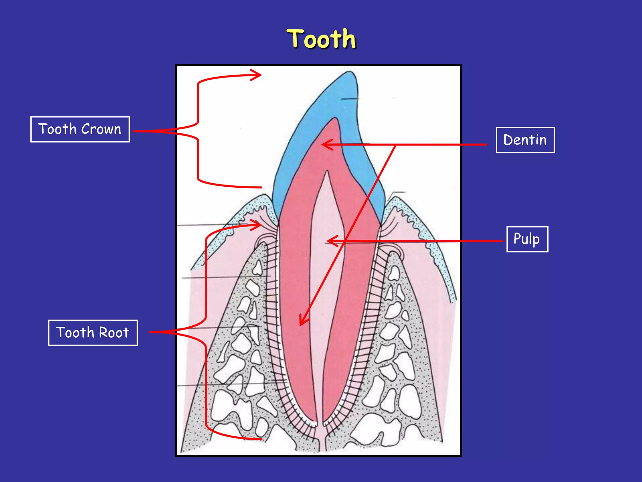
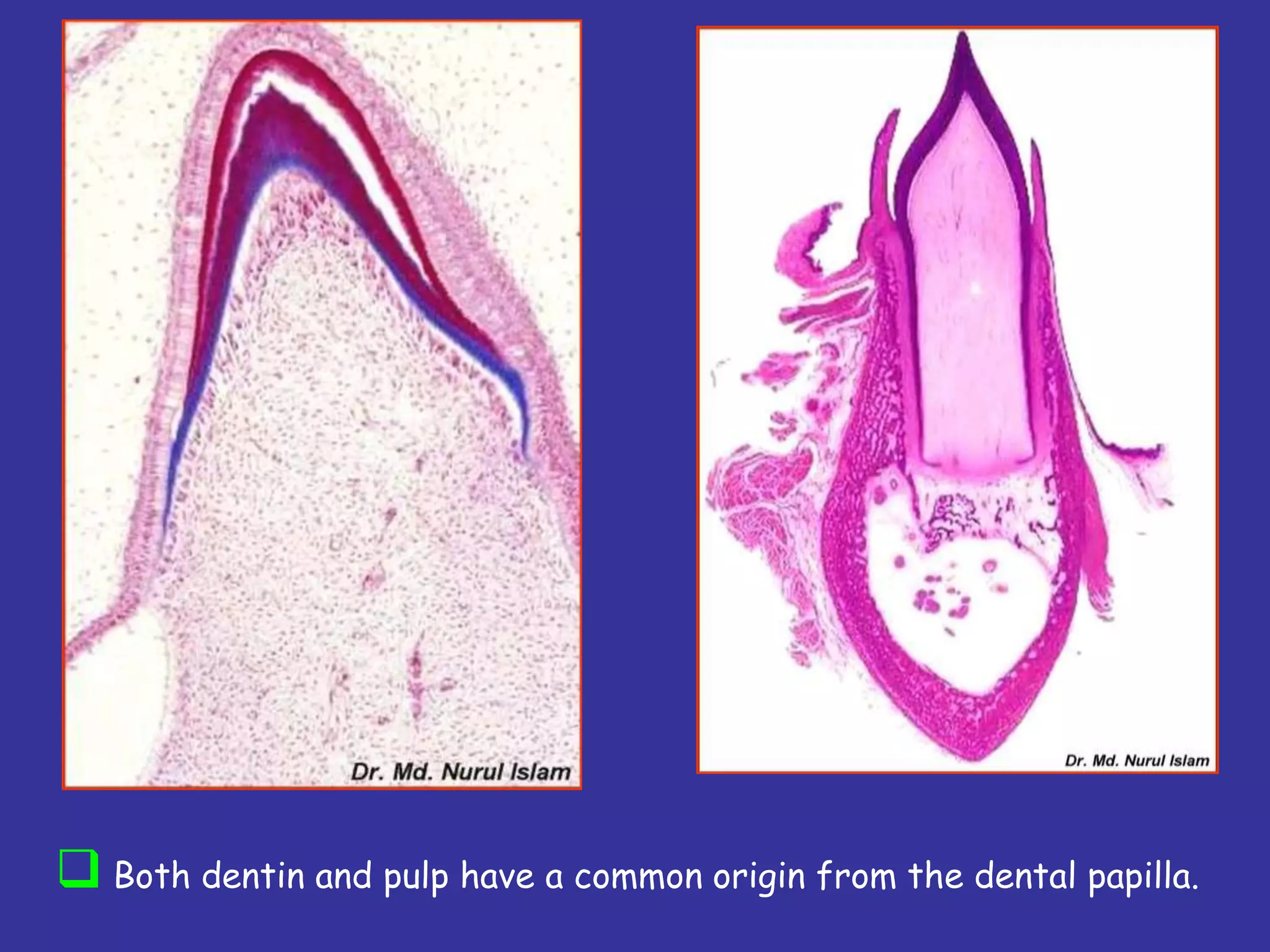
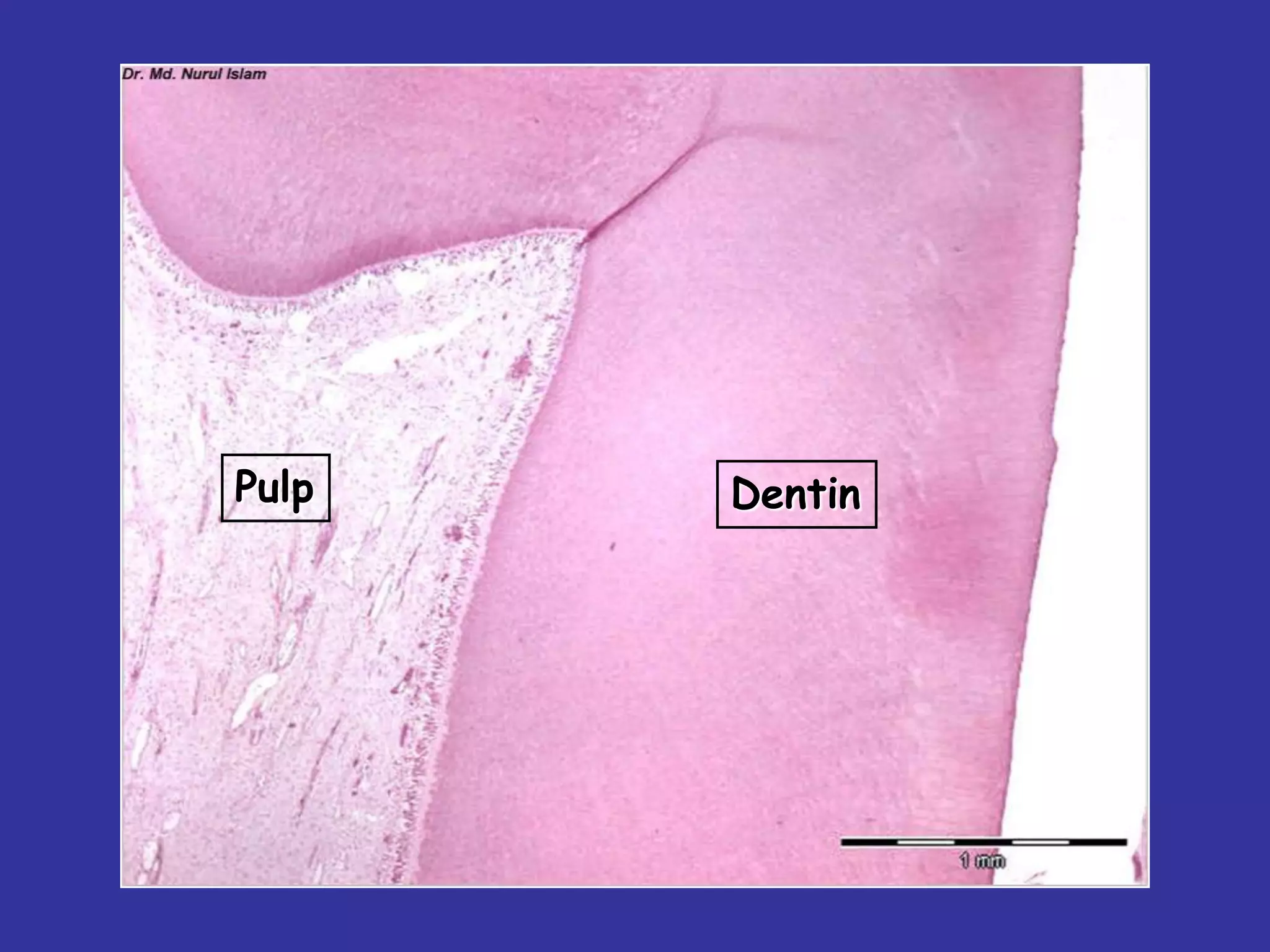
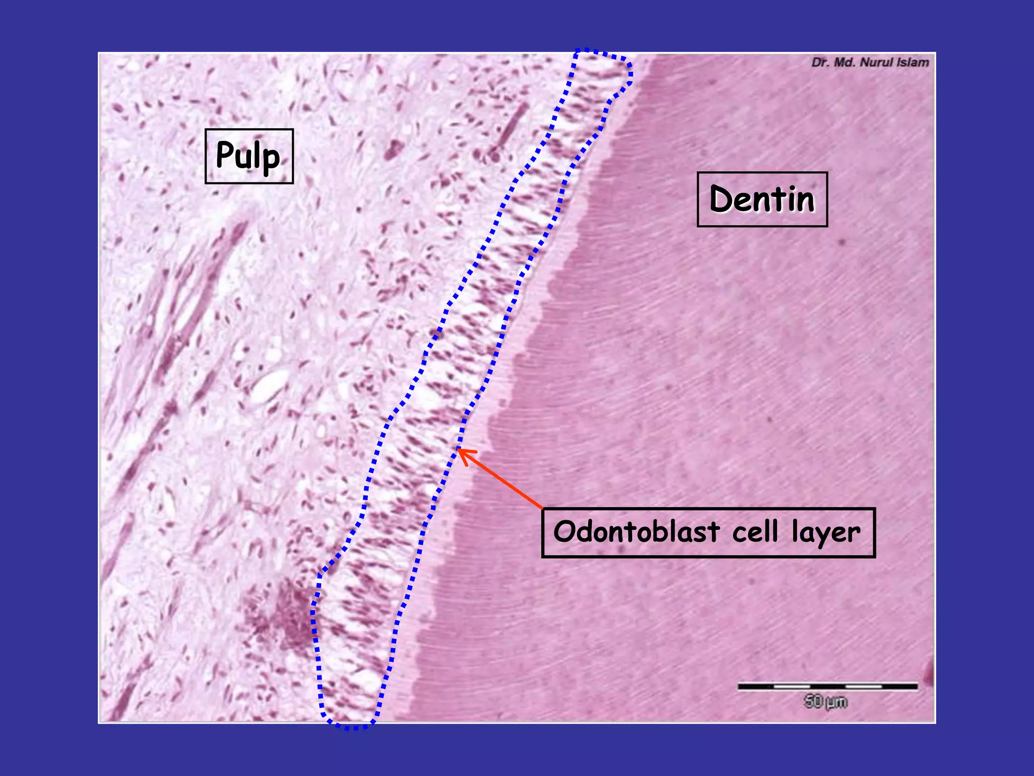
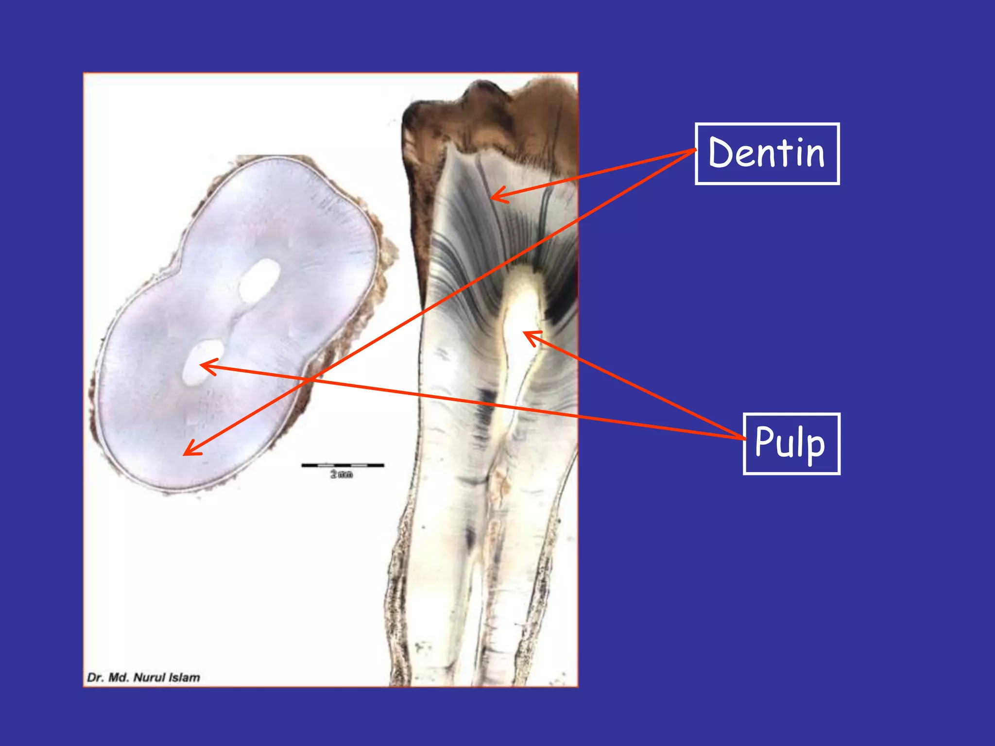
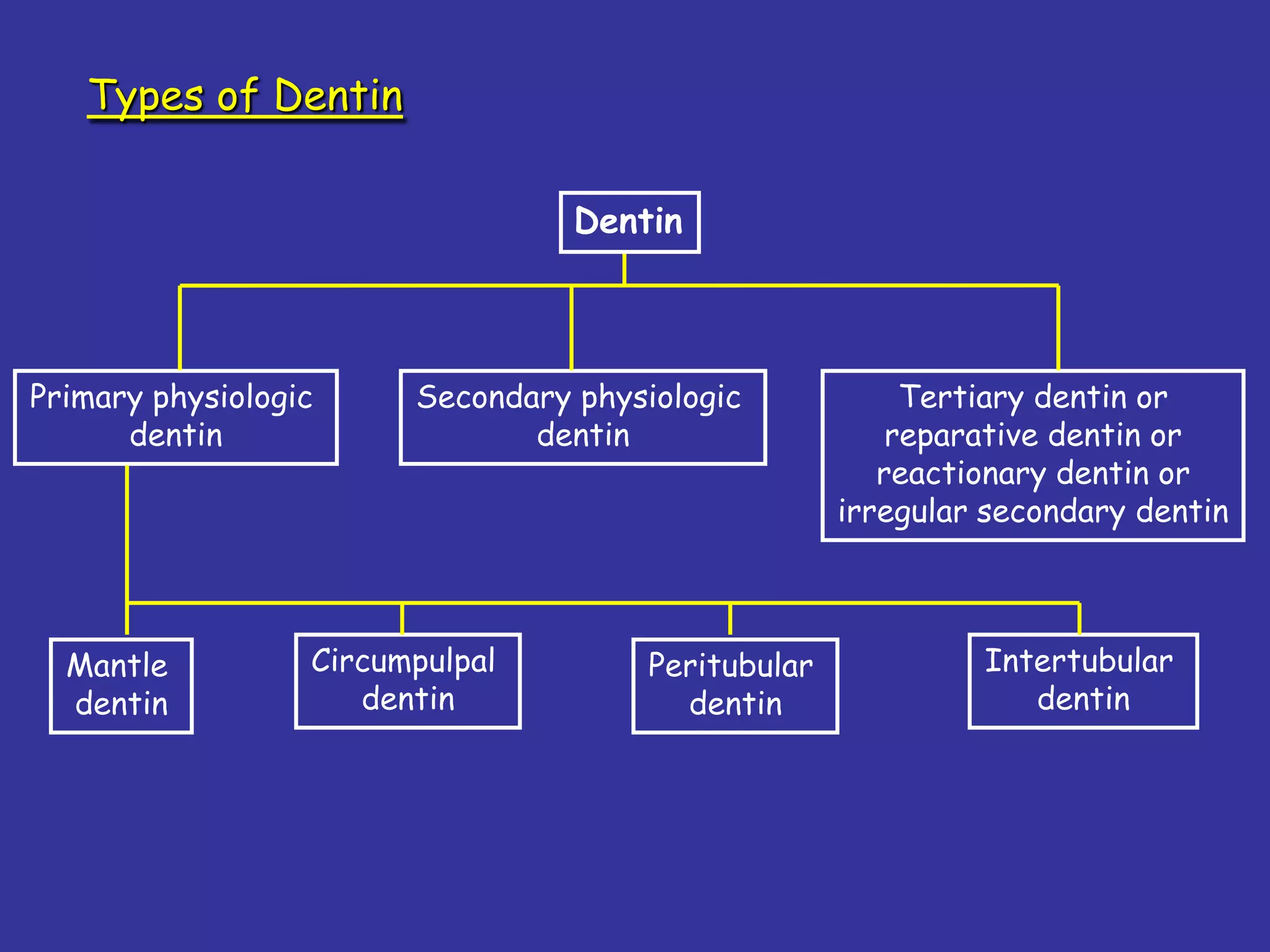
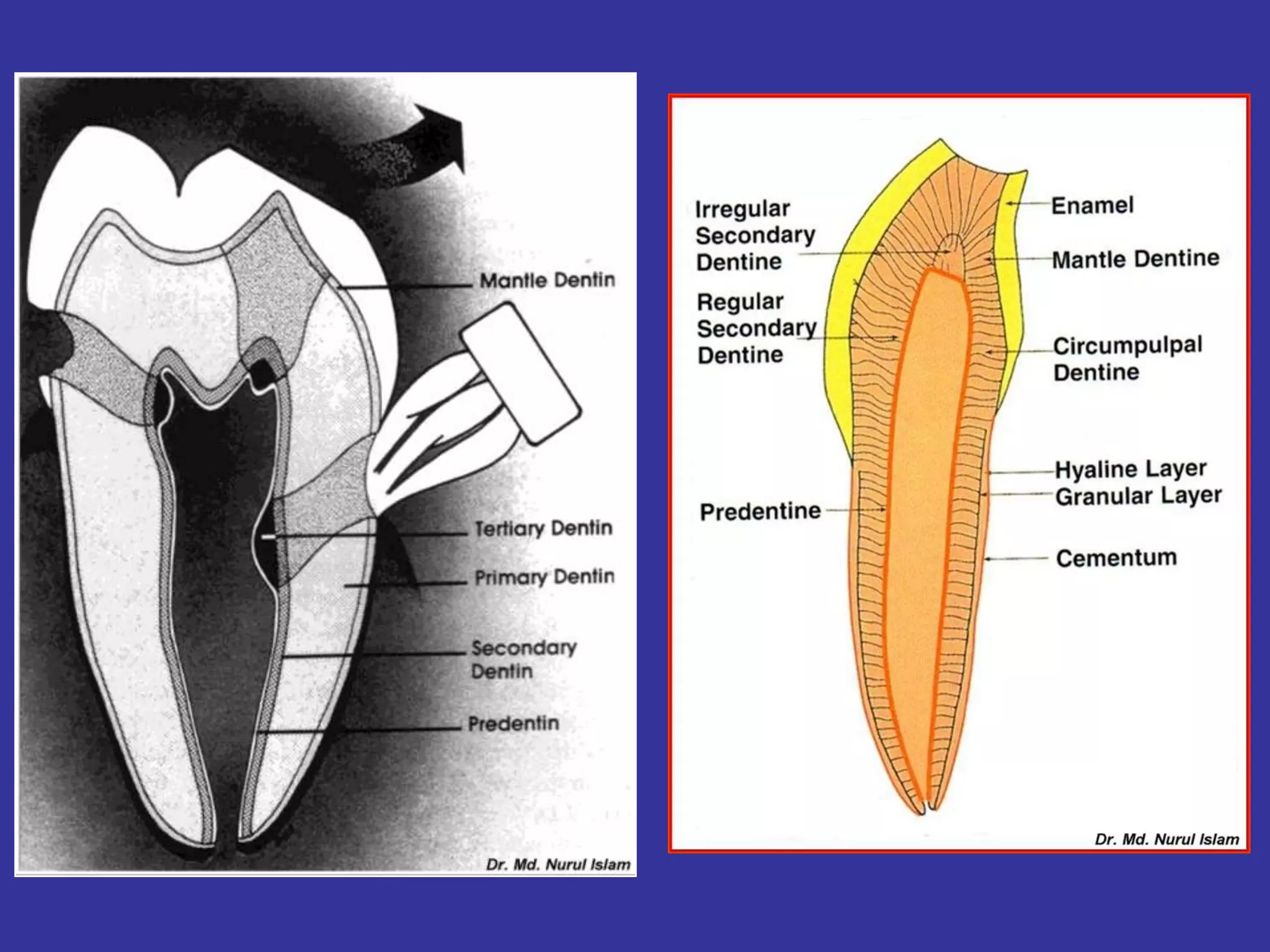
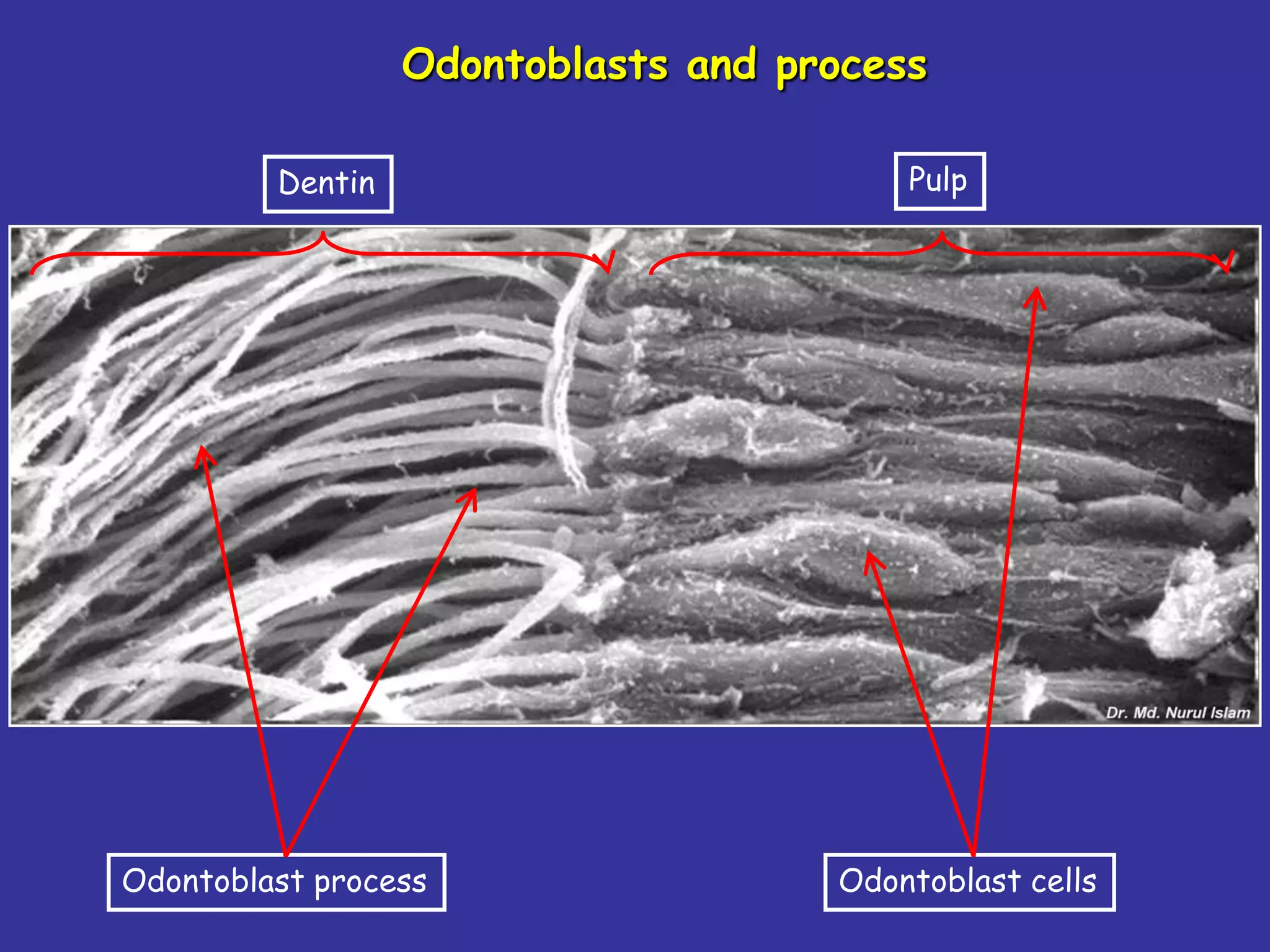
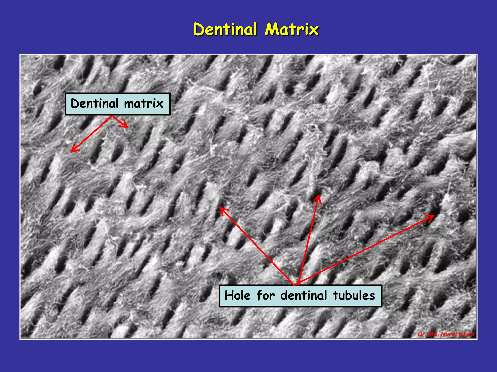
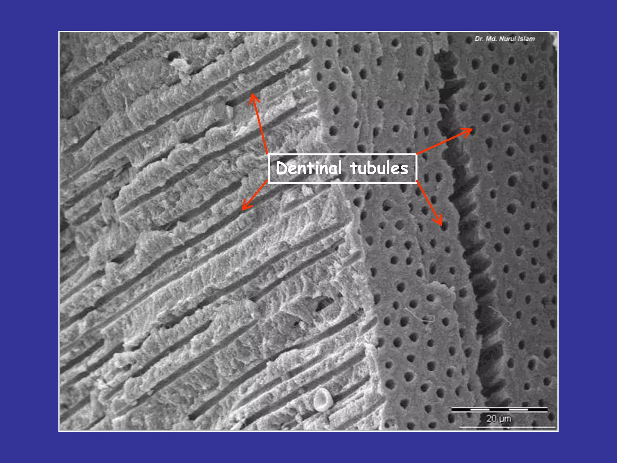
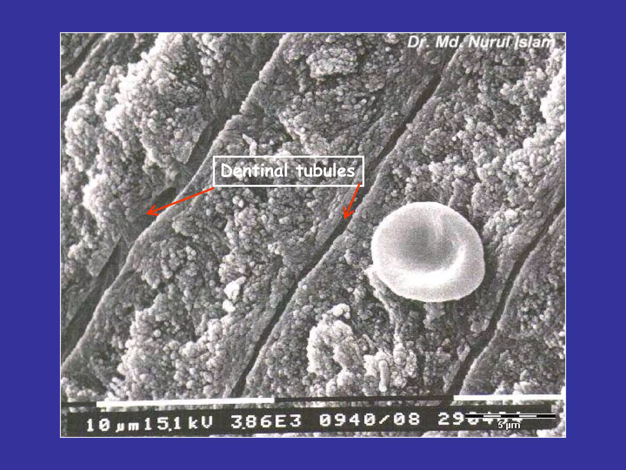
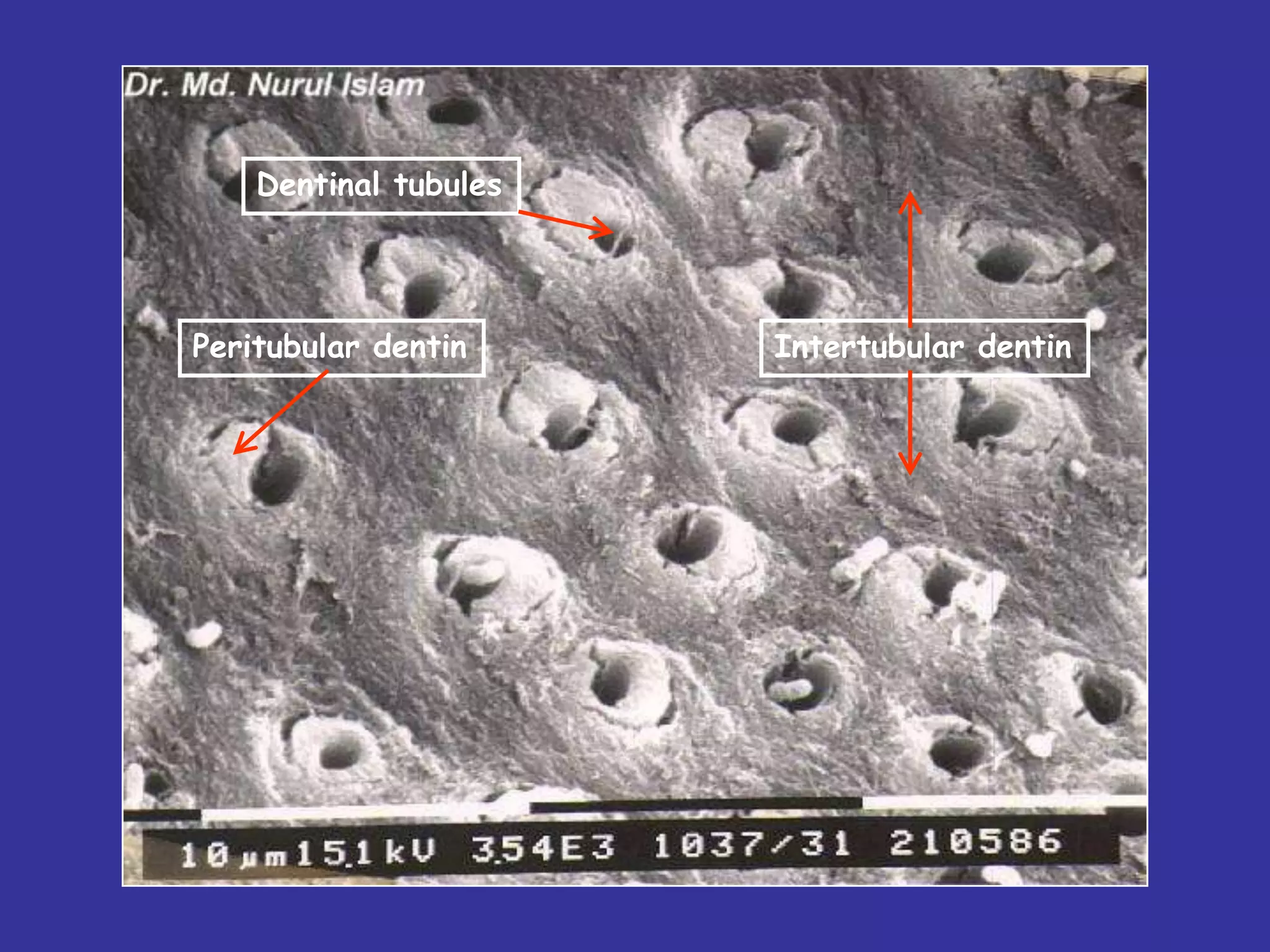
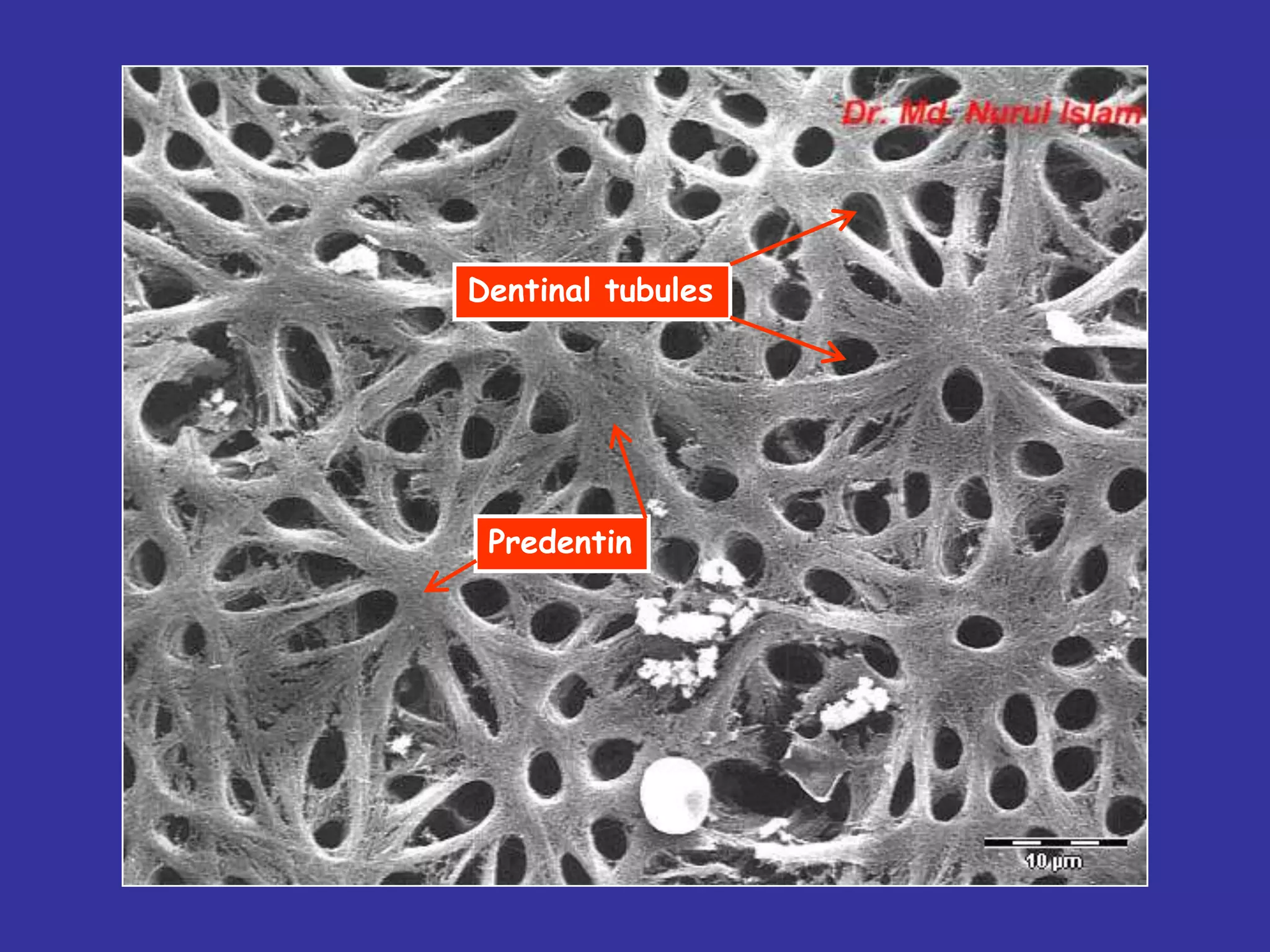
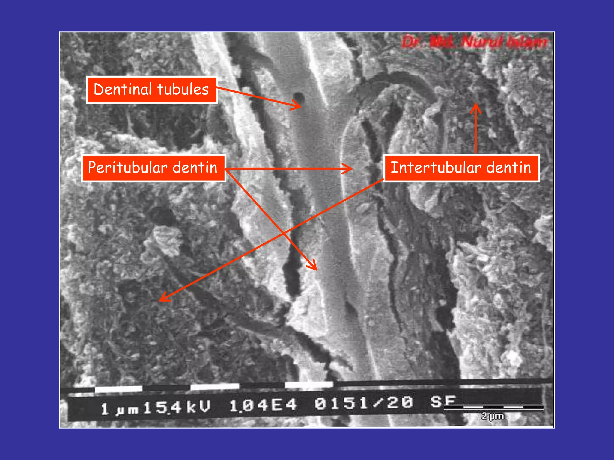
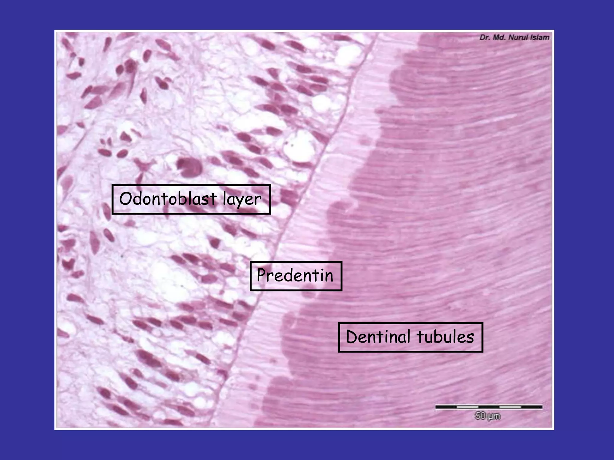
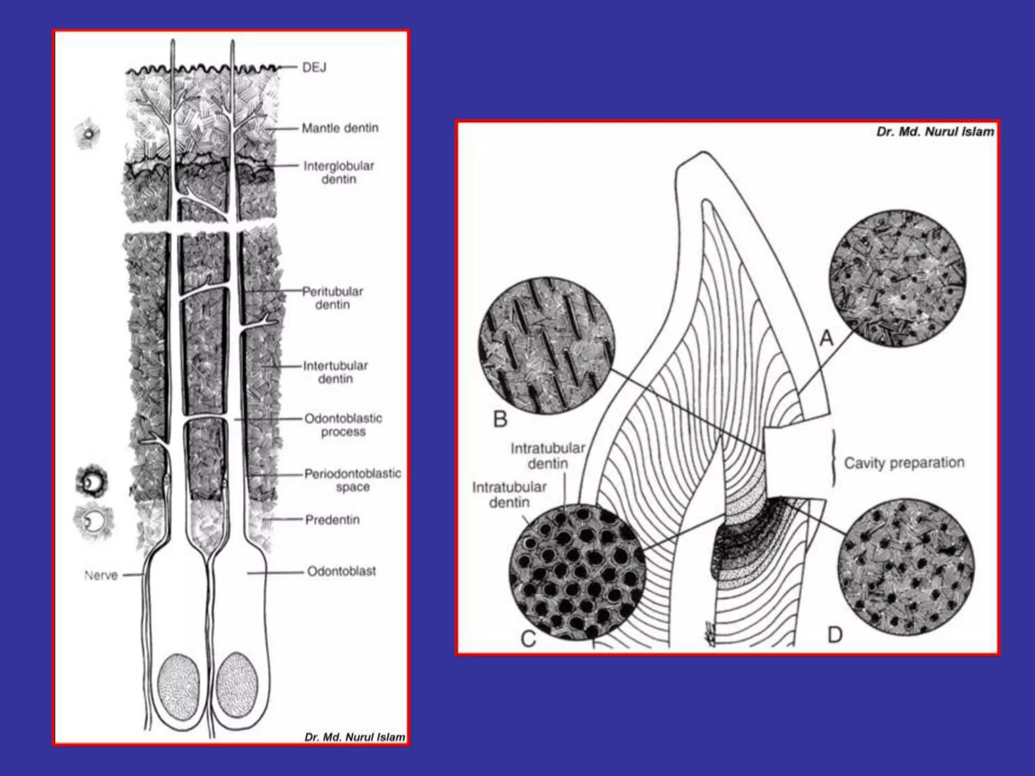
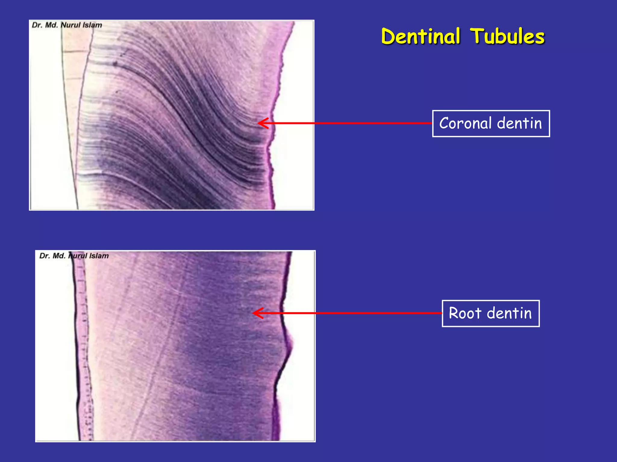
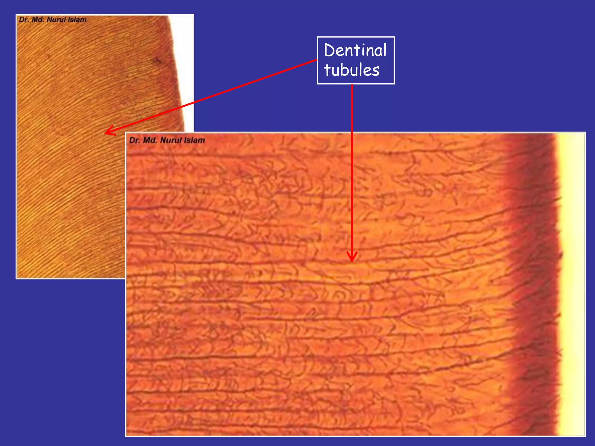
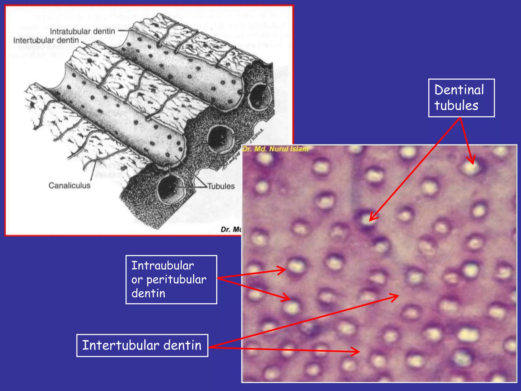
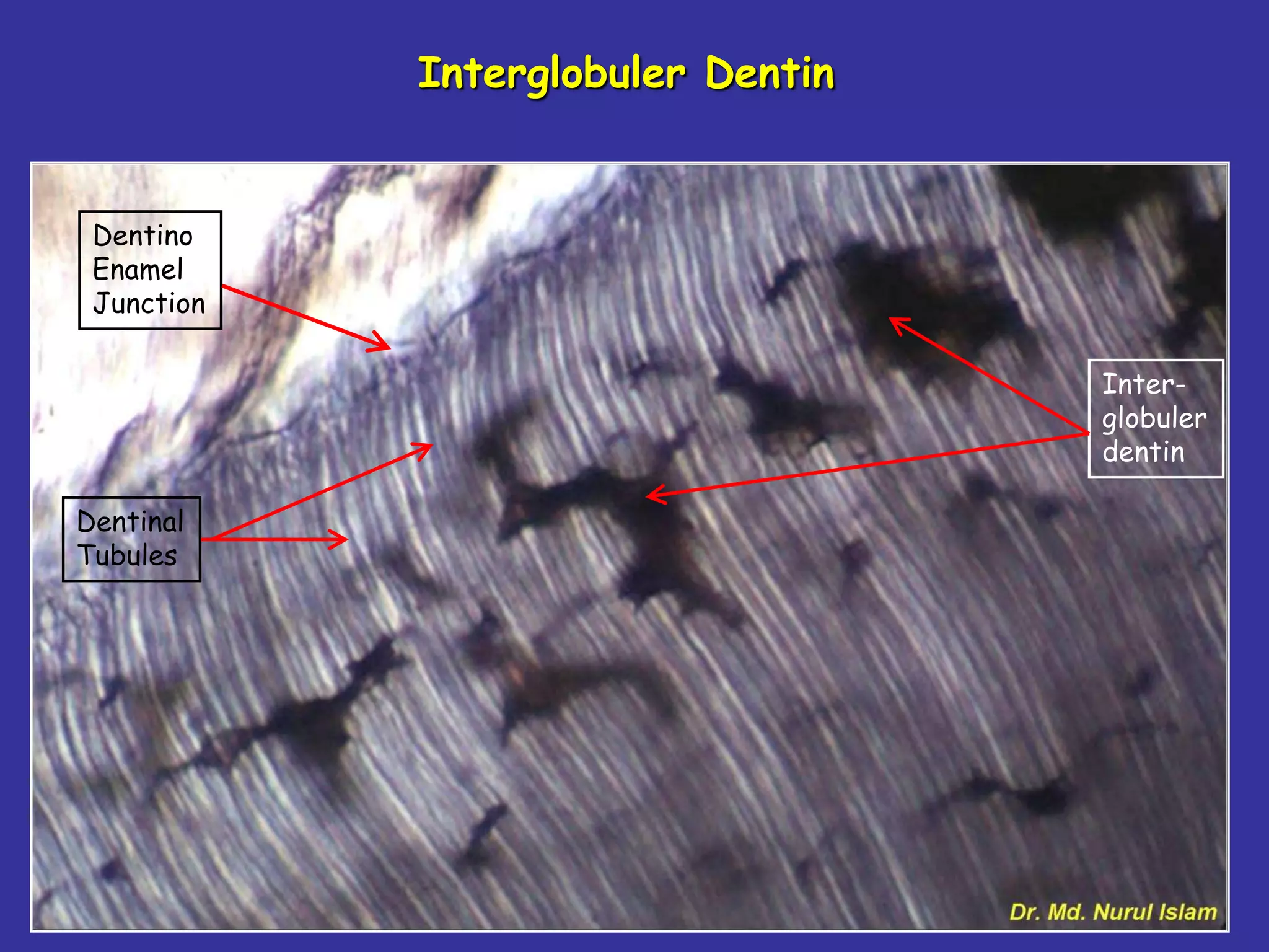
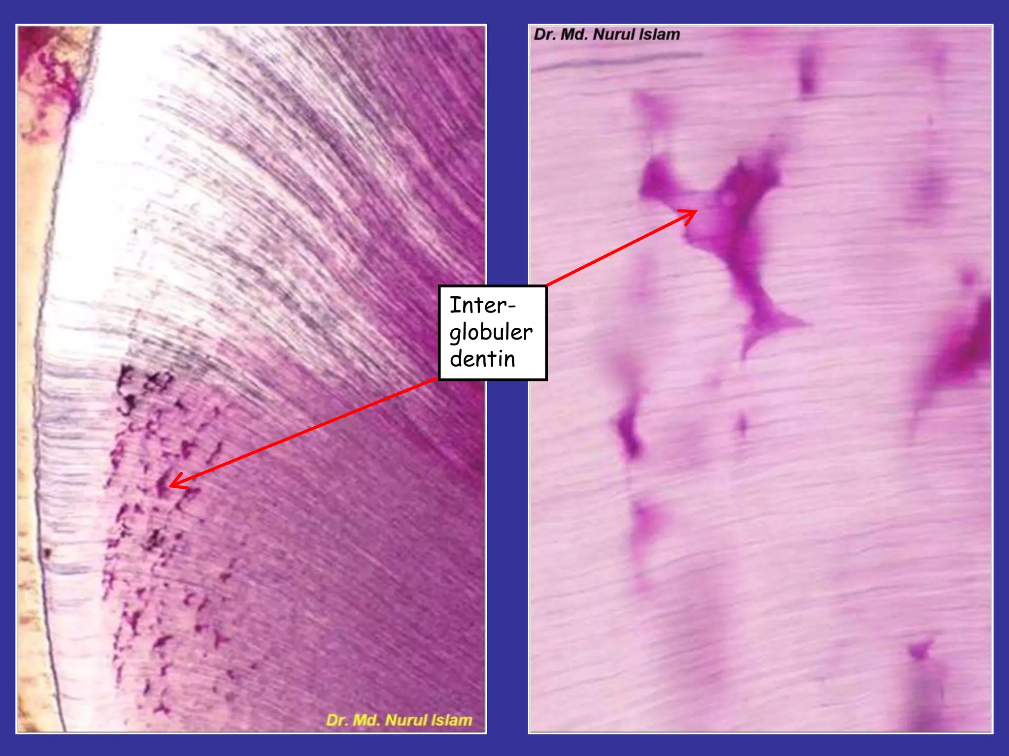
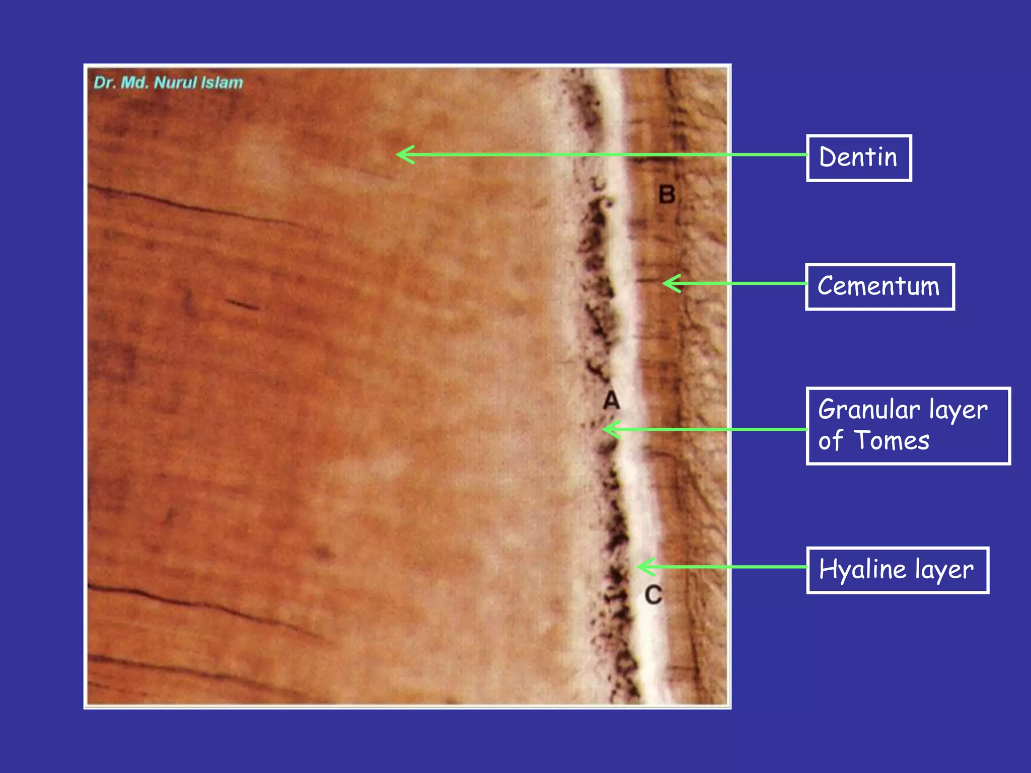
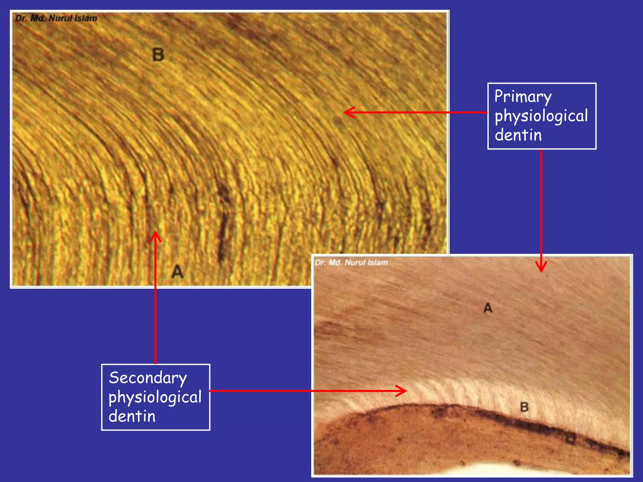
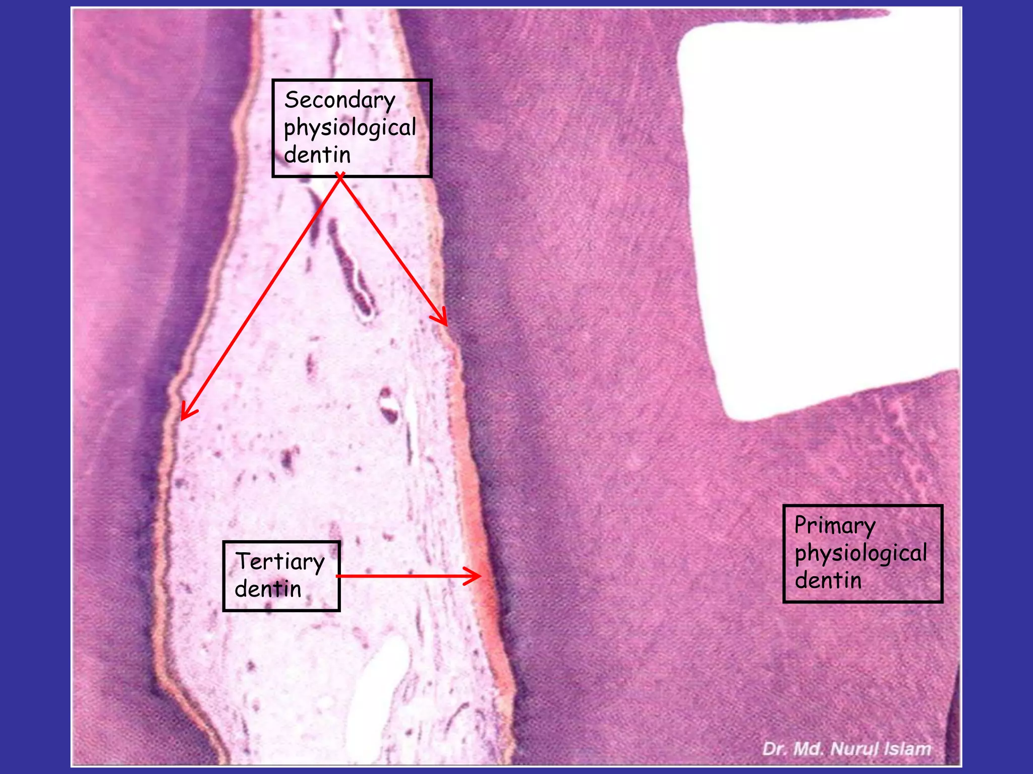
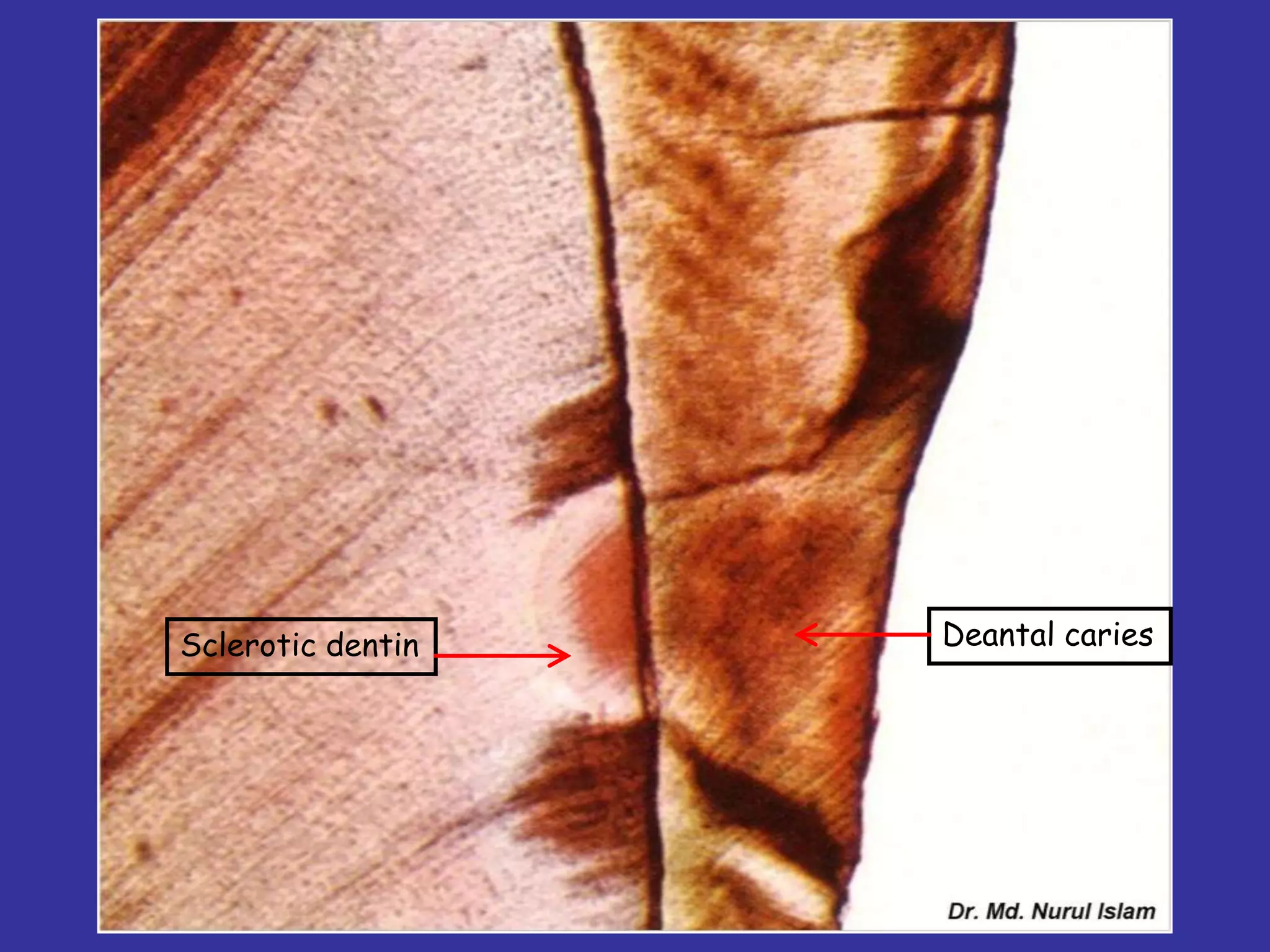
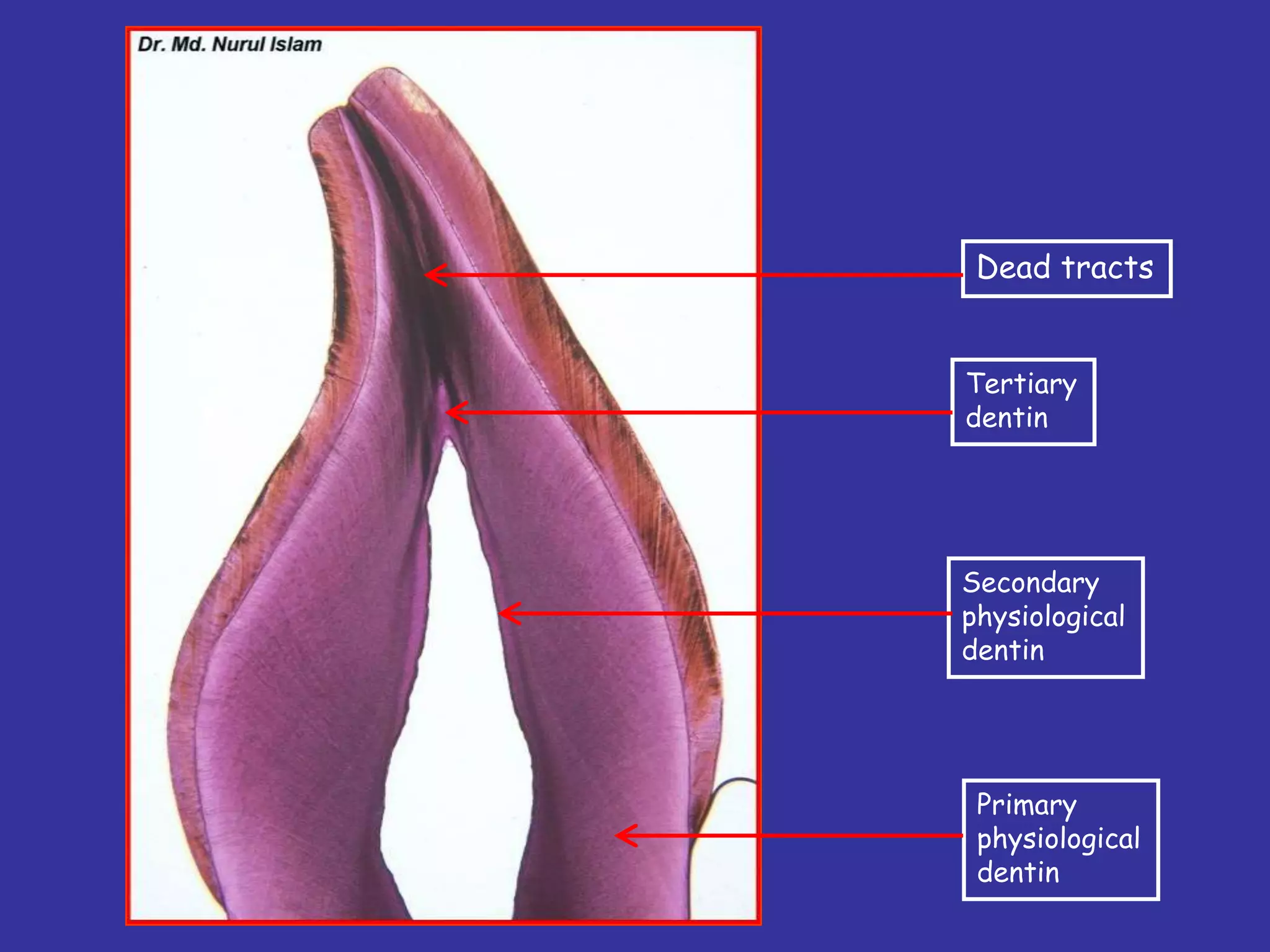
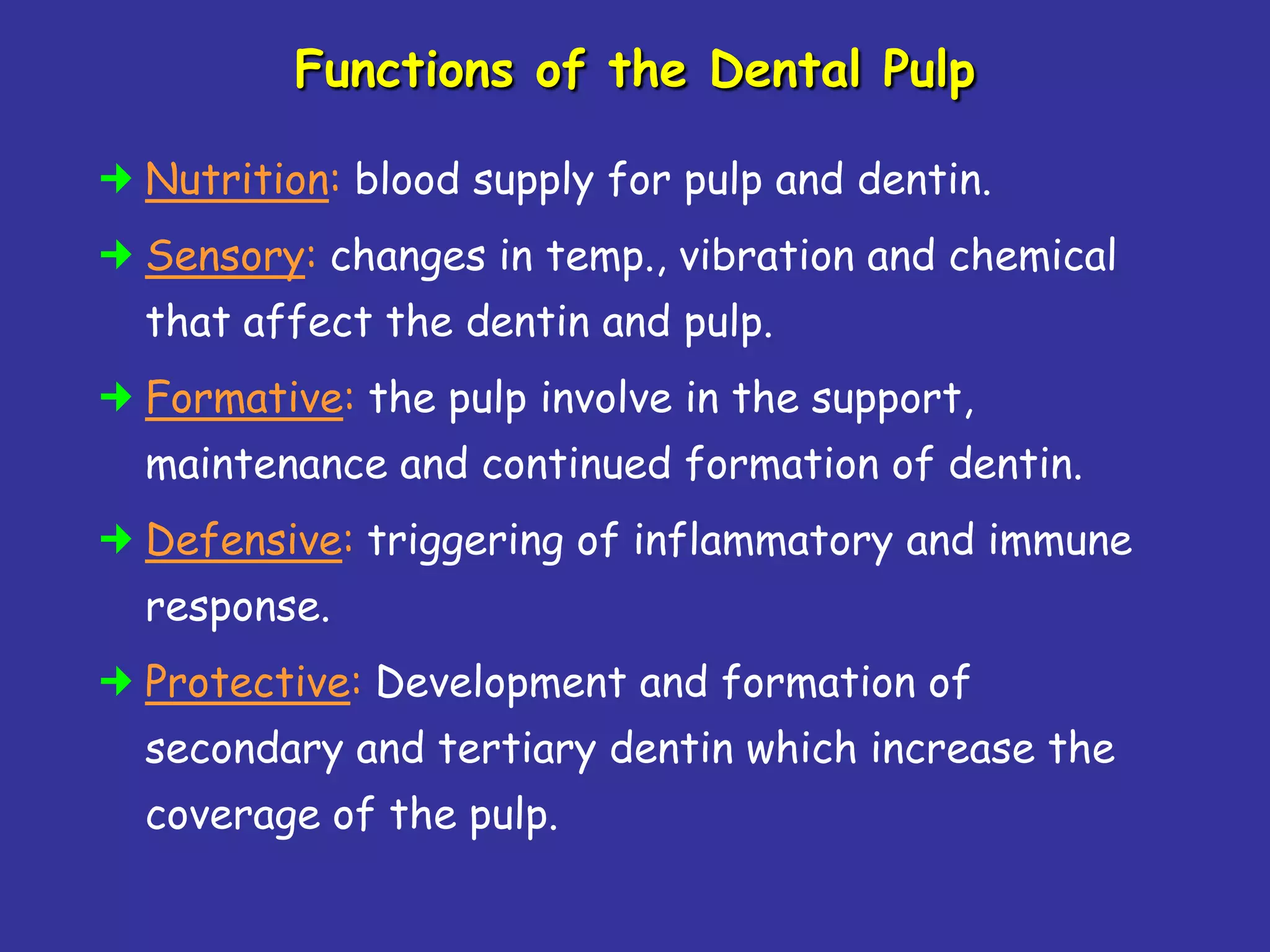
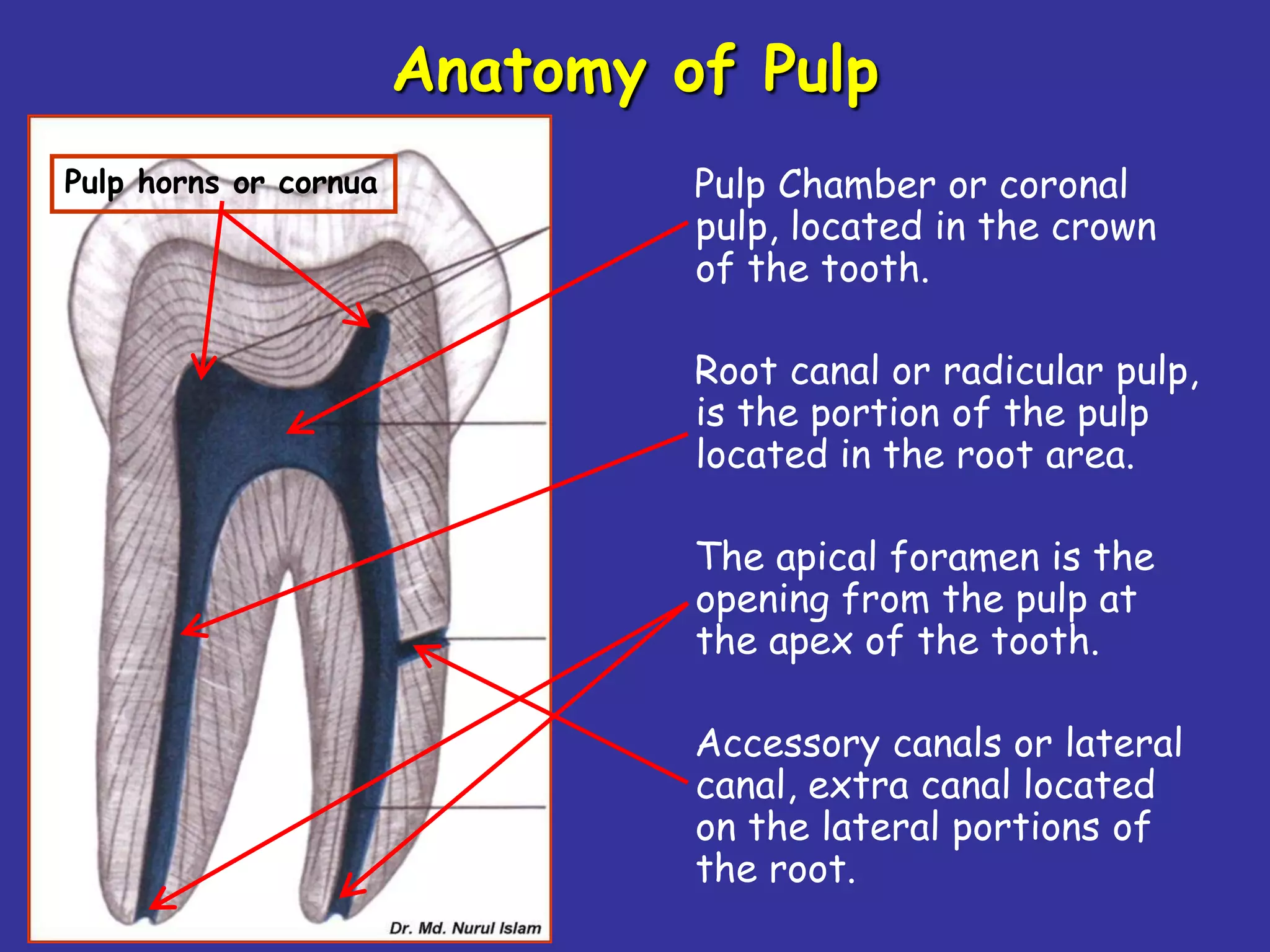
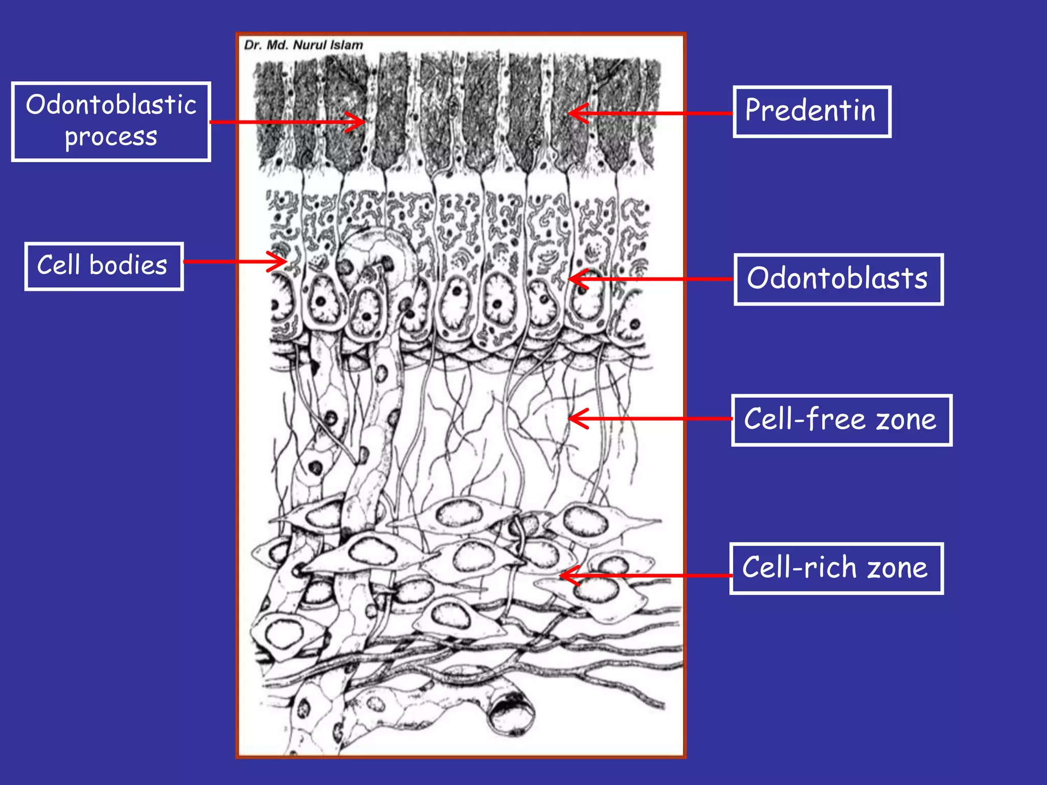
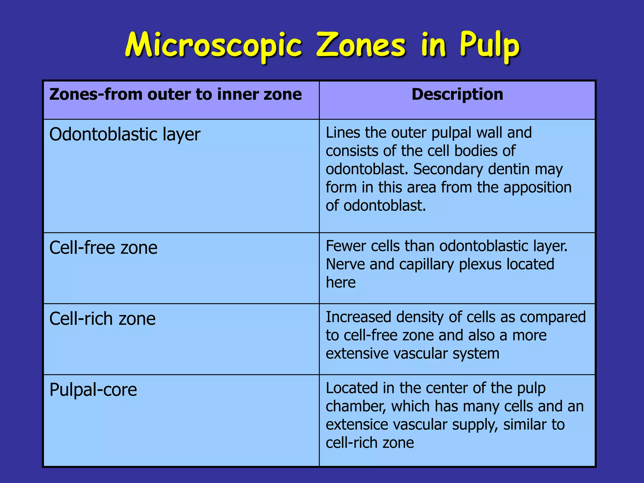
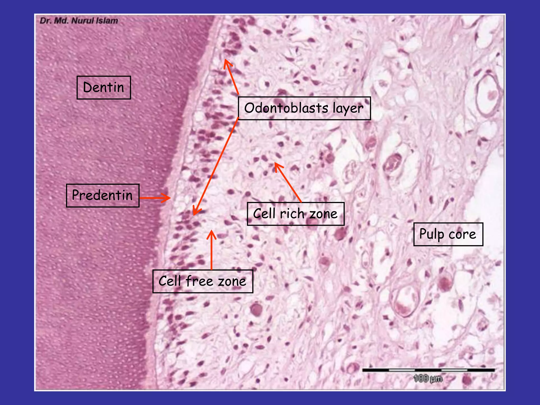
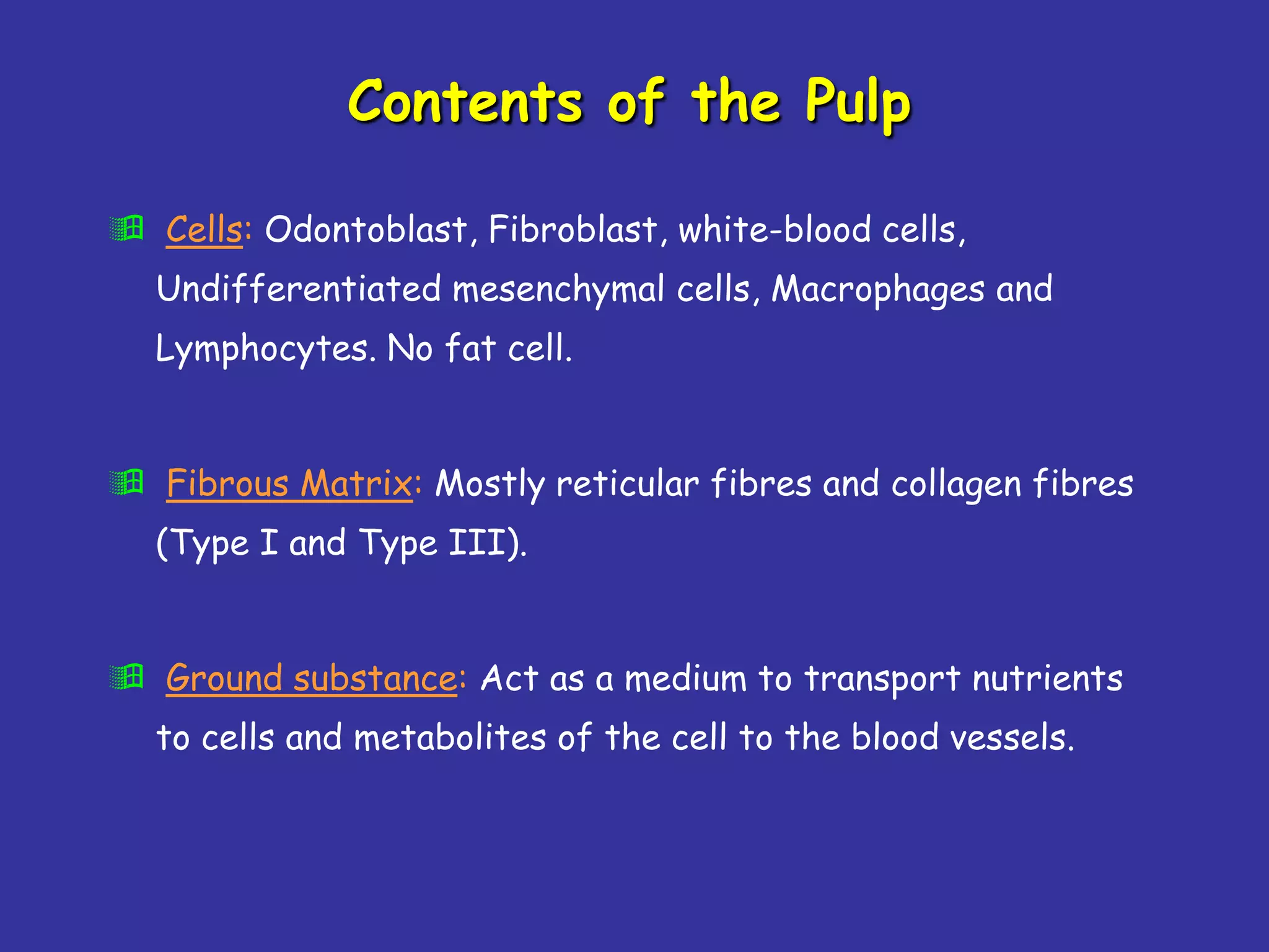
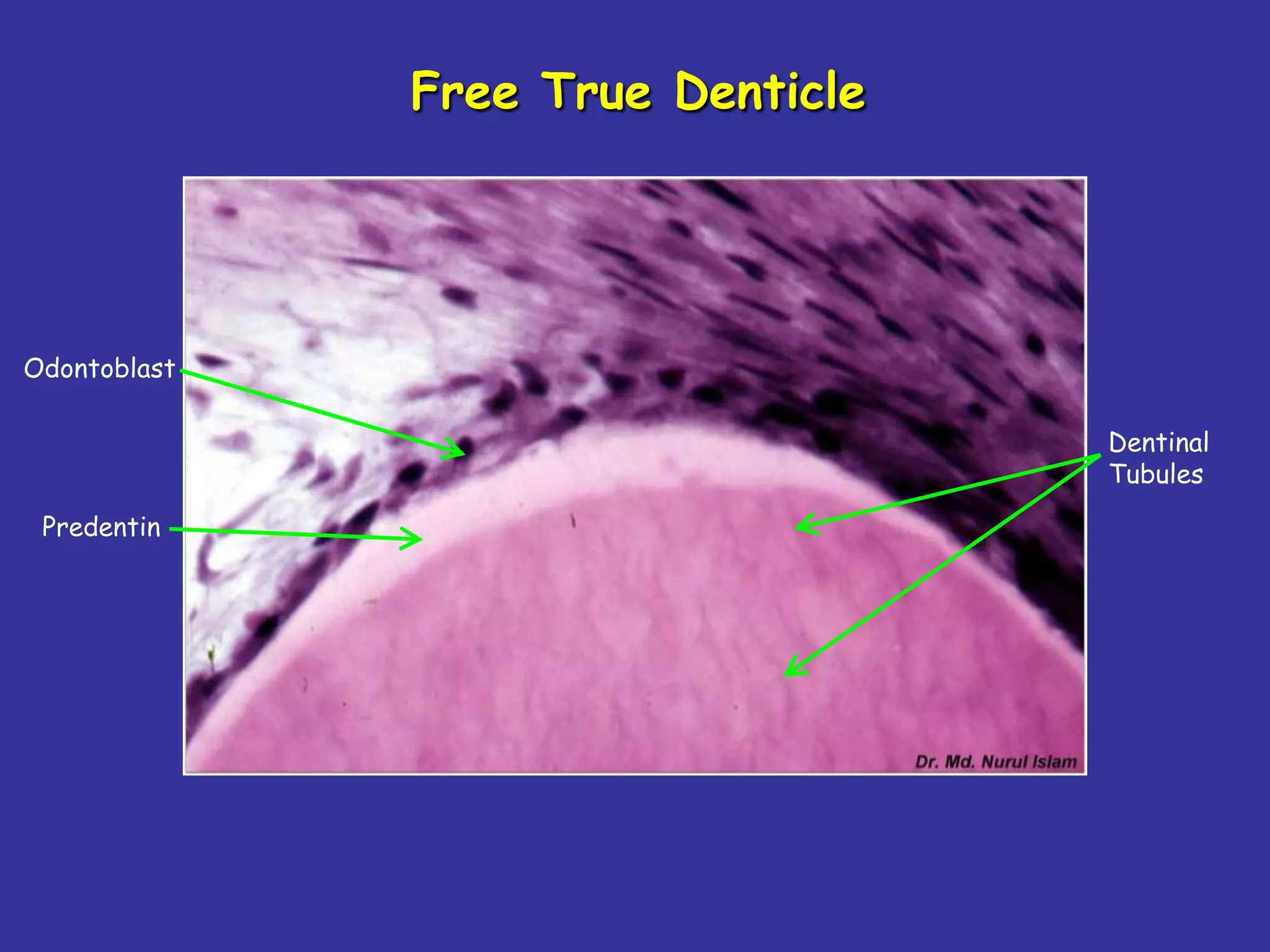
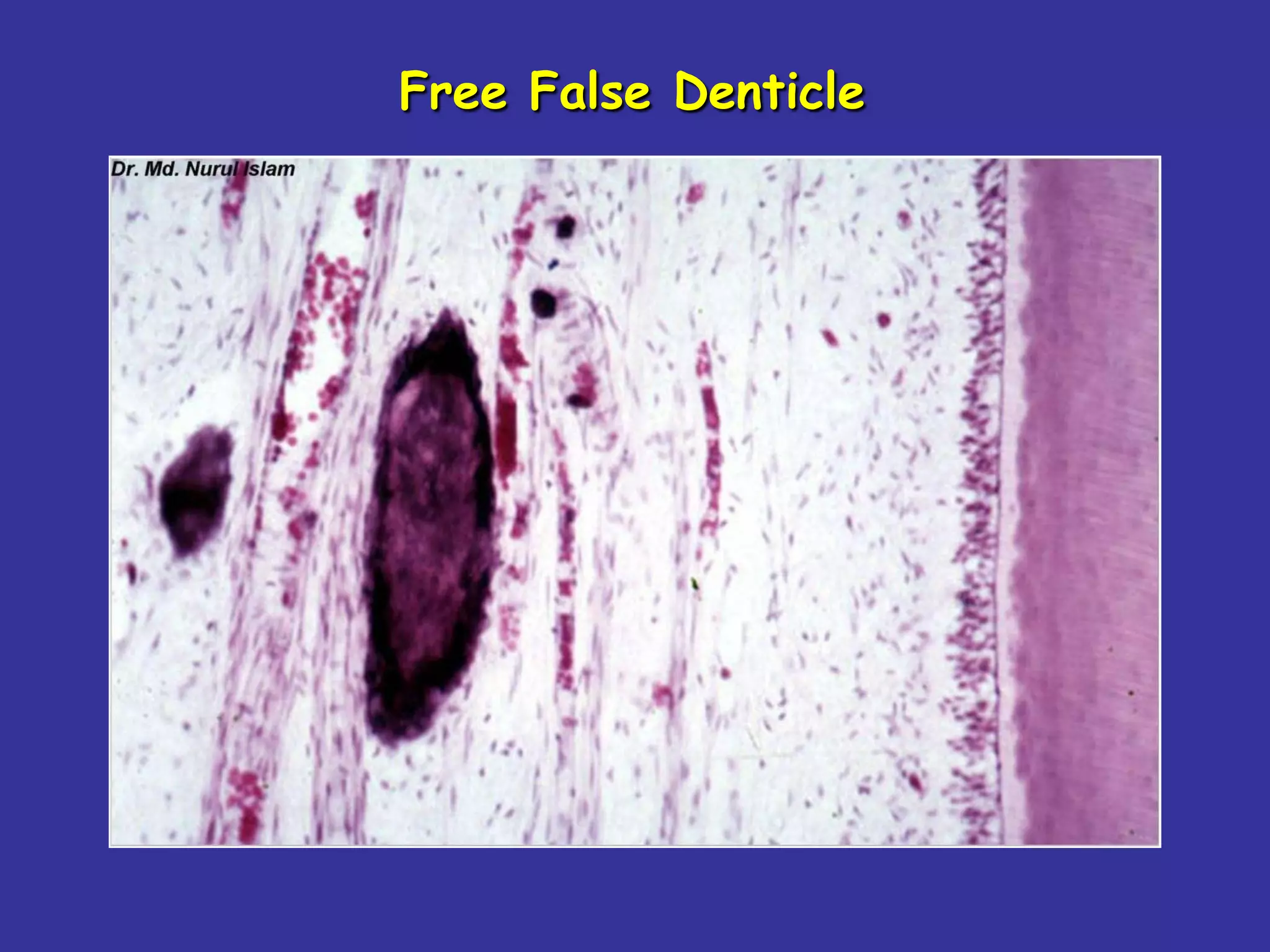
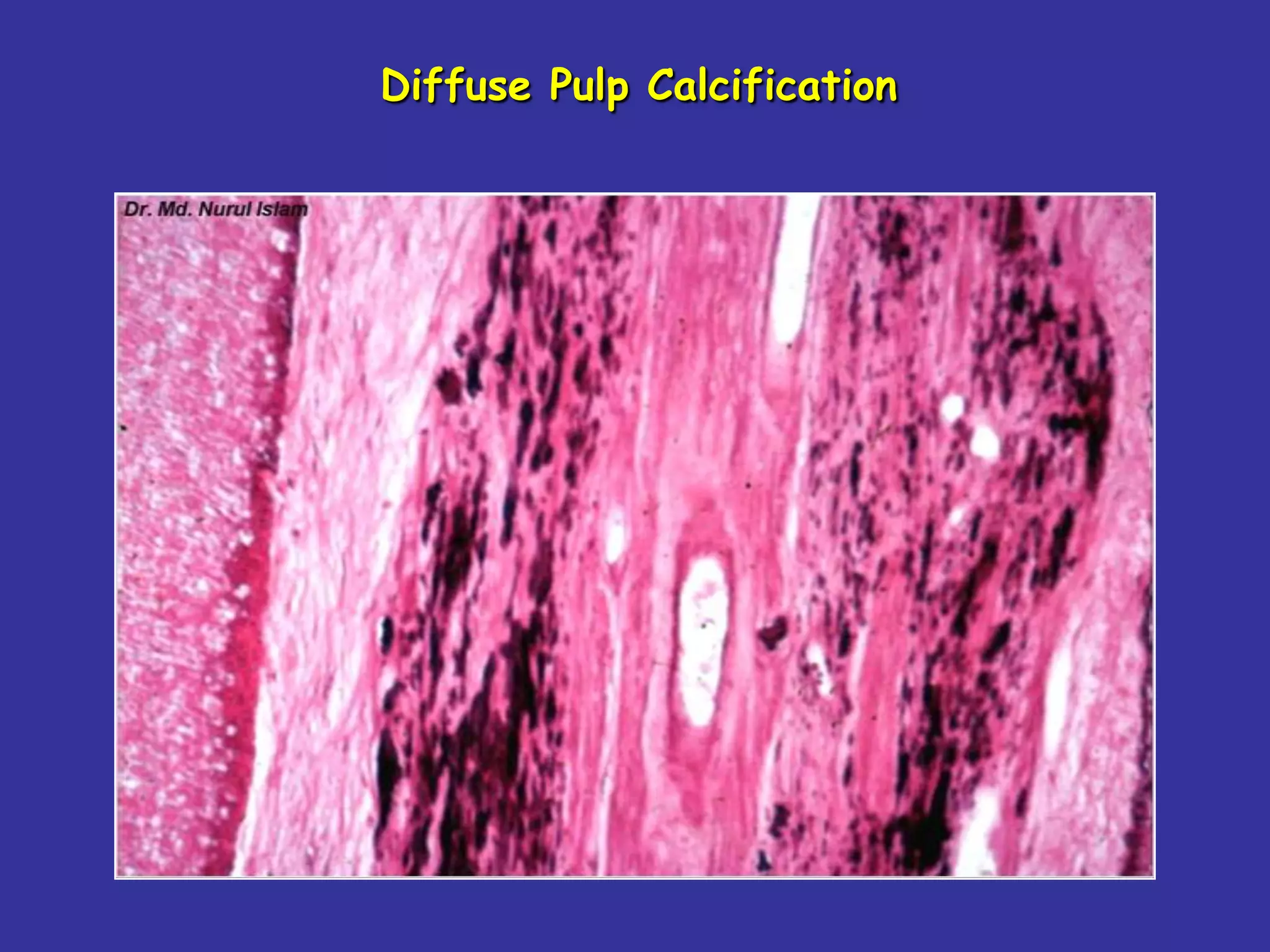


The document summarizes key aspects of the dentin-pulp complex. It describes how dentin and pulp have a common embryonic origin and are considered a single functional unit. It outlines the different types of dentin that form over time, including primary, secondary, and tertiary dentin. It also discusses the roles of odontoblasts and dentinal tubules. In less than 3 sentences, the document provides an overview of the embryological, histological, and functional relationship between dentin and pulp as a complex unit that forms over the life of a tooth.


![Pulp dentin complex[1]](https://cdn.slidesharecdn.com/ss_thumbnails/pulpdentincomplex1-140427062105-phpapp01-thumbnail.jpg?width=640&height=640&fit=bounds)


