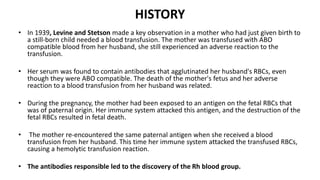
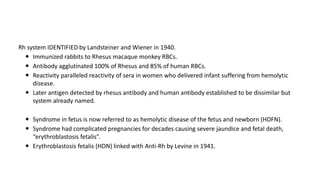
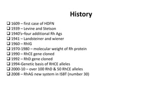
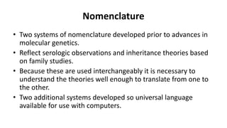
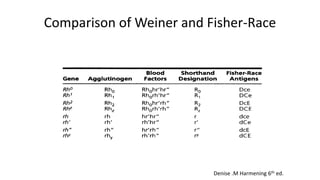
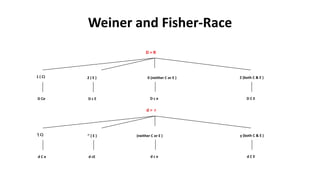
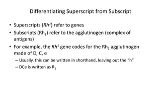
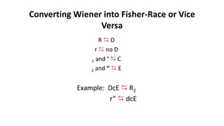
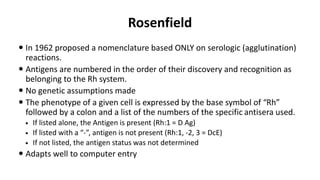
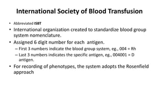
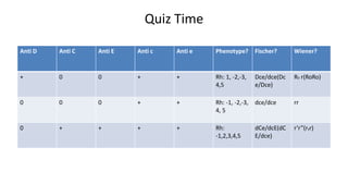
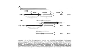
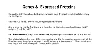
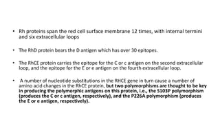
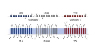
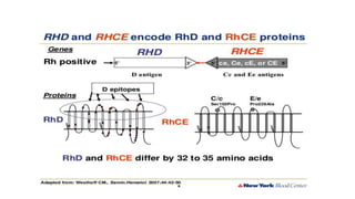
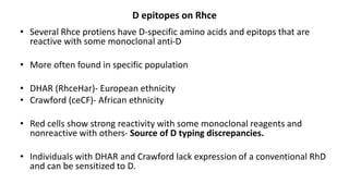
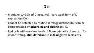
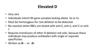
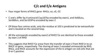
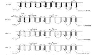
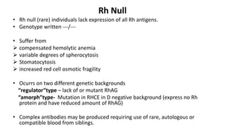
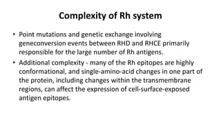
Rh BLOOD GROUP SYSTEM
Dr. Priyanka Roy
Discussion
• History
• Nomenclature
• Structure
• Genetics
• Variants of Rh
Introduction
• Rh is the most important blood group system after ABO in
transfusion medicine.
• Rh antigens especially D, are highly immunogenic and cause
HDFN and HTR.
• One of the most complex of all RBC blood group systems with
more than 60 different Rh antigens.
HISTORY
• In 1939, Levine and Stetson made a key observation in a mother who had just given birth to
a still-born child needed a blood transfusion. The mother was transfused with ABO
compatible blood from her husband, she still experienced an adverse reaction to the
transfusion.
• Her serum was found to contain antibodies that agglutinated her husband's RBCs, even
though they were ABO compatible. The death of the mother's fetus and her adverse
reaction to a blood transfusion from her husband was related.
• During the pregnancy, the mother had been exposed to an antigen on the fetal RBCs that
was of paternal origin. Her immune system attacked this antigen, and the destruction of the
fetal RBCs resulted in fetal death.
• The mother re-encountered the same paternal antigen when she received a blood
transfusion from her husband. This time her immune system attacked the transfused RBCs,
causing a hemolytic transfusion reaction.
• The antibodies responsible led to the discovery of the Rh blood group.
Rh system IDENTIFIED by Landsteiner and Wiener in 1940.
 Immunized rabbits to Rhesus macaque monkey RBCs.
 Antibody agglutinated 100% of Rhesus and 85% of human RBCs.
 Reactivity paralleled reactivity of sera in women who delivered infant suffering from hemolytic
disease.
 Later antigen detected by rhesus antibody and human antibody established to be dissimilar but
system already named.
 Syndrome in fetus is now referred to as hemolytic disease of the fetus and newborn (HDFN).
 Syndrome had complicated pregnancies for decades causing severe jaundice and fetal death,
“erythroblastosis fetalis”.
 Erythroblastosis fetalis (HDN) linked with Anti-Rh by Levine in 1941.
History
 1609 – first case of HDFN
 1939 – Levine and Stetson
 1940’s–four additional Rh Ags
 1941 – Landsteiner and wiener
 1960 – RhIG
 1970-1980 – molecular weight of Rh protein
 1990 – RhCE gene cloned
 1992 – RhD gene cloned
 1994-Genetic basis of RHCE alleles
 2000-10 – over 100 RhD & 50 RhCE alleles
 2008 – RhAG new system in ISBT (number 30)
Nomenclature
• Two systems of nomenclature developed prior to advances in
molecular genetics.
• Reflect serologic observations and inheritance theories based
on family studies.
• Because these are used interchangeably it is necessary to
understand the theories well enough to translate from one to
the other.
• Two additional systems developed so universal language
available for use with computers.
Rh nomenclature
Rh Terminology derived from 4 sets of investigators.
• Two of the terminologies are based on the postulated genetic
mechanisms of the Rh system.
• 3rd terminology describes only the presence or absence of a
given antigen.
• 4th is result of the effort of the International Society of Blood
Transfusion (ISBT) Working Party on Terminology for Red cell
Surface antigens.
Fisher-Race: CDE Terminology
• Fisher Race
– Suggested that antigens are determined by 3 pairs of genes which
occupy closely linked loci.
– Each gene complex carries D or its absence (d), C or c, E or e.
– Each gene (except d, which is an amorph) causes production of an
antigen.
– The order of loci on the gene appears to be “DCE” but many authors
prefer to use “CDE” to follow alphabet.
– Inherited from parents in linked fashion as haplotypes
– The gene d is assumed to be present when D is absent.
Fisher-Race
• Three loci carry the Rh genes are so closely linked that they
never separate but are passed from generation to generation
as a unit or gene complex.
Fisher-Race
 With the exception of d each allelic gene controls presence of
respective antigen on RBC.
 The gene complex DCe would cause production of the D, C and e
antigens on the red cells.
 If the same gene complex were on both paired chromsomes
(DCe/DCe) then only D, C and e would be present on the cells.
 If one chromsome carried DCe and the other was DcE this would
cause D, C, c, E and e antigens to be present on red blood cells.
 Each antigen except d is recognizable by testing red cells with
specific antiserum.
Wiener
 Postulated that TWO genes, one on each chromosome pair, controls
the entire express of Rh system.
 Each gene produces a structure on the red cell called an
agglutinogen (antigen).
 Eight (8) major alleles (agglutinogens): R0, R1, R2, Rz, r, r’, r” and ry.
 Each agglutinogen has 3 factors (antigens or epitopes)
 The three factors are the antigens expressed on the cell.
 For example the agglutinogen R0= Rh0 (D), hr’ (c), hr” (e)
 Each agglutinogen can be identified by its parts or factors that react
with specific antibodies (antiserums).
Weiner’s Theory
Weiner and Fisher-Race
 The two theories are the basis for the two notations currently used
for the Rh system.
 Immunohematologists use combinations of both systems when
recording most probable genotypes.
 We MUST be able to convert a Fisher-Race notation into Wiener
shorthand, i.e., Dce (Fisher-Race) is written R0.
 Given an individual’s phenotype we MUST determine all probable
genotypes and write them in both Fisher-Race and Wiener
notations.
 R1r is the most common D positive genotype.
 rr is the most common D negative genotype.
Comparison of Weiner and Fisher-Race
Denise .M Harmening 6th ed.
Weiner and Fisher-Race
1 ( C)
D Ce
2 ( E )
D c E
0 (neither C or E )
D c e
Z (both C & E )
D C E
‘( C)
d C e
‘’ ( E )
d cE
(neither C or E )
d c e
y (both C & E )
d C E
D = R
d = r
Differentiating Superscript from Subscript
• Superscripts (Rh1) refer to genes
• Subscripts (Rh1) refer to the agglutinogen (complex of
antigens)
• For example, the Rh1 gene codes for the Rh1 agglutinogen
made of D, C, e
– Usually, this can be written in shorthand, leaving out the “h”
– DCe is written as R1
Converting Wiener into Fisher-Race or Vice
Versa
R  D
r  no D
1 and ‘  C
2 and “  E
Example: DcE  R2
r”  dcE
Rosenfield
 In 1962 proposed a nomenclature based ONLY on serologic (agglutination)
reactions.
 Antigens are numbered in the order of their discovery and recognition as
belonging to the Rh system.
 No genetic assumptions made
 The phenotype of a given cell is expressed by the base symbol of “Rh”
followed by a colon and a list of the numbers of the specific antisera used.
 If listed alone, the Antigen is present (Rh:1 = D Ag)
 If listed with a “-”, antigen is not present (Rh:1, -2, 3 = DcE)
 If not listed, the antigen status was not determined
 Adapts well to computer entry
Comparison of Three Systems
International Society of Blood Transfusion
• Abbreviated ISBT
• International organization created to standardize blood group
system nomenclature.
• Assigned 6 digit number for each antigen.
– First 3 numbers indicate the blood group system, eg., 004 = Rh
– Last 3 numbers indicates the specific antigen, eg., 004001 = D
antigen.
• For recording of phenotypes, the system adopts the Rosenfield
approach
Frequency
Antigen Beenu et al whites blacks
D 93.39% 85% 92%
C 84.76% 68% 27%
c 52.82% 80% 96%
E 17.9% 29% 22%
e 98.3% 98% 98%
R1R1 –most common in India
R0 r – rare in India but common in blacks
Quiz Time
Anti D Anti C Anti E Anti c Anti e
+ 0 0 + +
0 0 0 + +
0 + + + +
Quiz Time
Anti D Anti C Anti E Anti c Anti e Phenotype? Fischer? Wiener?
+ 0 0 + + Rh: 1, -2,-3,
4,5
Dce/dce(Dc
e/Dce)
R0 r(RoRo)
0 0 0 + + Rh: -1, -2,-3,
4, 5
dce/dce rr
0 + + + + Rh:
-1,2,3,4,5
dCe/dcE(dC
E/dce)
r’r”(ryr)
Expression of Rh Antigens
• Rh antigens - expressed as part of a protein complex in the RBC membrane.
• Complex only expressed in cells of the erythroid line so Rh antigens only expressed
in RBCs.
• Composition of the complex is unknown, but it is thought to be a tetramer,
consisting of two molecules of Rh-associated glycoprotein (RhAG) and two
molecules of Rh proteins.
• The Rh proteins may be RhD (carrying the D antigen) or RhCE (carrying the C or c
antigen and the E or e antigen).
• It is unknown whether both RhCE and RhD can be in a single complex, but in D-
negative individuals the complex would only contain RhCE.
• RhAG must be present to direct the Rh antigens to the RBC membrane. If it is
missing, none of the Rh antigens are expressed.
• RHAG is related to the Rh proteins, sharing about 35% of their primary sequence
and is the same type of transmembrane protein. However, it is not polymorphic and
does not carry Rh antigens itself
G. Daniels THE RH BLOOD-GROUP SYSTEM 603
Chromosomal Structure
• Two genes designated RHD and RHCE encode the Rh proteins
closely linked near 3 end of chromosome 1p36.11
• Both genes are oriented in tail to tail arrangement- 5-RHD-3 - 3-
RHCE-5- centromer with a blood group irrelevant gene, TMEM50A,
overlapping the 3 end of RHCE and another gene RSRP1 completely
overlapping RHD gene but in opposite orientation.
• RHD results from RHCE in duplication event.
• They are 97% identical, each has 10 exons, and they are the result
of gene duplication on chromosome 1p34–36
Genes & Expressed Proteins
• Rh-positive individuals have both genes, whereas most Rh-negative individuals have only
the RHCE gene
• Rh and RhCE are 417-amino-acid, nonglycosylated proteins
• One protein carries the D antigen, and the other carries various combinations of the CE
antigens (ce,cE,Ce,or CE).
• RhD differs from RhCE by 32–35 aminoacids, depending on which form of RhCE is present.
• This relatively large degree of difference explains why D is the most immunogenic of all the
blood group proteins ,because most other blood group antigen polymorphisms result from
only single aminoacid changes in the respective protein
• Rh proteins span the red cell surface membrane 12 times, with internal termini
and six extracellular loops
• The RhD protein bears the D antigen which has over 30 epitopes.
• The RhCE protein carries the epitope for the C or c antigen on the second extracellular
loop, and the epitope for the E or e antigen on the fourth extracellular loop.
• A number of nucleotide substitutions in the RHCE gene in turn cause a number of
amino acid changes in the RhCE protein, but two polymorphisms are thought to be key
in producing the polymorphic antigens on this protein, i.e., the S103P polymorphism
(produces the C or c antigen, respectively), and the P226A polymorphism (produces
the E or e antigen, respectively).
RhAG (Rh Associated Glycoprotein)
• Gene on chromosome 6
• Similar to Rh, but glycosylated
• Coexpressor required for successful expression of Rh antigens
• ISBT no. 030
• Antigens associated:
Duclos
Ola
DSLK
RHAG4
Basis for Antigen Expression
• Rh-positive and Rh-negative refer to the presence or absence of the D antigen, respectively.
• The Rh-negative phenotype occurs in 15-17% of Whites, but is not as common in other ethnic populations
and is very rare in Asia.
• Absence of D on the red cells of people of Europeans- caused by a complete deletion of the RHD gene
• Deletion of the RHD gene is associated with being“Rh-negative” in all populations, but inactive or silence
d RHD is also a cause of D-negative phenotypes in Asians or Africans.
• D-negative phenotypes in Asians occur with a frequency of <1%, and most carry mutations in RHD genes
associated with RHCE∗Ce, indicating that they probably originated on a DCe(R1) haplotype.
• Only 3–7% of South African blacks are D-negative, but 66% have RHD genes that contain a 37 bp internal
duplication, which results in a premature stop codon. Additionally, 15% of D-negative phenotypes in
Africans result from a hybrid RHDIIIa-CE-D gene that does not encode D epitopes.
• This is important when designing polymerase chain reaction (PCR) based methods to predict the D status
of the fetus and the possibility of HDFN. The population being tested, and the different molecular events
responsible for D-negative phenotypes (i.e.,gene deletion or gene mutation) must be considered.
Ceppellini Effect
• Less D antigen is expressed when C antigen is expressed.
• Red cells from Dce/Ce individual express significantly fewer D
antigen sites than red cells from a Dce/ce individual.
• Important to choose red cells with same Rh phenotype when
performing serial Anti Dtitres in antenatal screening setting
because significantly different titres can be obtained if red cells
differ in their underlying zygosity.
Variants of Rh D
• Weak D
• C in trans to D
• Del
• Elevated D antigen
• Partial D
Quantitative Qualitative
Weak Expression of D (Quantitative)
0.2–1% of whites-
• reduced expression of the D antigen, characterized by weaker
than expected reactivity with anti-D typing reagents
• failure of such red cells to agglutinate directly with anti-D
reagents, requiring the use of an indirect antiglobulin test for
detection.
The basis of weak expression of D – heterogeneous.
Primarily associated with the presence of point mutations in
RHD.
• The mutations encode aminoacid changes- intracellular or in
the trans membrane regions of RhD rather than on the outer
surface of the red cell
• These mutations primarily affect - efficiency of insertion
therefore, the quantity of protein in the membrane ,and they
may not affect the expression of D epitopes.
• This explains why most individuals with a weak-D phenotype
can safely receive D-positive blood and do not make anti-D.
• In genaral donor labelled as D+ and as recipient they are
typed as D-.
• Over 80 different mutations known to cause weak-D expression (Type1
through Type 84).
•The long history of transfusing patients who have weak-D red cells with
D-positive blood suggests that weak-D Types 1, 2, and 3(which represent
the majority of whites with weak D) are unlikely to make anti-D.
RBC with normal
amounts of D antigen
Weak D (Du)
Weak D
Position Effect (C in Trans to D)
• C trans - position effect;
• The D gene is in trans to the C gene, eg., C and D are on OPPOSITE sides:
Dce/dCe
• C and D antigen arrangement causes steric hindrance which results in
weakening or suppression of D expression.
Position Effect
C in trans position to D:
D c e / d C e
C in cis position to D:
D C e / d c e
Weak D
NO weak D
Qualitative variation in the Rh D Antigen
Partial D
• Some individuals lack part of the normal D antigen, such that they make anti-D to
the missing part on exposure to the whole D antigen, thus they are called ‘Partial
D’
• Caused by hybrid genes - Missing portions of RHD are replaced by corresponding
portions of RHCE in the great majority of cases
• Can generate new antigens (eg, BARC, Dw, FPTT, DAK, Evans, Rh32)
D Mosaic/Partial D
• If the patient is transfused with D positive red cells, they may
develop an anti-D alloantibody* to the part of the antigen
(epitope) that is missing
*alloantibody- antibody produced with specificity other than self antigen
Missing
portion
RBC RBC
• In contrast to the single amino-acid changes that cause weak D
which are predicted to be cytoplasmic or transmembrane in
location, those that cause partial-D phenotypes are often
predicted to be located on the extracellular loops of the
protein
D VI Phenotype
• The most important partial D in routine tests
• Guidelines for blood transfusion services from AABB, BCSH
mention that the DVI phenotype must be identified in donors
• At the same time, the DVI phenotype should not be detected
in patients
• Patients with DVI phenotype should be typed as D negative
and they should receive D negative blood
D epitopes on Rhce
• Several Rhce protiens have D-specific amino acids and epitops that are
reactive with some monoclonal anti-D
• More often found in specific population
• DHAR (RhceHar)- European ethnicity
• Crawford (ceCF)- African ethnicity
• Red cells show strong reactivity with some monoclonal reagents and
nonreactive with others- Source of D typing discrepancies.
• Individuals with DHAR and Crawford lack expression of a conventional RhD
and can be sensitized to D.
D el
• In Asians(10–30% of D-negative) - very weak form of D
expression (Del)
• Cannot be detected by routine serology methods but can be
demonstrated by adsorbing and eluting anti-D.
• Red cells with very low levels of D are primarily of concern for
donor testing- stimulated anti-D in D-negative recipients.
Elevated D
• Very rare
• Individuals inherit Rh gene complex lacking alleles Ee or Cc
• Must be homozygous for rare deletion to be detected.
• No reaction when RBCs are tested with anti-E, anti-e, anti-C or anti-
c
• Requires transfusion of other D-deletion red cells, because these
individuals may produce antibodies with single or separate
specificities.
• Written as D- - or -D-
C/c and E/e Antigens
• Four major forms of RHCE gene- RHCe,-ce,-cE,-CE
• C and c differ by 4 aminacid Csys16Trp encoded by exon1, and Ile60Leu,
Ser68Asn, and Ser103Pro encoded by exon 2
• Of those four amino acids, only the residue at 103 is predicted to be extracellular
and is located on the second loop.
• All the aminoacids encoded by exon2 of RHCE∗Ce are identical to those encoded
by exon 2 of RHD
• This suggests that RHCE∗Ce arose from the transfer of exon 2 from RHD in to an
RHCE∗ce gene, respectively. The sharing of exon 2 encoded aminoacids by RhD,
RhCe, and RhCE accounts for the expression of the G antigen on red cells that are
D or C positive.
Altered CE
• Cw and Cx are low-incidence antigens that result from single
amino-acid changes (Gln41Arg and Ala36Thr, respectively)
predicted to be located on the first extracellular loop of RhCE
• These antigens are more common in Finns(4%) and are most
often present on RhCe
• Cw is also associated with the deletion phenotype DCw
• V and VS antigens (expressed on RBCs of more than 30% of blacks)-
Leu245Val substitution located in the predicted eighth trans
membrane segment of Rhce
• V–VS+ phenotype- Gly336Cys change on the 245Val background
• V+ and VS+ are associated with weak and altered expression of e.
Variants of e
• e antigen-second in complexity to D because variant expression has
frequently been observed.
• The e antigen is altered on many red cells from African blacks
Variants of E
• EI, EII, and EIII
• Result from a point mutation (EI)or gene conversion
replacement of RhcE aminoacids with RhD residues (EII and
EIII) with concurrent loss of some E epitope expression.
• EIV red cells-aminoacid substitution in an intracellular domain,
donot lack E epitopes but have reduced E expression
Variants of c
• Infrequent
• Very rare RH:-26 results from a Gly96Ser transmembrane
aminoacid change that abolishes Rh26 and weakens c
expression
G Antigen
• Serine at position 103 of the Rh polypeptides
• Coded by either RHD or RHCE
• Almost invariably present on red cells possessing either C or D
• Antibodies against G appear superficially to be anti-C+D which can
not be separated.
• Antibody can be adsorbed by either D-C+ or D+C- red cells.
• Presence of Anti G explain why a D negative person who was
transfused with D-C+ blood, or a D Negative woman delivered a D-
C+ child, can subsequently appear to have made anti D.
Contd…
• D– persons immunized by C–D+ red cells sometimes appear to
have made anti-C as well as anti-D
• D– persons who are exposed to C+D– red cells develop
antibodies appearing to contain an anti-D component
• Differentiating anti-G from anti-D is important in obstetrics
• Women with anti-G but not anti-D are candidates for RhIg
Rh Null
• Rh null (rare) individuals lack expression of all Rh antigens.
• Genotype written ---/---
• Suffer from
 compensated hemolytic anemia
 variable degrees of spherocytosis
 Stomatocytosis
 increased red cell osmotic fragility
• Ocurrs on two different genetic backgrounds
“regulator”type – lack of or mutant RhAG
“amorph”type- Mutation in RHCE in D negative background (express no Rh
protein and have reduced amount of RhAG)
• Complex antibodies may be produced requiring use of rare, autologous or
compatible blood from siblings.
Cw
• Variant Rh antigen
• Low frequency antigen found in only 1-2% of Whites and rare
in Blacks
• Most individuals who are C+ are Cw+
• Antibodies to these antigens can be naturally occuring and may
play a role in HFDN and HTR
LW
• Discovered at same time as Rh antigen.
• LW detected on cells of Rhesus monkeys and human rbcs in
same proportion as D antigen.
– Thought to be the same antigen but discovered differences.
– Named LW in honor of Landsteiner and Wiener.
• Rare individuals lack LW yet have normal Rh antigens.
• Can form allo anti-LW.
– Reacts more strongly with D pos than D neg cells.
– Keep in mind when D pos individual appears to have anti-D (anti LW)
Complexity of Rh system
• Point mutations and genetic exchange involving
geneconversion events between RHD and RHCE primarily
responsible for the large number of Rh antigens.
• Additional complexity - many of the Rh epitopes are highly
conformational, and single-amino-acid changes in one part of
the protein, including changes within the transmembrane
regions, can affect the expression of cell-surface-exposed
antigen epitopes.
Rh System Continues to Grow
• Last decade has led to abundance of information detailing
genetic diversity of the RH locus.
• Has exceeded all estimates predicted by serology.
• Well over 100 RHD and more than 50 different RHCE have been
documented.
• New alleles are still being discovered.
Refrences
• Rossi Principles of Transfusion Medicine 5th edition.
• AABB Technical manual 19th edition.
• G. Daniels Blood groups on red cells, platelets and neutrophils
p603 chapter 37.
• Laura Dean; Blood groups and red cell antigens, 2005 edition.
Thank You

Rh Blood Group PowerPoint Presentation..

  • 1.
    Rh BLOOD GROUPSYSTEM Dr. Priyanka Roy
  • 2.
    Discussion • History • Nomenclature •Structure • Genetics • Variants of Rh
  • 3.
    Introduction • Rh isthe most important blood group system after ABO in transfusion medicine. • Rh antigens especially D, are highly immunogenic and cause HDFN and HTR. • One of the most complex of all RBC blood group systems with more than 60 different Rh antigens.
  • 4.
    HISTORY • In 1939,Levine and Stetson made a key observation in a mother who had just given birth to a still-born child needed a blood transfusion. The mother was transfused with ABO compatible blood from her husband, she still experienced an adverse reaction to the transfusion. • Her serum was found to contain antibodies that agglutinated her husband's RBCs, even though they were ABO compatible. The death of the mother's fetus and her adverse reaction to a blood transfusion from her husband was related. • During the pregnancy, the mother had been exposed to an antigen on the fetal RBCs that was of paternal origin. Her immune system attacked this antigen, and the destruction of the fetal RBCs resulted in fetal death. • The mother re-encountered the same paternal antigen when she received a blood transfusion from her husband. This time her immune system attacked the transfused RBCs, causing a hemolytic transfusion reaction. • The antibodies responsible led to the discovery of the Rh blood group.
  • 5.
    Rh system IDENTIFIEDby Landsteiner and Wiener in 1940.  Immunized rabbits to Rhesus macaque monkey RBCs.  Antibody agglutinated 100% of Rhesus and 85% of human RBCs.  Reactivity paralleled reactivity of sera in women who delivered infant suffering from hemolytic disease.  Later antigen detected by rhesus antibody and human antibody established to be dissimilar but system already named.  Syndrome in fetus is now referred to as hemolytic disease of the fetus and newborn (HDFN).  Syndrome had complicated pregnancies for decades causing severe jaundice and fetal death, “erythroblastosis fetalis”.  Erythroblastosis fetalis (HDN) linked with Anti-Rh by Levine in 1941.
  • 6.
    History  1609 –first case of HDFN  1939 – Levine and Stetson  1940’s–four additional Rh Ags  1941 – Landsteiner and wiener  1960 – RhIG  1970-1980 – molecular weight of Rh protein  1990 – RhCE gene cloned  1992 – RhD gene cloned  1994-Genetic basis of RHCE alleles  2000-10 – over 100 RhD & 50 RhCE alleles  2008 – RhAG new system in ISBT (number 30)
  • 7.
    Nomenclature • Two systemsof nomenclature developed prior to advances in molecular genetics. • Reflect serologic observations and inheritance theories based on family studies. • Because these are used interchangeably it is necessary to understand the theories well enough to translate from one to the other. • Two additional systems developed so universal language available for use with computers.
  • 8.
    Rh nomenclature Rh Terminologyderived from 4 sets of investigators. • Two of the terminologies are based on the postulated genetic mechanisms of the Rh system. • 3rd terminology describes only the presence or absence of a given antigen. • 4th is result of the effort of the International Society of Blood Transfusion (ISBT) Working Party on Terminology for Red cell Surface antigens.
  • 9.
    Fisher-Race: CDE Terminology •Fisher Race – Suggested that antigens are determined by 3 pairs of genes which occupy closely linked loci. – Each gene complex carries D or its absence (d), C or c, E or e. – Each gene (except d, which is an amorph) causes production of an antigen. – The order of loci on the gene appears to be “DCE” but many authors prefer to use “CDE” to follow alphabet. – Inherited from parents in linked fashion as haplotypes – The gene d is assumed to be present when D is absent.
  • 10.
    Fisher-Race • Three locicarry the Rh genes are so closely linked that they never separate but are passed from generation to generation as a unit or gene complex.
  • 11.
    Fisher-Race  With theexception of d each allelic gene controls presence of respective antigen on RBC.  The gene complex DCe would cause production of the D, C and e antigens on the red cells.  If the same gene complex were on both paired chromsomes (DCe/DCe) then only D, C and e would be present on the cells.  If one chromsome carried DCe and the other was DcE this would cause D, C, c, E and e antigens to be present on red blood cells.  Each antigen except d is recognizable by testing red cells with specific antiserum.
  • 12.
    Wiener  Postulated thatTWO genes, one on each chromosome pair, controls the entire express of Rh system.  Each gene produces a structure on the red cell called an agglutinogen (antigen).  Eight (8) major alleles (agglutinogens): R0, R1, R2, Rz, r, r’, r” and ry.  Each agglutinogen has 3 factors (antigens or epitopes)  The three factors are the antigens expressed on the cell.  For example the agglutinogen R0= Rh0 (D), hr’ (c), hr” (e)  Each agglutinogen can be identified by its parts or factors that react with specific antibodies (antiserums).
  • 13.
  • 14.
    Weiner and Fisher-Race The two theories are the basis for the two notations currently used for the Rh system.  Immunohematologists use combinations of both systems when recording most probable genotypes.  We MUST be able to convert a Fisher-Race notation into Wiener shorthand, i.e., Dce (Fisher-Race) is written R0.  Given an individual’s phenotype we MUST determine all probable genotypes and write them in both Fisher-Race and Wiener notations.  R1r is the most common D positive genotype.  rr is the most common D negative genotype.
  • 15.
    Comparison of Weinerand Fisher-Race Denise .M Harmening 6th ed.
  • 16.
    Weiner and Fisher-Race 1( C) D Ce 2 ( E ) D c E 0 (neither C or E ) D c e Z (both C & E ) D C E ‘( C) d C e ‘’ ( E ) d cE (neither C or E ) d c e y (both C & E ) d C E D = R d = r
  • 17.
    Differentiating Superscript fromSubscript • Superscripts (Rh1) refer to genes • Subscripts (Rh1) refer to the agglutinogen (complex of antigens) • For example, the Rh1 gene codes for the Rh1 agglutinogen made of D, C, e – Usually, this can be written in shorthand, leaving out the “h” – DCe is written as R1
  • 18.
    Converting Wiener intoFisher-Race or Vice Versa R  D r  no D 1 and ‘  C 2 and “  E Example: DcE  R2 r”  dcE
  • 19.
    Rosenfield  In 1962proposed a nomenclature based ONLY on serologic (agglutination) reactions.  Antigens are numbered in the order of their discovery and recognition as belonging to the Rh system.  No genetic assumptions made  The phenotype of a given cell is expressed by the base symbol of “Rh” followed by a colon and a list of the numbers of the specific antisera used.  If listed alone, the Antigen is present (Rh:1 = D Ag)  If listed with a “-”, antigen is not present (Rh:1, -2, 3 = DcE)  If not listed, the antigen status was not determined  Adapts well to computer entry
  • 20.
  • 21.
    International Society ofBlood Transfusion • Abbreviated ISBT • International organization created to standardize blood group system nomenclature. • Assigned 6 digit number for each antigen. – First 3 numbers indicate the blood group system, eg., 004 = Rh – Last 3 numbers indicates the specific antigen, eg., 004001 = D antigen. • For recording of phenotypes, the system adopts the Rosenfield approach
  • 22.
    Frequency Antigen Beenu etal whites blacks D 93.39% 85% 92% C 84.76% 68% 27% c 52.82% 80% 96% E 17.9% 29% 22% e 98.3% 98% 98% R1R1 –most common in India R0 r – rare in India but common in blacks
  • 23.
    Quiz Time Anti DAnti C Anti E Anti c Anti e + 0 0 + + 0 0 0 + + 0 + + + +
  • 24.
    Quiz Time Anti DAnti C Anti E Anti c Anti e Phenotype? Fischer? Wiener? + 0 0 + + Rh: 1, -2,-3, 4,5 Dce/dce(Dc e/Dce) R0 r(RoRo) 0 0 0 + + Rh: -1, -2,-3, 4, 5 dce/dce rr 0 + + + + Rh: -1,2,3,4,5 dCe/dcE(dC E/dce) r’r”(ryr)
  • 25.
    Expression of RhAntigens • Rh antigens - expressed as part of a protein complex in the RBC membrane. • Complex only expressed in cells of the erythroid line so Rh antigens only expressed in RBCs. • Composition of the complex is unknown, but it is thought to be a tetramer, consisting of two molecules of Rh-associated glycoprotein (RhAG) and two molecules of Rh proteins. • The Rh proteins may be RhD (carrying the D antigen) or RhCE (carrying the C or c antigen and the E or e antigen). • It is unknown whether both RhCE and RhD can be in a single complex, but in D- negative individuals the complex would only contain RhCE. • RhAG must be present to direct the Rh antigens to the RBC membrane. If it is missing, none of the Rh antigens are expressed. • RHAG is related to the Rh proteins, sharing about 35% of their primary sequence and is the same type of transmembrane protein. However, it is not polymorphic and does not carry Rh antigens itself
  • 26.
    G. Daniels THERH BLOOD-GROUP SYSTEM 603
  • 27.
    Chromosomal Structure • Twogenes designated RHD and RHCE encode the Rh proteins closely linked near 3 end of chromosome 1p36.11 • Both genes are oriented in tail to tail arrangement- 5-RHD-3 - 3- RHCE-5- centromer with a blood group irrelevant gene, TMEM50A, overlapping the 3 end of RHCE and another gene RSRP1 completely overlapping RHD gene but in opposite orientation. • RHD results from RHCE in duplication event. • They are 97% identical, each has 10 exons, and they are the result of gene duplication on chromosome 1p34–36
  • 29.
    Genes & ExpressedProteins • Rh-positive individuals have both genes, whereas most Rh-negative individuals have only the RHCE gene • Rh and RhCE are 417-amino-acid, nonglycosylated proteins • One protein carries the D antigen, and the other carries various combinations of the CE antigens (ce,cE,Ce,or CE). • RhD differs from RhCE by 32–35 aminoacids, depending on which form of RhCE is present. • This relatively large degree of difference explains why D is the most immunogenic of all the blood group proteins ,because most other blood group antigen polymorphisms result from only single aminoacid changes in the respective protein
  • 30.
    • Rh proteinsspan the red cell surface membrane 12 times, with internal termini and six extracellular loops • The RhD protein bears the D antigen which has over 30 epitopes. • The RhCE protein carries the epitope for the C or c antigen on the second extracellular loop, and the epitope for the E or e antigen on the fourth extracellular loop. • A number of nucleotide substitutions in the RHCE gene in turn cause a number of amino acid changes in the RhCE protein, but two polymorphisms are thought to be key in producing the polymorphic antigens on this protein, i.e., the S103P polymorphism (produces the C or c antigen, respectively), and the P226A polymorphism (produces the E or e antigen, respectively).
  • 33.
    RhAG (Rh AssociatedGlycoprotein) • Gene on chromosome 6 • Similar to Rh, but glycosylated • Coexpressor required for successful expression of Rh antigens • ISBT no. 030 • Antigens associated: Duclos Ola DSLK RHAG4
  • 34.
    Basis for AntigenExpression • Rh-positive and Rh-negative refer to the presence or absence of the D antigen, respectively. • The Rh-negative phenotype occurs in 15-17% of Whites, but is not as common in other ethnic populations and is very rare in Asia. • Absence of D on the red cells of people of Europeans- caused by a complete deletion of the RHD gene • Deletion of the RHD gene is associated with being“Rh-negative” in all populations, but inactive or silence d RHD is also a cause of D-negative phenotypes in Asians or Africans. • D-negative phenotypes in Asians occur with a frequency of <1%, and most carry mutations in RHD genes associated with RHCE∗Ce, indicating that they probably originated on a DCe(R1) haplotype. • Only 3–7% of South African blacks are D-negative, but 66% have RHD genes that contain a 37 bp internal duplication, which results in a premature stop codon. Additionally, 15% of D-negative phenotypes in Africans result from a hybrid RHDIIIa-CE-D gene that does not encode D epitopes. • This is important when designing polymerase chain reaction (PCR) based methods to predict the D status of the fetus and the possibility of HDFN. The population being tested, and the different molecular events responsible for D-negative phenotypes (i.e.,gene deletion or gene mutation) must be considered.
  • 35.
    Ceppellini Effect • LessD antigen is expressed when C antigen is expressed. • Red cells from Dce/Ce individual express significantly fewer D antigen sites than red cells from a Dce/ce individual. • Important to choose red cells with same Rh phenotype when performing serial Anti Dtitres in antenatal screening setting because significantly different titres can be obtained if red cells differ in their underlying zygosity.
  • 36.
    Variants of RhD • Weak D • C in trans to D • Del • Elevated D antigen • Partial D Quantitative Qualitative
  • 37.
    Weak Expression ofD (Quantitative) 0.2–1% of whites- • reduced expression of the D antigen, characterized by weaker than expected reactivity with anti-D typing reagents • failure of such red cells to agglutinate directly with anti-D reagents, requiring the use of an indirect antiglobulin test for detection. The basis of weak expression of D – heterogeneous. Primarily associated with the presence of point mutations in RHD.
  • 38.
    • The mutationsencode aminoacid changes- intracellular or in the trans membrane regions of RhD rather than on the outer surface of the red cell • These mutations primarily affect - efficiency of insertion therefore, the quantity of protein in the membrane ,and they may not affect the expression of D epitopes. • This explains why most individuals with a weak-D phenotype can safely receive D-positive blood and do not make anti-D. • In genaral donor labelled as D+ and as recipient they are typed as D-.
  • 39.
    • Over 80different mutations known to cause weak-D expression (Type1 through Type 84). •The long history of transfusing patients who have weak-D red cells with D-positive blood suggests that weak-D Types 1, 2, and 3(which represent the majority of whites with weak D) are unlikely to make anti-D.
  • 40.
    RBC with normal amountsof D antigen Weak D (Du) Weak D
  • 41.
    Position Effect (Cin Trans to D) • C trans - position effect; • The D gene is in trans to the C gene, eg., C and D are on OPPOSITE sides: Dce/dCe • C and D antigen arrangement causes steric hindrance which results in weakening or suppression of D expression.
  • 42.
    Position Effect C intrans position to D: D c e / d C e C in cis position to D: D C e / d c e Weak D NO weak D
  • 43.
    Qualitative variation inthe Rh D Antigen Partial D • Some individuals lack part of the normal D antigen, such that they make anti-D to the missing part on exposure to the whole D antigen, thus they are called ‘Partial D’ • Caused by hybrid genes - Missing portions of RHD are replaced by corresponding portions of RHCE in the great majority of cases • Can generate new antigens (eg, BARC, Dw, FPTT, DAK, Evans, Rh32)
  • 44.
    D Mosaic/Partial D •If the patient is transfused with D positive red cells, they may develop an anti-D alloantibody* to the part of the antigen (epitope) that is missing *alloantibody- antibody produced with specificity other than self antigen Missing portion RBC RBC
  • 45.
    • In contrastto the single amino-acid changes that cause weak D which are predicted to be cytoplasmic or transmembrane in location, those that cause partial-D phenotypes are often predicted to be located on the extracellular loops of the protein
  • 47.
    D VI Phenotype •The most important partial D in routine tests • Guidelines for blood transfusion services from AABB, BCSH mention that the DVI phenotype must be identified in donors • At the same time, the DVI phenotype should not be detected in patients • Patients with DVI phenotype should be typed as D negative and they should receive D negative blood
  • 48.
    D epitopes onRhce • Several Rhce protiens have D-specific amino acids and epitops that are reactive with some monoclonal anti-D • More often found in specific population • DHAR (RhceHar)- European ethnicity • Crawford (ceCF)- African ethnicity • Red cells show strong reactivity with some monoclonal reagents and nonreactive with others- Source of D typing discrepancies. • Individuals with DHAR and Crawford lack expression of a conventional RhD and can be sensitized to D.
  • 49.
    D el • InAsians(10–30% of D-negative) - very weak form of D expression (Del) • Cannot be detected by routine serology methods but can be demonstrated by adsorbing and eluting anti-D. • Red cells with very low levels of D are primarily of concern for donor testing- stimulated anti-D in D-negative recipients.
  • 50.
    Elevated D • Veryrare • Individuals inherit Rh gene complex lacking alleles Ee or Cc • Must be homozygous for rare deletion to be detected. • No reaction when RBCs are tested with anti-E, anti-e, anti-C or anti- c • Requires transfusion of other D-deletion red cells, because these individuals may produce antibodies with single or separate specificities. • Written as D- - or -D-
  • 51.
    C/c and E/eAntigens • Four major forms of RHCE gene- RHCe,-ce,-cE,-CE • C and c differ by 4 aminacid Csys16Trp encoded by exon1, and Ile60Leu, Ser68Asn, and Ser103Pro encoded by exon 2 • Of those four amino acids, only the residue at 103 is predicted to be extracellular and is located on the second loop. • All the aminoacids encoded by exon2 of RHCE∗Ce are identical to those encoded by exon 2 of RHD • This suggests that RHCE∗Ce arose from the transfer of exon 2 from RHD in to an RHCE∗ce gene, respectively. The sharing of exon 2 encoded aminoacids by RhD, RhCe, and RhCE accounts for the expression of the G antigen on red cells that are D or C positive.
  • 53.
    Altered CE • Cwand Cx are low-incidence antigens that result from single amino-acid changes (Gln41Arg and Ala36Thr, respectively) predicted to be located on the first extracellular loop of RhCE • These antigens are more common in Finns(4%) and are most often present on RhCe • Cw is also associated with the deletion phenotype DCw
  • 54.
    • V andVS antigens (expressed on RBCs of more than 30% of blacks)- Leu245Val substitution located in the predicted eighth trans membrane segment of Rhce • V–VS+ phenotype- Gly336Cys change on the 245Val background • V+ and VS+ are associated with weak and altered expression of e. Variants of e • e antigen-second in complexity to D because variant expression has frequently been observed. • The e antigen is altered on many red cells from African blacks
  • 55.
    Variants of E •EI, EII, and EIII • Result from a point mutation (EI)or gene conversion replacement of RhcE aminoacids with RhD residues (EII and EIII) with concurrent loss of some E epitope expression. • EIV red cells-aminoacid substitution in an intracellular domain, donot lack E epitopes but have reduced E expression
  • 56.
    Variants of c •Infrequent • Very rare RH:-26 results from a Gly96Ser transmembrane aminoacid change that abolishes Rh26 and weakens c expression
  • 58.
    G Antigen • Serineat position 103 of the Rh polypeptides • Coded by either RHD or RHCE • Almost invariably present on red cells possessing either C or D • Antibodies against G appear superficially to be anti-C+D which can not be separated. • Antibody can be adsorbed by either D-C+ or D+C- red cells. • Presence of Anti G explain why a D negative person who was transfused with D-C+ blood, or a D Negative woman delivered a D- C+ child, can subsequently appear to have made anti D.
  • 59.
    Contd… • D– personsimmunized by C–D+ red cells sometimes appear to have made anti-C as well as anti-D • D– persons who are exposed to C+D– red cells develop antibodies appearing to contain an anti-D component • Differentiating anti-G from anti-D is important in obstetrics • Women with anti-G but not anti-D are candidates for RhIg
  • 60.
    Rh Null • Rhnull (rare) individuals lack expression of all Rh antigens. • Genotype written ---/--- • Suffer from  compensated hemolytic anemia  variable degrees of spherocytosis  Stomatocytosis  increased red cell osmotic fragility • Ocurrs on two different genetic backgrounds “regulator”type – lack of or mutant RhAG “amorph”type- Mutation in RHCE in D negative background (express no Rh protein and have reduced amount of RhAG) • Complex antibodies may be produced requiring use of rare, autologous or compatible blood from siblings.
  • 61.
    Cw • Variant Rhantigen • Low frequency antigen found in only 1-2% of Whites and rare in Blacks • Most individuals who are C+ are Cw+ • Antibodies to these antigens can be naturally occuring and may play a role in HFDN and HTR
  • 62.
    LW • Discovered atsame time as Rh antigen. • LW detected on cells of Rhesus monkeys and human rbcs in same proportion as D antigen. – Thought to be the same antigen but discovered differences. – Named LW in honor of Landsteiner and Wiener. • Rare individuals lack LW yet have normal Rh antigens. • Can form allo anti-LW. – Reacts more strongly with D pos than D neg cells. – Keep in mind when D pos individual appears to have anti-D (anti LW)
  • 63.
    Complexity of Rhsystem • Point mutations and genetic exchange involving geneconversion events between RHD and RHCE primarily responsible for the large number of Rh antigens. • Additional complexity - many of the Rh epitopes are highly conformational, and single-amino-acid changes in one part of the protein, including changes within the transmembrane regions, can affect the expression of cell-surface-exposed antigen epitopes.
  • 64.
    Rh System Continuesto Grow • Last decade has led to abundance of information detailing genetic diversity of the RH locus. • Has exceeded all estimates predicted by serology. • Well over 100 RHD and more than 50 different RHCE have been documented. • New alleles are still being discovered.
  • 65.
    Refrences • Rossi Principlesof Transfusion Medicine 5th edition. • AABB Technical manual 19th edition. • G. Daniels Blood groups on red cells, platelets and neutrophils p603 chapter 37. • Laura Dean; Blood groups and red cell antigens, 2005 edition.
  • 66.