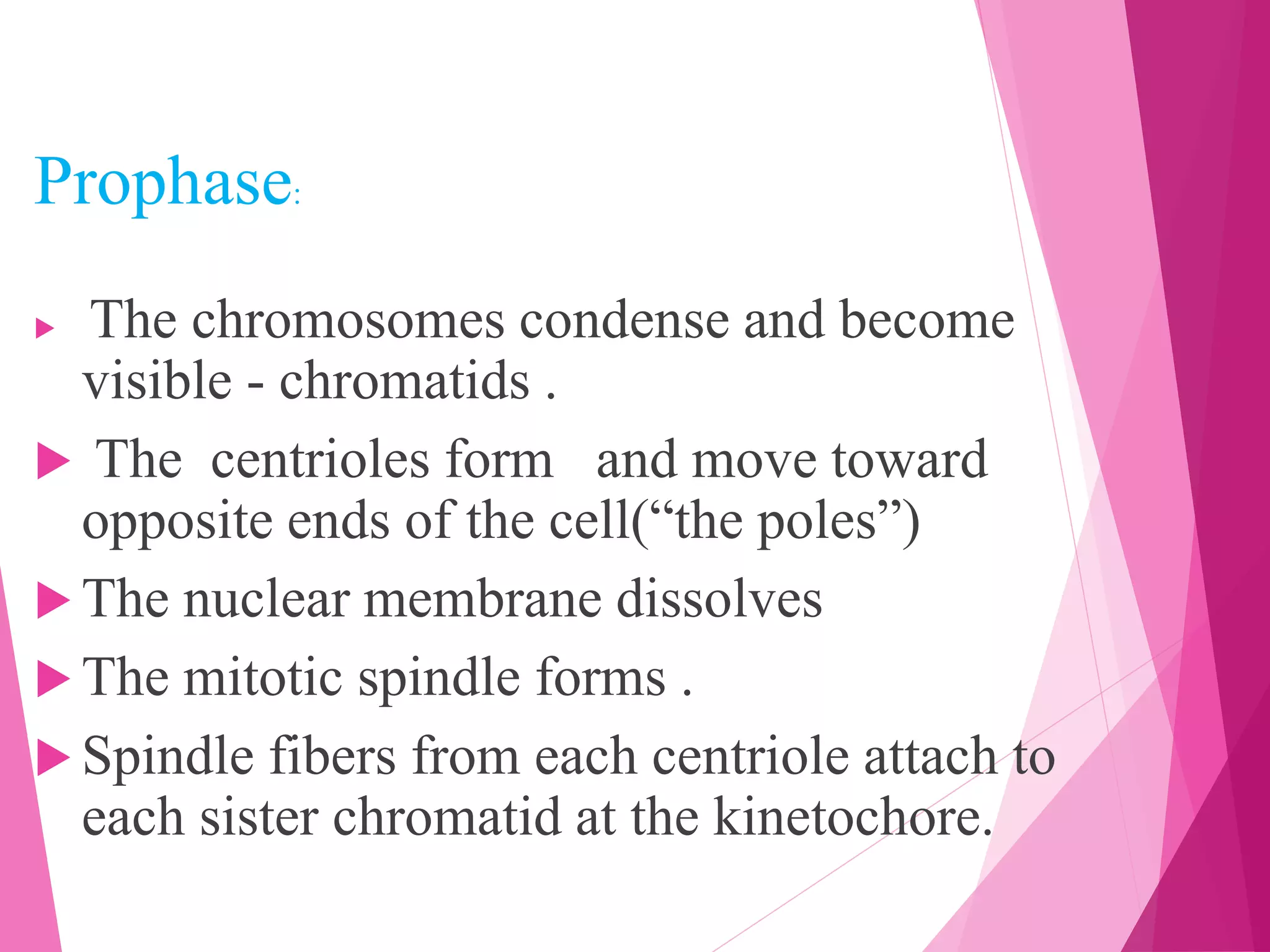
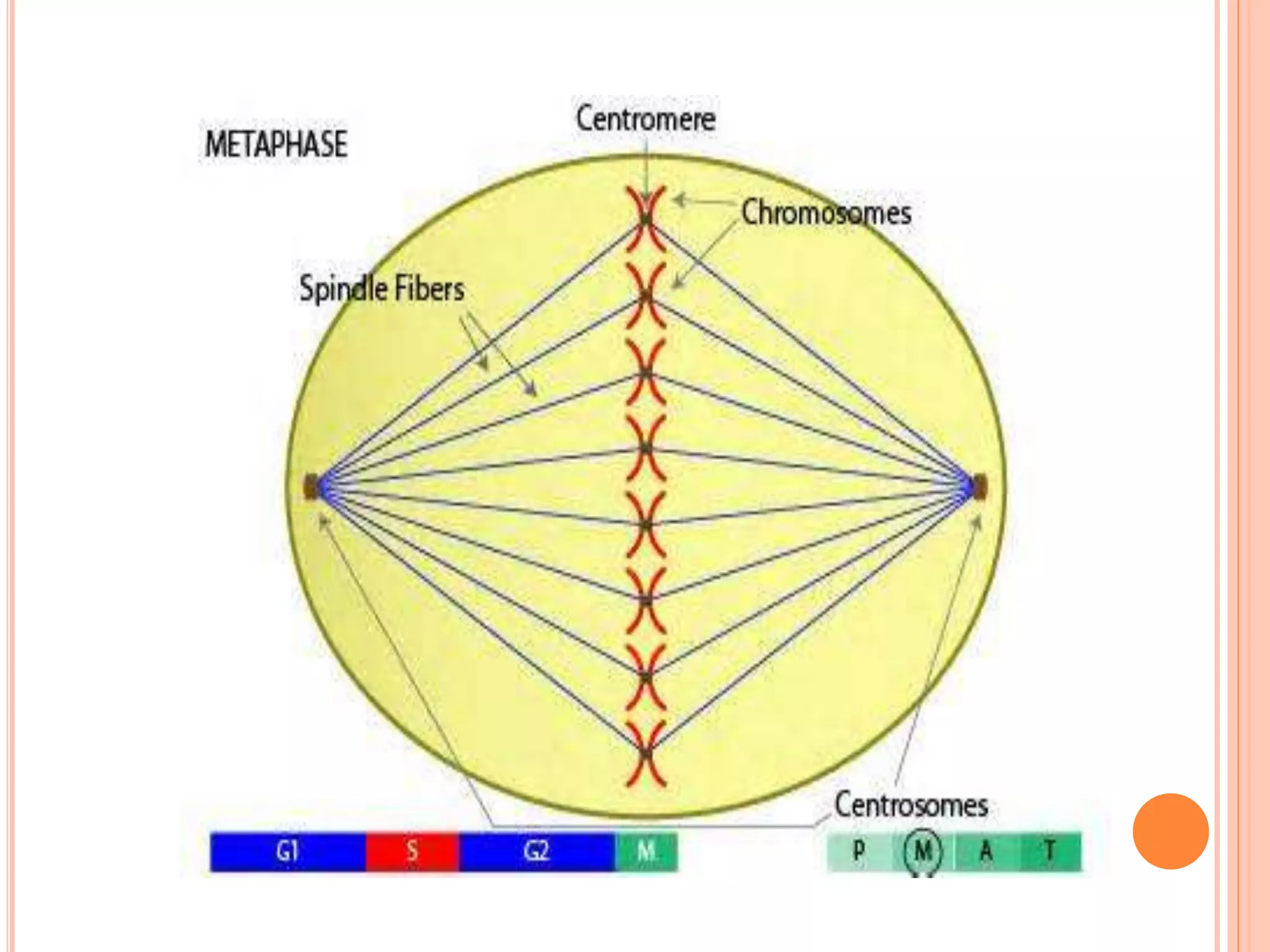
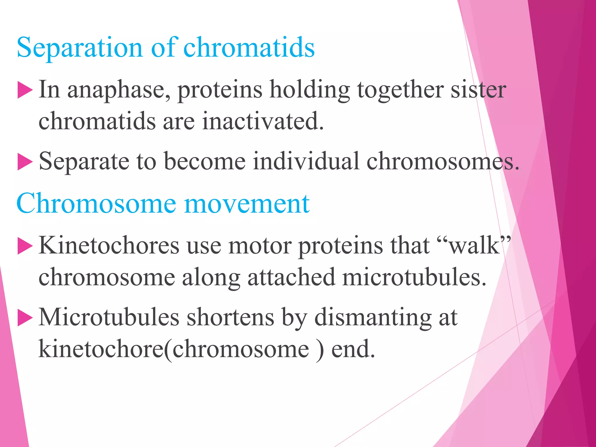
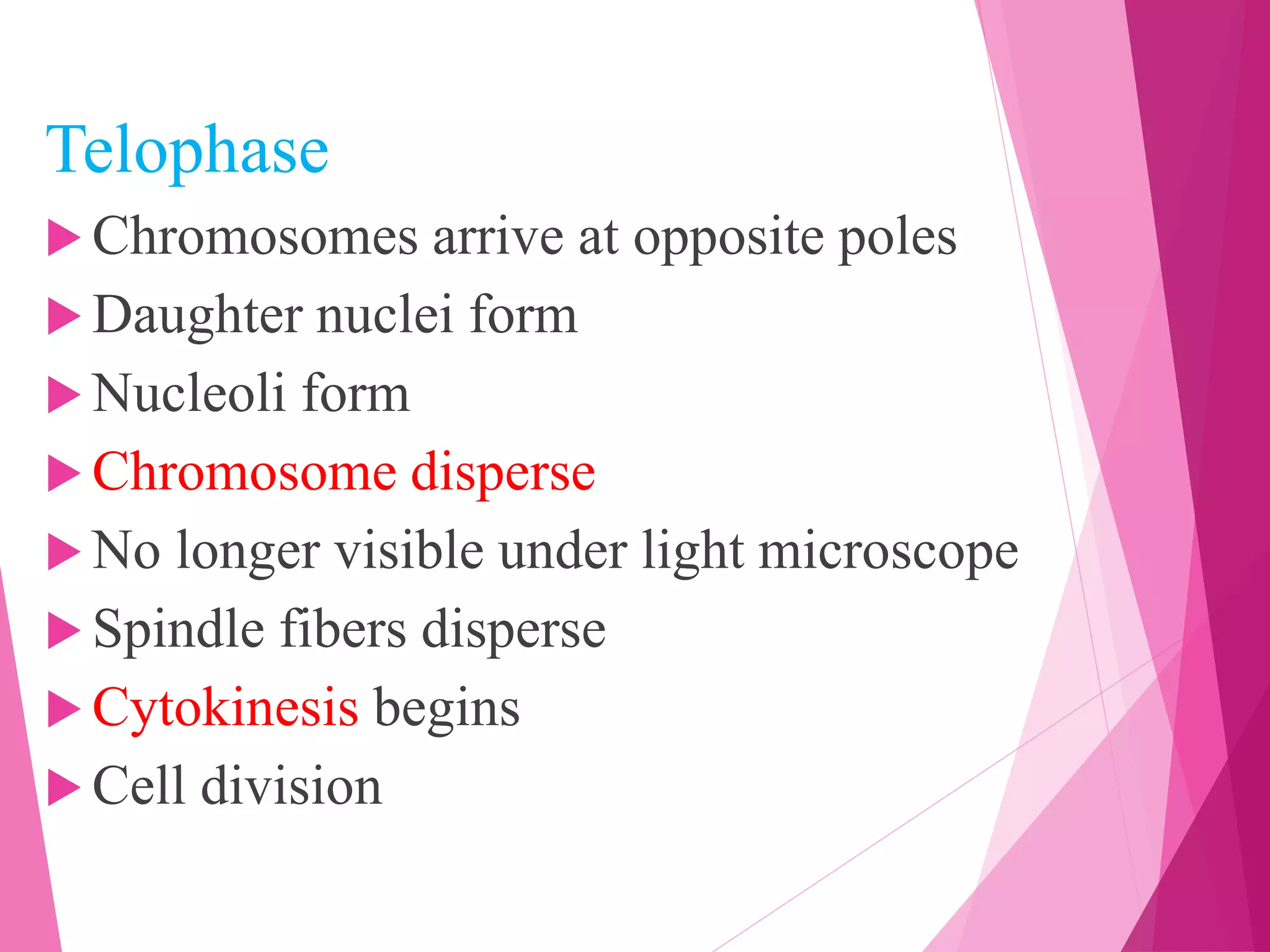
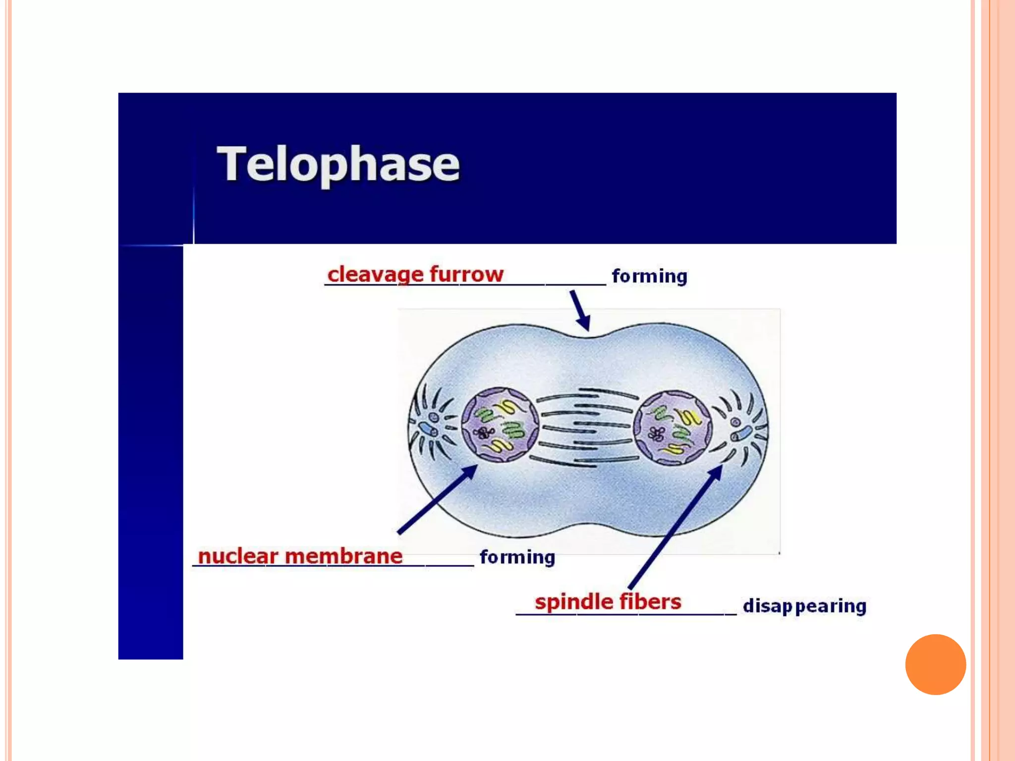
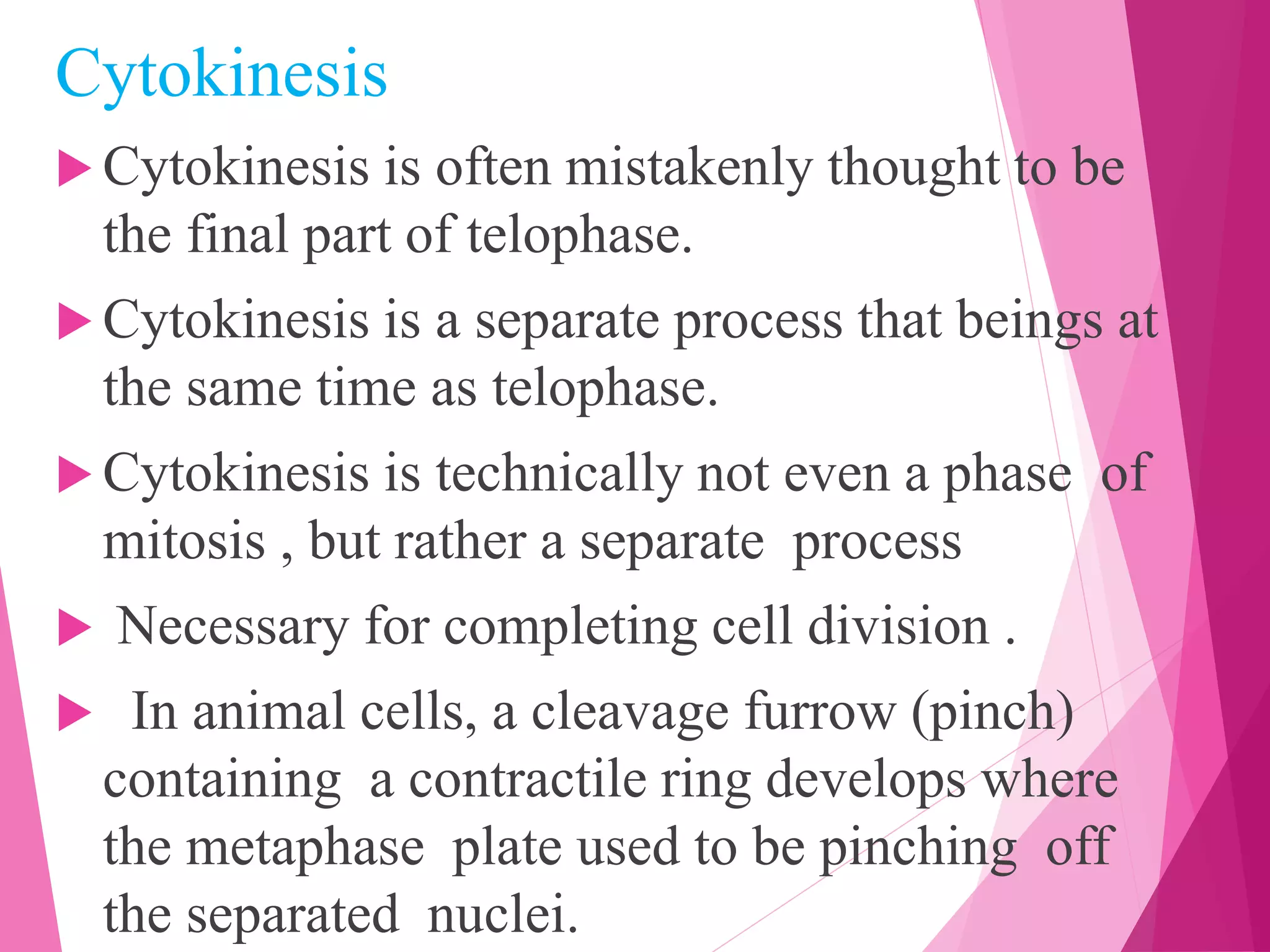
Mitotic cell division
RINI JOSEPH
Definition
 Mitosis is a process of nuclear division in
eukaryotic cells that occurs when a parent
cell divides to produce two identical daughter
cells .
 During cell division , mitosis refers
specifically to the separation of the
duplicated genetic material carried in the
nucleus .
Phases of mitosis
Mitosis consists of four basic phases :
1. Prophase
2. Metaphase
3. Anaphase
4. Telophase
EARLY PROPHASE :
In early prophase , the cell starts to break
down some structures and built others up
setting the stage for division of the
chromosomes .
» The chromosomes start to condense.
» Mitotic spindle begin to from –
microtubules , strong fibers that are part of the
cells “skeletal ”.
» The nucleolus a part of the nucleus where
ribosomes are ,made disappears .
Prophase:
 The chromosomes condense and become
visible - chromatids .
 The centrioles form and move toward
opposite ends of the cell(“the poles”)
 The nuclear membrane dissolves
 The mitotic spindle forms .
 Spindle fibers from each centriole attach to
each sister chromatid at the kinetochore.
 Creating kinetochores
 Microtubules attach at kinetochores
 Connect centromeres to centrioles
 Chromosomes begins moving.
Metaphase:
 Chromosomes align along middle of cell
 Metaphase plate
 Meta=middle
 Spindle fibers coordinate movement
 Helps to ensures chromosomes separate properly
 So each new nucleus receives only one copy of
each chromosome
Anaphase
 Sister chromatids separate at kinetochores
 Move to opposite poles
 Pulled at centromeres
 Pulled by motor proteins “waking” along
microtubules
 Increased production of ATP by mitochondria
 Poles move farther apart
 Polar microtubules lengthen
Separation of chromatids
 In anaphase, proteins holding together sister
chromatids are inactivated.
 Separate to become individual chromosomes.
Chromosome movement
 Kinetochores use motor proteins that “walk”
chromosome along attached microtubules.
 Microtubules shortens by dismanting at
kinetochore(chromosome ) end.
Telophase
 Chromosomes arrive at opposite poles
 Daughter nuclei form
 Nucleoli form
 Chromosome disperse
 No longer visible under light microscope
 Spindle fibers disperse
 Cytokinesis begins
 Cell division
Cytokinesis
 Cytokinesis is often mistakenly thought to be
the final part of telophase.
 Cytokinesis is a separate process that beings at
the same time as telophase.
 Cytokinesis is technically not even a phase of
mitosis , but rather a separate process
 Necessary for completing cell division .
 In animal cells, a cleavage furrow (pinch)
containing a contractile ring develops where
the metaphase plate used to be pinching off
the separated nuclei.
 In both animal and plants cells, cell division
is also driven by vesicles derived from the
golgi apparatus, which move along
microtubules to the middle of the cell
 In plant cell this structure coalesces into a
cell plate at the centre of phargmoplast and
develops into a cell wall, separating the two
nuclei.
Mitotic cell division

Mitotic cell division

  • 1.
  • 2.
    Definition  Mitosis isa process of nuclear division in eukaryotic cells that occurs when a parent cell divides to produce two identical daughter cells .  During cell division , mitosis refers specifically to the separation of the duplicated genetic material carried in the nucleus .
  • 4.
    Phases of mitosis Mitosisconsists of four basic phases : 1. Prophase 2. Metaphase 3. Anaphase 4. Telophase
  • 5.
    EARLY PROPHASE : Inearly prophase , the cell starts to break down some structures and built others up setting the stage for division of the chromosomes . » The chromosomes start to condense. » Mitotic spindle begin to from – microtubules , strong fibers that are part of the cells “skeletal ”. » The nucleolus a part of the nucleus where ribosomes are ,made disappears .
  • 6.
    Prophase:  The chromosomescondense and become visible - chromatids .  The centrioles form and move toward opposite ends of the cell(“the poles”)  The nuclear membrane dissolves  The mitotic spindle forms .  Spindle fibers from each centriole attach to each sister chromatid at the kinetochore.
  • 7.
     Creating kinetochores Microtubules attach at kinetochores  Connect centromeres to centrioles  Chromosomes begins moving.
  • 8.
    Metaphase:  Chromosomes alignalong middle of cell  Metaphase plate  Meta=middle  Spindle fibers coordinate movement  Helps to ensures chromosomes separate properly  So each new nucleus receives only one copy of each chromosome
  • 10.
    Anaphase  Sister chromatidsseparate at kinetochores  Move to opposite poles  Pulled at centromeres  Pulled by motor proteins “waking” along microtubules  Increased production of ATP by mitochondria  Poles move farther apart  Polar microtubules lengthen
  • 12.
    Separation of chromatids In anaphase, proteins holding together sister chromatids are inactivated.  Separate to become individual chromosomes. Chromosome movement  Kinetochores use motor proteins that “walk” chromosome along attached microtubules.  Microtubules shortens by dismanting at kinetochore(chromosome ) end.
  • 13.
    Telophase  Chromosomes arriveat opposite poles  Daughter nuclei form  Nucleoli form  Chromosome disperse  No longer visible under light microscope  Spindle fibers disperse  Cytokinesis begins  Cell division
  • 15.
    Cytokinesis  Cytokinesis isoften mistakenly thought to be the final part of telophase.  Cytokinesis is a separate process that beings at the same time as telophase.  Cytokinesis is technically not even a phase of mitosis , but rather a separate process  Necessary for completing cell division .  In animal cells, a cleavage furrow (pinch) containing a contractile ring develops where the metaphase plate used to be pinching off the separated nuclei.
  • 16.
     In bothanimal and plants cells, cell division is also driven by vesicles derived from the golgi apparatus, which move along microtubules to the middle of the cell  In plant cell this structure coalesces into a cell plate at the centre of phargmoplast and develops into a cell wall, separating the two nuclei.