What is CellDivision?
• Cell division can be defined as a process by which a cell distributes its genetic material and cytoplasm and
gives rise to new daughter cells. It is a part of the larger cell cycle and has a direct role in cell
reproduction.
• In well-developed organisms, there are two types of cell division observed, mitosis and meiosis. These are
very complex processes that are carried out through different phases.
• However, if simplified, mitosis can be defined as the exact duplication of a cell where the daughter cells
will have the same genetic information as the parent cell.
• In meiosis, the daughter cells will only have half of the genetic information of the original cell.
• The common end phase in both processes is cytokinesis and the division of the cytoplasm.
3.
Cell Division- Mitosisand Meiosis
The two well-documented types of cell division are:
1.Mitosis
2. Meiosis
3. Binary Fission
4.
Mitosis
• It isthe type of cell division where one cell divides to produce two genetically identical daughter cells.
• A great majority of cell divisions that take place in our body is mitosis.
• The process is integral to an organism's body growth and development, and it takes place throughout
the organism's lifetime.
• For some single-celled organisms such as yeast, mitotic cell division is the only way they can reproduce.
5.
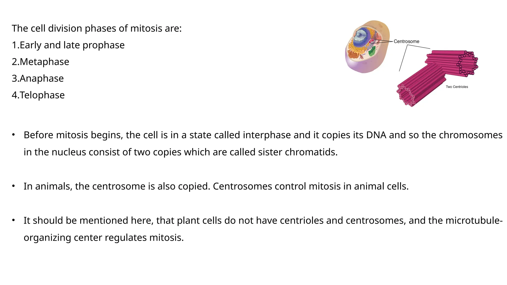
The cell divisionphases of mitosis are:
1.Early and late prophase
2.Metaphase
3.Anaphase
4.Telophase
• Before mitosis begins, the cell is in a state called interphase and it copies its DNA and so the chromosomes
in the nucleus consist of two copies which are called sister chromatids.
• In animals, the centrosome is also copied. Centrosomes control mitosis in animal cells.
• It should be mentioned here, that plant cells do not have centrioles and centrosomes, and the microtubule-
organizing center regulates mitosis.
6.
Early and LateProphase
• In the early prophase, the cell initiates cell division by breaking down some cell components and building
other components and then the chromosome division starts.
• In this stage, the chromosomes start to condense which helps them to separate easily in later stages
• Afterwards, the mitotic spindle starts to form, a structure made of microtubules. It organizes the
chromosomes and moves them around during mitosis. The mitotic spindle grows between the
centrosomes of the cell as they move towards different poles.
• The nucleolus then disappears which is a sign that the nucleus is getting ready to break down.
7.
• In thelate prophase which is also called prometaphase, the mitotic spindle starts to organize the
chromosomes.
• Once the chromosomes finish condensing, they form a compact structure.
• Then the nuclear envelope breaks down and the chromosomes are released.
• At the end of the prophase, the mitotic spindle grows, and some microtubules start to capture and
organize chromosomes.
8.
Metaphase
• Metaphase startswhen the mitotic spindle organizes all chromosomes and lines them up in the middle of
the cell to divide.
• All chromosomes align at the metaphase plate.
• At this stage of metaphase, the two kinetochores of each chromosome should be attached to
microtubules from opposite spindle poles. Before proceeding forward to anaphase, the cell will check if all
kinetochores are properly attached to microtubules and it is called spindle checkpoint.
• The spindle checkpoint ensures that the sister chromatids are split equally into two daughter cells.
9.
Anaphase
• In thisstage, the sister chromatids separate from each other and move towards the opposite poles of the
cell. The protein glue that holds them breaks and allows them to separate.
• Microtubules that are not attached to chromosomes elongate and push apart. In doing so they separate
the poles and makes the cell longer. These processes are controlled by motor proteins and these proteins
carry the chromosomes and microtubules as they move.
10.
Telophase
• In thisstage, the cell is almost divided and starts to re-establish its normal cellular structures as
cytokinesis takes place.
• The mitotic spindle breaks down into its building blocks and two new nuclei are formed, one for each set
of chromosomes.
• The nuclear membrane and the nucleoli then reappear and the chromosomes begin to de- condense to
return to their normal form.
11.
Cytokinesis
• In animalcells, cytokinesis is contractile. There's a pinch-like formation within the cell which divides it in
two like a coin purse with a 'drawstring'. The "drawstring" is a band of actin protein filaments. The pinch
crease is called the cleavage furrow.
• Plant cells can't be divided like this as they have a rigid cell wall and are too stiff. A cell plate forms down
the middle of the cell which splits the daughter cells.
12.
Meiosis
• In meiosis,a single cell divides twice to produce four cells that contain half of the original amount of
genetic material. It can be observed in sperm cells in males and egg cells in females.
There are 9 meiotic cell division phases. These are discussed below:
Interphase
• Similar to mitosis the genetic material of the cell is copied and two identical sets of chromosomes are
formed.
• The centrosomes and the centrioles are also copied and in this phase, the microtubules extend from
centrosomes.
13.
Prophase I
• Thetwo sets of chromosomes condense into an X-shaped formation
• Each chromosome consists of two sister chromatids which contain identical genetic information.
• All chromosomes pair up. For example, both copies of chromosome 1 and both copies of chromosome 2
are together.
• The chromosome pairs may then exchange parts of DNA through crossing over or recombination.
• In the end, in this stage, the nuclear membrane dissolves and releases the chromosomes.
• The meiotic spindle which consists of microtubules and other proteins extends across the cell.
14.
1. Leptotene: Inthis stage, the initiation of chromosome condensation takes place and it attains a composite form.
2. Zygotene: In this stage, the homologous chromosome pairs, the process is caleled chromosomal synapsis. It is followed by
the generation of a composite composition called synaptonemal complex.
3. Pachytene: In this stage, the crossing over of non-sister chromatoids of homologous chromose takes place. The
chromosome stay associated at the crosssing over sites.
4. Diplotene: It marks the synaptonemal complex dissolution and seperation of the homologous chromosomes except at the
crossing over sites. The formation of X-shaped compositions takes place at the time of seperation called chiasmata.
5. Diakinesis: It is signified by the end of chiasmata and assembly of the meiotic spindle to distinguish the homologous
chromosomes. The disappearance of nucleolus takes place and the nuclear envelopes dissociates.
15.
Metaphase I
• Thechromosome pairs align next to each other along the center of the cell.
• The centrioles move at the opposite poles of the cell and the meiotic spindles extend from them. Their
fibers attach to one chromosome of each pair.
Anaphase I
• The chromosome pairs are then separated by the meiotic spindle and move one chromosome to opposite
poles of the cell.
• In meiosis, the sister chromatids of the cell stay together.
16.
Telophase I andCytokinesis
• The chromosomes move to opposite poles of a cell and each pole has a full set of chromosomes.
• A nuclear membrane starts to form around each set of chromosomes to form two new nuclei.
• Cytokinesis takes place and two daughter cells are produced.
17.
Meiosis II
Prophase II
•At the end of meiosis, there are two daughter cells with 23 chromosomes
• The chromosomes condense again and form visible X-shaped structures
• The nuclear membrane will dissolve releasing the chromosomes.
• The centrioles duplicate and the meiotic spindle is formed.
Metaphase II
• Similar to metaphase I, the sister chromatid align along the center of the cell
• The centrioles move to opposite poles of the daughter cells.
• Meiotic spindle fibers attach to individual sister chromatids.
18.
Anaphase II
• Thesister chromatids are separated and moved to opposite poles by the meiotic spindle and they become
individual chromosomes.
Telophase II and Cytokinesis
• The chromosomes move to opposite poles of the cell and each pole has a full set of chromosomes.
• A nuclear membrane starts to form again and two new cell nuclei are formed.
• Cytokinesis takes place.
• Once cytokinesis is completed there are four new cells, with a haploid set of chromosomes
• In males, all four cells are sperm cells
• In females, one new is an egg cell and the others are polar bodies
Editor's Notes
#5 Centrosomes are membrane-free organelles that serve as main microtubule-organizing centres in distinct eukaryotic lineages