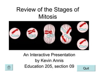
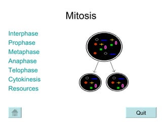
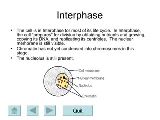
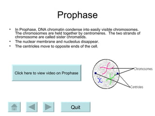
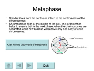
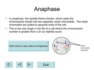
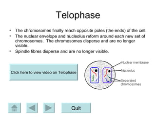
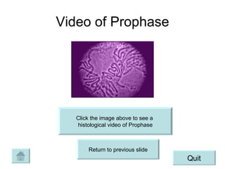
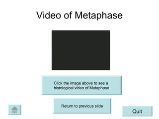
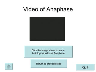
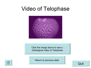
Review of the Stages of Mitosis An Interactive Presentation by Kevin Annis Education 205, section 09 Quit
Mitosis Interphase Prophase Metaphase Anaphase Telophase Cytokinesis Resources Quit
Interphase The cell is in Interphase for most of its life cycle.  In Interphase, the cell “prepares” for division by obtaining nutrients and growing, copying its DNA, and replicating its centrioles.  The nuclear membrane is still visible. Chromatin has not yet condensed into chromosomes in this stage. The nucleolus is still present. Quit
Prophase In Prophase, DNA chromatin condense into easily visible chromosomes.  The chromosomes are held together by centromeres.  The two strands of chromosome are called sister chromatids. The nuclear membrane and nucleolus disappear. The centrioles move to opposite ends of the cell. Quit Click here to view video on Prophase
Metaphase Spindle fibres from the centrioles attach to the centromeres of the chromosomes Chromosomes align at the middle of the cell. This organization helps to ensure that in the next phase, when the chromosomes are separated, each new nucleus will receive only one copy of each chromosome. Quit Click here to view video of Metaphase
Anaphase In anaphase, the spindle fibres shorten, which splits the chromosome strand into two separate, sister chromatids.  The sister chromatids are pulled to opposite ends of the cell. This is the only stage in the life of a cell where the chromosome number is greater than a 2n (or diploid) count. Quit Click here to view video of Anaphase
Telophase The chromosomes finally reach opposite poles (the ends) of the cell. The nuclear envelope and nucleolus reform around each new set of chromosomes.  The chromosomes disperse and are no longer visible. Spindle fibres disperse and are no longer visible. Quit Click here to view video on Telophase
Citokinesis Citokinesis marks the end of mitosis in the cell cycle.  It is where the cell officially splits into two separate cells, called daughter cells.  Each daughter cell now has the same number of chromosomes as the original cell before mitosis.  The cell splits when a furrow forms that pinches the cell in two. Quit Click on the link below to see video of the  overall process of Mitosis http://www.sp.uconn.edu/~bi107vc/images/anim/mitosis.gif
Video of Prophase Click the image above to see a  histological video of Prophase Return to previous slide Quit
Video of Metaphase Click the image above to see a  histological video of Metaphase Return to previous slide Quit
Video of Anaphase Click the image above to see a  histological video of Anaphase Return to previous slide Quit
Video of Telophase Click the image above to see a  histological video of Telophase Return to previous slide Quit
References Interphase through Telophase images: http://library.thinkquest.org/C0118084/Gene/Chromosomal_Inheritance/StagesMitosis.htm Cytokinesis image: http://citruscollege.edu/pic/46/c08_08a.jpg Information obtained from: http://www.biology.arizona.edu/Cell_bio/tutorials/cell_cycle/cells3.html Quit
Concept Map Quit
About the Author Kevin Annis is a junior at Grand Valley State University.  He is majoring in biology and is pursuing a minor in chemistry.  Currently he is considering getting a second minor in psychology.  He plans on teaching biology at the high school level when he completes college.  He most enjoys the summer months, where he spends most any free time golfing or fishing. Quit Click here to send me an e-mail

Mitosis Ppt

  • 1.
    Review of theStages of Mitosis An Interactive Presentation by Kevin Annis Education 205, section 09 Quit
  • 2.
    Mitosis Interphase ProphaseMetaphase Anaphase Telophase Cytokinesis Resources Quit
  • 3.
    Interphase The cellis in Interphase for most of its life cycle. In Interphase, the cell “prepares” for division by obtaining nutrients and growing, copying its DNA, and replicating its centrioles. The nuclear membrane is still visible. Chromatin has not yet condensed into chromosomes in this stage. The nucleolus is still present. Quit
  • 4.
    Prophase In Prophase,DNA chromatin condense into easily visible chromosomes. The chromosomes are held together by centromeres. The two strands of chromosome are called sister chromatids. The nuclear membrane and nucleolus disappear. The centrioles move to opposite ends of the cell. Quit Click here to view video on Prophase
  • 5.
    Metaphase Spindle fibresfrom the centrioles attach to the centromeres of the chromosomes Chromosomes align at the middle of the cell. This organization helps to ensure that in the next phase, when the chromosomes are separated, each new nucleus will receive only one copy of each chromosome. Quit Click here to view video of Metaphase
  • 6.
    Anaphase In anaphase,the spindle fibres shorten, which splits the chromosome strand into two separate, sister chromatids. The sister chromatids are pulled to opposite ends of the cell. This is the only stage in the life of a cell where the chromosome number is greater than a 2n (or diploid) count. Quit Click here to view video of Anaphase
  • 7.
    Telophase The chromosomesfinally reach opposite poles (the ends) of the cell. The nuclear envelope and nucleolus reform around each new set of chromosomes. The chromosomes disperse and are no longer visible. Spindle fibres disperse and are no longer visible. Quit Click here to view video on Telophase
  • 8.
    Citokinesis Citokinesis marksthe end of mitosis in the cell cycle. It is where the cell officially splits into two separate cells, called daughter cells. Each daughter cell now has the same number of chromosomes as the original cell before mitosis. The cell splits when a furrow forms that pinches the cell in two. Quit Click on the link below to see video of the overall process of Mitosis http://www.sp.uconn.edu/~bi107vc/images/anim/mitosis.gif
  • 9.
    Video of ProphaseClick the image above to see a histological video of Prophase Return to previous slide Quit
  • 10.
    Video of MetaphaseClick the image above to see a histological video of Metaphase Return to previous slide Quit
  • 11.
    Video of AnaphaseClick the image above to see a histological video of Anaphase Return to previous slide Quit
  • 12.
    Video of TelophaseClick the image above to see a histological video of Telophase Return to previous slide Quit
  • 13.
    References Interphase throughTelophase images: http://library.thinkquest.org/C0118084/Gene/Chromosomal_Inheritance/StagesMitosis.htm Cytokinesis image: http://citruscollege.edu/pic/46/c08_08a.jpg Information obtained from: http://www.biology.arizona.edu/Cell_bio/tutorials/cell_cycle/cells3.html Quit
  • 14.
  • 15.
    About the AuthorKevin Annis is a junior at Grand Valley State University. He is majoring in biology and is pursuing a minor in chemistry. Currently he is considering getting a second minor in psychology. He plans on teaching biology at the high school level when he completes college. He most enjoys the summer months, where he spends most any free time golfing or fishing. Quit Click here to send me an e-mail