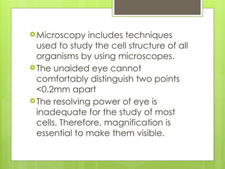
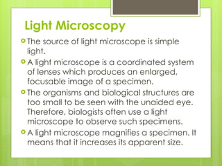
MICROSCOPY
Microscopy includes techniques
used to study the cell structure of all
organisms by using microscopes.
The unaided eye cannot
comfortably distinguish two points
<0.2mm apart
The resolving power of eye is
inadequate for the study of most
cells. Therefore, magnification is
essential to make them visible.
Light Microscopy
 The source of light microscope is simple
light.
 A light microscope is a coordinated system
of lenses which produces an enlarged,
focusable image of a specimen.
 The organisms and biological structures are
too small to be seen with the unaided eye.
Therefore, biologists often use a light
microscope to observe such specimens.
 A light microscope magnifies a specimen. It
means that it increases its apparent size.
Principles of light microscopy
 Principle. The light microscope is an instrument
for visualizing fine detail of an object. It does this
by creating a magnified image through the use
of a series of glass lenses, which first focus a
beam of light onto or through an object, and
convex objective lenses to enlarge the image
formed.
 In the majority of light microscopes, the image is
viewed directly through binocular eyepieces
that act as a secondary lens in the form of a
magnifying glass to observe the projected
image.
 For light
microscopy,
visible light is
passed through
the specimen and
then through a
series of lenses
that bend the
light in a manner
that results in
magnification of
the organisms
present in the
specimen
Application of Light Microscopy
 Light microscopes play an important role in many
research laboratories. Biologists use microscopes to
observe objects and details at a cellular level to learn
more about the building blocks of all organisms.
 Microscopes are used when studying light and optics
to learn how light refracts through converging lenses
and how a combination of different lenses with
varying focal lengths affects the properties of the
image.
 Microscopes are used in schools as part of the
technology section of the curriculum. Microscopes
provide students with an understanding of real cells
and their supporting structures.
 Microscopes are used in forensics to help solve
many crimes. Often times, there will be human
evidences left on the crime scene. This allows
forensic scientists to examine the evidence under
a microscope and match the results with a
database to find the culprit.
 Mineralogists also use light microscopy, typically
with a special preparation of a sample called
thin section. Thin sections are very thin slices of a
rock. The sample needs to be thin enough for
light to travel through from the light source to the
user's eye. The thin section will allow the shape of
different crystal grains to be seen.
Terms Commonly used in Microscopy
1. Magnification
Magnification is the capacity of an optical
instrument to increase the size of an object than its
original size.
 Magnification of a microscope is the product of
multiplying the individual magnification of the
ocular (eye piece) and objectives.
Magnification power of a microscope = Objective
magnification x ocular magnification
 E.g. If the eye piece is 10X and objective is 40X
then the specimen image is magnified 400 times.
i.e. 10X x 40X = 400X
2. Resolution/ Resolving power
 Effective magnification depends on the resolving
power of a microscope.
The resolving power is the ability to distinguish two
adjacent objects as separate and distinct images
rather than as a single blurred image.
 The minimum capacity of a lens to differentiate
between two adjacent points is called resolution
power of the lens.
 Resolution is the ability to distinguish two points as
separate points. Our naked eye can distinguish
two points. Such points have at least 0.1mm
distance. Therefore, resolution of naked eye is
0.1mm.
3. Contrast
 Contrast is the difference between the lightest
and darkest parts of an image.
 The ability to distinguish detail also depends
on contrast.
 Therefore, many specimens examined with a
light microscope are stained with artificial
dyes. These dyes increase the contrast and
make the specimen more visible.
 Contrast refers to the darkness of the
background relative to the specimen. Lighter
specimens are easier to see on darker
backgrounds.

MICROSCOPY, Light Microscopy, Magnification, Resolution, Contrast.pptx

  • 1.
  • 2.
    Microscopy includes techniques usedto study the cell structure of all organisms by using microscopes. The unaided eye cannot comfortably distinguish two points <0.2mm apart The resolving power of eye is inadequate for the study of most cells. Therefore, magnification is essential to make them visible.
  • 3.
    Light Microscopy  Thesource of light microscope is simple light.  A light microscope is a coordinated system of lenses which produces an enlarged, focusable image of a specimen.  The organisms and biological structures are too small to be seen with the unaided eye. Therefore, biologists often use a light microscope to observe such specimens.  A light microscope magnifies a specimen. It means that it increases its apparent size.
  • 4.
    Principles of lightmicroscopy  Principle. The light microscope is an instrument for visualizing fine detail of an object. It does this by creating a magnified image through the use of a series of glass lenses, which first focus a beam of light onto or through an object, and convex objective lenses to enlarge the image formed.  In the majority of light microscopes, the image is viewed directly through binocular eyepieces that act as a secondary lens in the form of a magnifying glass to observe the projected image.
  • 5.
     For light microscopy, visiblelight is passed through the specimen and then through a series of lenses that bend the light in a manner that results in magnification of the organisms present in the specimen
  • 7.
    Application of LightMicroscopy  Light microscopes play an important role in many research laboratories. Biologists use microscopes to observe objects and details at a cellular level to learn more about the building blocks of all organisms.  Microscopes are used when studying light and optics to learn how light refracts through converging lenses and how a combination of different lenses with varying focal lengths affects the properties of the image.  Microscopes are used in schools as part of the technology section of the curriculum. Microscopes provide students with an understanding of real cells and their supporting structures.
  • 8.
     Microscopes areused in forensics to help solve many crimes. Often times, there will be human evidences left on the crime scene. This allows forensic scientists to examine the evidence under a microscope and match the results with a database to find the culprit.  Mineralogists also use light microscopy, typically with a special preparation of a sample called thin section. Thin sections are very thin slices of a rock. The sample needs to be thin enough for light to travel through from the light source to the user's eye. The thin section will allow the shape of different crystal grains to be seen.
  • 9.
    Terms Commonly usedin Microscopy 1. Magnification Magnification is the capacity of an optical instrument to increase the size of an object than its original size.  Magnification of a microscope is the product of multiplying the individual magnification of the ocular (eye piece) and objectives. Magnification power of a microscope = Objective magnification x ocular magnification  E.g. If the eye piece is 10X and objective is 40X then the specimen image is magnified 400 times. i.e. 10X x 40X = 400X
  • 11.
    2. Resolution/ Resolvingpower  Effective magnification depends on the resolving power of a microscope. The resolving power is the ability to distinguish two adjacent objects as separate and distinct images rather than as a single blurred image.  The minimum capacity of a lens to differentiate between two adjacent points is called resolution power of the lens.  Resolution is the ability to distinguish two points as separate points. Our naked eye can distinguish two points. Such points have at least 0.1mm distance. Therefore, resolution of naked eye is 0.1mm.
  • 13.
    3. Contrast  Contrastis the difference between the lightest and darkest parts of an image.  The ability to distinguish detail also depends on contrast.  Therefore, many specimens examined with a light microscope are stained with artificial dyes. These dyes increase the contrast and make the specimen more visible.  Contrast refers to the darkness of the background relative to the specimen. Lighter specimens are easier to see on darker backgrounds.