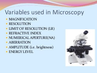
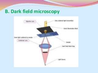
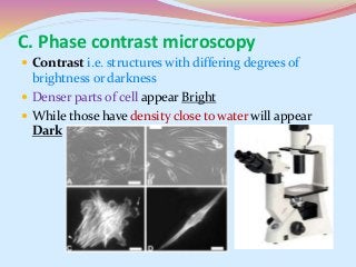
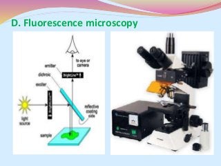
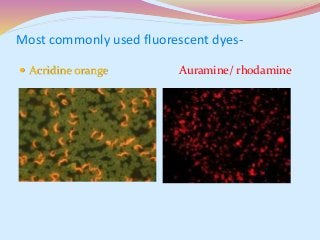
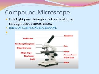
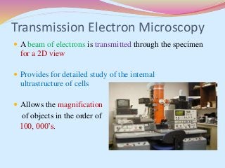
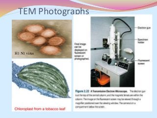
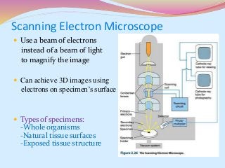
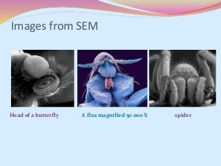
Raveendra Kumar Vidyarthi
(Ph.D Scholar)
Supervisor- Dr. Rajeshwar K.K. Arya
Department of Pharmaceutical Sciences
Bhimtal Campus, K.U. Nainital
Contents
 Introduction to microscopy
 Microscope
 Variables used in microscopy
 Principle of microscopy
 Types of microscopy
 Types of Microscope
 Uses
Introduction to Microscopy
 The science of investigating small objects using a
microscope is called microscopy
 Anton van Leeuwenhoek is generally
credited with bringing the microscope
to the attention of biologists
 1661 - He discovered bacteria, free-living
parasitic microbes, sperm cells, blood cells, microscopic
nematodes etc.
Microscope
 A microscope (Greek: micron = small and scopos = aim)
 A device that transmits light through several lenses to
produce an enlarged image of a microscopic specimen
 MICROSCOPE - An instrument for viewing
objects that are too small to be seen by the
naked or unaided eye
Variables used in Microscopy
 MAGNIFICATION
 RESOLUTION
 LIMIT OF RESOLUTION (LR)
 REFRACTIVE INDEX
 NUMERICAL APERTURE(NA)
 ABERRATION
 AMPLITUDE (i.e. brightness)
 ENERGY LEVEL
Magnification
 Degree of enlargement
 No of times the length, breadth or
diameter, of an object is multiplied
 Also known as Resolving power
 The capacity of a microscope to distinguish images of
two pointed objects lying very close to each other.
Resolution
Limit of Resolution
 Limit of Resolution is described mathematically by an
equation developed in the 1870s by Ernst Abbe,
 Where, d= minimal distance (d) between two objects
λ= wavelength of light
(n sin ѳ)= numerical aperture of the lens
Resolving power of std LM=200nm
Numerical aperture
 Ratio of diameter of lens to its focal length
NA = n Sin θ
n = refractive index,
θ = angle of aperture
 The numerical aperture with respect to a point P
depends on the half-angle θ of the maximum cone of
light that can enter or exit the lens.
Aberration
 Chromatic aberration
 Correction of aberration – Achromatic
objective and Apochromatic objectives
Principle of Microscopy
 The principle of Microscopy is Refraction.
 Refractive index is the light bending ability of
a medium
 The light may bend in air so much
that it misses the small high-magnification
lens.
 Immersion oil is used to keep light
away from bending.
RI of Air- 1.0
RI of Water -1.3
RI of Glass(avg)- 1.5
RI of Oil- 1.52
TYPES OF LIGHT MICROSCOPY
 Bright Field Microscopy
 Dark Field Microscopy
 Phase Contrast Microscopy
 Fluorescent Microscopy
 Confocal Microscopy
A. Bright field microscopy
B. Dark field microscopy
This method used for unstained preparation
 Contrast i.e. structures with differing degrees of
brightness or darkness
 Denser parts of cell appear Bright
 While those have density close to water will appear
Dark
C. Phase contrast microscopy
D. Fluorescence microscopy
Most commonly used fluorescent dyes-
 Acridine orange Auramine/ rhodamine
Types of Microscope
 Light microscope (simple and Compound)
 Electron microscope ( SEM, TEM)
 Scanning Probe microscope
e.g. Scanning tunneling microscope &
Atomic force microscope
Compound Microscope
 Lets light pass through an object and then
through two or more lenses.
 PARTS OF COMPOUND MICROSCOPE

Transmission Electron Microscopy
 A beam of electrons is transmitted through the specimen
for a 2D view
 Provides for detailed study of the internal
ultrastructure of cells
 Allows the magnification
of objects in the order of
100, 000’s.
TEM Photographs
H1 N1 virus
Scanning Electron Microscope
 Use a beam of electrons
instead of a beam of light
to magnify the image
 Can achieve 3D images using
electrons on specimen’s surface
 Types of specimens:
-Whole organisms
-Natural tissue surfaces
-Exposed tissue structure
Images from SEM
Head of a butterfly A flea magnified 50 000 X spider
USES OF MICROSCOPE
 1. Drawing Sketches With Camera Lucida
It is used to draw clear, simple and exactly proportionate
outline sketches of the objects under study.
 2. Micrometry - the field of science in which microscopic
objects like chromosomes, cells, etc, are measured with the help
of the compound microscope with the help of micrometer.
 In pharmaceutical science- evaluation of nanoparticles,
microemulsions, microspheres, powder microscopy, etc
References
 http://nobelprize.org/educational/physics/microscop
es/1.html
 www.googlescholars.com
 J. M. Willey, L.M. Sherwood, C. J. Woolverton, “ Prescott’s
Microbiology” 7th edition, Mcgraw Hill publication. 2014 p.p 22-41.
Light and Scanning Electron Microscopy By RK Vidyarthi

Light and Scanning Electron Microscopy By RK Vidyarthi

  • 1.
    Raveendra Kumar Vidyarthi (Ph.DScholar) Supervisor- Dr. Rajeshwar K.K. Arya Department of Pharmaceutical Sciences Bhimtal Campus, K.U. Nainital
  • 2.
    Contents  Introduction tomicroscopy  Microscope  Variables used in microscopy  Principle of microscopy  Types of microscopy  Types of Microscope  Uses
  • 3.
    Introduction to Microscopy The science of investigating small objects using a microscope is called microscopy  Anton van Leeuwenhoek is generally credited with bringing the microscope to the attention of biologists  1661 - He discovered bacteria, free-living parasitic microbes, sperm cells, blood cells, microscopic nematodes etc.
  • 4.
    Microscope  A microscope(Greek: micron = small and scopos = aim)  A device that transmits light through several lenses to produce an enlarged image of a microscopic specimen  MICROSCOPE - An instrument for viewing objects that are too small to be seen by the naked or unaided eye
  • 5.
    Variables used inMicroscopy  MAGNIFICATION  RESOLUTION  LIMIT OF RESOLUTION (LR)  REFRACTIVE INDEX  NUMERICAL APERTURE(NA)  ABERRATION  AMPLITUDE (i.e. brightness)  ENERGY LEVEL
  • 6.
    Magnification  Degree ofenlargement  No of times the length, breadth or diameter, of an object is multiplied  Also known as Resolving power  The capacity of a microscope to distinguish images of two pointed objects lying very close to each other. Resolution
  • 7.
    Limit of Resolution Limit of Resolution is described mathematically by an equation developed in the 1870s by Ernst Abbe,  Where, d= minimal distance (d) between two objects λ= wavelength of light (n sin ѳ)= numerical aperture of the lens Resolving power of std LM=200nm
  • 8.
    Numerical aperture  Ratioof diameter of lens to its focal length NA = n Sin θ n = refractive index, θ = angle of aperture  The numerical aperture with respect to a point P depends on the half-angle θ of the maximum cone of light that can enter or exit the lens.
  • 9.
    Aberration  Chromatic aberration Correction of aberration – Achromatic objective and Apochromatic objectives
  • 10.
    Principle of Microscopy The principle of Microscopy is Refraction.  Refractive index is the light bending ability of a medium  The light may bend in air so much that it misses the small high-magnification lens.  Immersion oil is used to keep light away from bending. RI of Air- 1.0 RI of Water -1.3 RI of Glass(avg)- 1.5 RI of Oil- 1.52
  • 11.
    TYPES OF LIGHTMICROSCOPY  Bright Field Microscopy  Dark Field Microscopy  Phase Contrast Microscopy  Fluorescent Microscopy  Confocal Microscopy
  • 12.
    A. Bright fieldmicroscopy
  • 13.
    B. Dark fieldmicroscopy
  • 14.
    This method usedfor unstained preparation
  • 15.
     Contrast i.e.structures with differing degrees of brightness or darkness  Denser parts of cell appear Bright  While those have density close to water will appear Dark C. Phase contrast microscopy
  • 16.
  • 17.
    Most commonly usedfluorescent dyes-  Acridine orange Auramine/ rhodamine
  • 18.
    Types of Microscope Light microscope (simple and Compound)  Electron microscope ( SEM, TEM)  Scanning Probe microscope e.g. Scanning tunneling microscope & Atomic force microscope
  • 19.
    Compound Microscope  Letslight pass through an object and then through two or more lenses.  PARTS OF COMPOUND MICROSCOPE 
  • 20.
    Transmission Electron Microscopy A beam of electrons is transmitted through the specimen for a 2D view  Provides for detailed study of the internal ultrastructure of cells  Allows the magnification of objects in the order of 100, 000’s.
  • 21.
  • 22.
    Scanning Electron Microscope Use a beam of electrons instead of a beam of light to magnify the image  Can achieve 3D images using electrons on specimen’s surface  Types of specimens: -Whole organisms -Natural tissue surfaces -Exposed tissue structure
  • 23.
    Images from SEM Headof a butterfly A flea magnified 50 000 X spider
  • 24.
    USES OF MICROSCOPE 1. Drawing Sketches With Camera Lucida It is used to draw clear, simple and exactly proportionate outline sketches of the objects under study.  2. Micrometry - the field of science in which microscopic objects like chromosomes, cells, etc, are measured with the help of the compound microscope with the help of micrometer.  In pharmaceutical science- evaluation of nanoparticles, microemulsions, microspheres, powder microscopy, etc
  • 25.
    References  http://nobelprize.org/educational/physics/microscop es/1.html  www.googlescholars.com J. M. Willey, L.M. Sherwood, C. J. Woolverton, “ Prescott’s Microbiology” 7th edition, Mcgraw Hill publication. 2014 p.p 22-41.