Whose discovery ofmicrobes in sealed
flasks of broth that had been boiled
revived the theory of spontaneous
generation?
Robert Hooke
Francesco
Redi
Antony van
Leeuwenhoek
John
Needham
Lazzaro
Spallazani
Louis Pasteur John Tyndall
4.
Whose discovery ofmicrobes in sealed
flasks of broth that had been boiled
revived the theory of spontaneous
generation?
Robert Hooke
Francesco
Redi
Antony van
Leeuwenhoek
John
Needham
Lazzaro
Spallazani
Louis Pasteur John Tyndall
5.
Who helped disprovespontaneous generation by
demonstrating that boiled nutrient broths would
remain sterile as long as microbes in the air could
not settle onto the surface of the liquid?
Robert Hooke
Francesco
Redi
Antony van
Leeuwenhoek
John
Needham
Lazzaro
Spallazani
Louis Pasteur John Tyndall
6.
Who helped disprovespontaneous generation by
demonstrating that boiled nutrient broths would
remain sterile as long as microbes in the air could
not settle onto the surface of the liquid?
Robert Hooke
Francesco
Redi
Antony van
Leeuwenhoek
John
Needham
Lazzaro
Spallazani
Louis Pasteur John Tyndall
7.
Whose experiments usingboiling as a method of
sterilizing broths lead him to suggest that some
microbes exist in two forms, one that is
susceptible to heat and one that is not?
Robert Hooke
Francesco
Redi
Antony van
Leeuwenhoek
John
Needham
Lazzaro
Spallazani
Louis Pasteur John Tyndall
8.
Whose experiments usingboiling as a method of
sterilizing broths lead him to suggest that some
microbes exist in two forms, one that is
susceptible to heat and one that is not?
Robert Hooke
Francesco
Redi
Antony van
Leeuwenhoek
John
Needham
Lazzaro
Spallazani
Louis Pasteur John Tyndall
9.
This researcher sealedboiled nutrient broths in
glass flasks in two ways, with corks and with
fused-necks, showing that only broths exposed to
air after boiling contained microbes.
Robert Hooke
Francesco
Redi
Antony van
Leeuwenhoek
John
Needham
Lazzaro
Spallazani
Louis Pasteur John Tyndall
10.
This researcher sealedboiled nutrient broths in
glass flasks in two ways, with corks and with
fused-necks, showing that only broths exposed to
air after boiling contained microbes.
Robert Hooke
Francesco
Redi
Antony van
Leeuwenhoek
John
Needham
Lazzaro
Spallazani
Louis Pasteur John Tyndall
11.
Who viewed anddescribed the actions
of microbial “animalcules” using a
homemade microscope?
Robert Hooke
Francesco
Redi
Antony van
Leeuwenhoek
John
Needham
Lazzaro
Spallazani
Louis Pasteur John Tyndall
12.
Who viewed anddescribed the actions
of microbial “animalcules” using a
homemade microscope?
Robert Hooke
Francesco
Redi
Antony van
Leeuwenhoek
John
Needham
Lazzaro
Spallazani
Louis Pasteur John Tyndall
13.
Whose early challengeto spontaneous
generation involved covering rotting meat
with gauze, preventing flies from landing
and depositing eggs?
Robert Hooke
Francesco
Redi
Antony van
Leeuwenhoek
John
Needham
Lazzaro
Spallazani
Louis Pasteur John Tyndall
14.
Whose early challengeto spontaneous
generation involved covering rotting meat
with gauze, preventing flies from landing
and depositing eggs?
Robert Hooke
Francesco
Redi
Antony van
Leeuwenhoek
John
Needham
Lazzaro
Spallazani
Louis Pasteur John Tyndall
15.
Who described the“microbial
mushroom” of bread mold using a
homemade microscope?
Robert Hooke
Francesco
Redi
Antony van
Leeuwenhoek
John
Needham
Lazzaro
Spallazani
Louis Pasteur John Tyndall
16.
Who described the“microbial
mushroom” of bread mold using a
homemade microscope?
Robert Hooke
Francesco
Redi
Antony van
Leeuwenhoek
John
Needham
Lazzaro
Spallazani
Louis Pasteur John Tyndall
17.
Who
invented a
method of
usingheat
to sterilize
food that is
still in use
today?
Oliver Wendell Holmes & Ignaz
Semmelweiss
Joseph Lister
Louis Pasteur
Robert Koch
Antony van Leeuwenhoek
18.
Who
invented a
method of
usingheat
to sterilize
food that is
still in use
today?
Oliver Wendell Holmes & Ignaz
Semmelweiss
Joseph Lister
Louis Pasteur
Robert Koch
Antony van Leeuwenhoek
Who made
over 250
microscopes,
including
somethat
were capable
of magnifying
specimens to
300 times
normal size?
Oliver Wendell Holmes & Ignaz
Semmelweiss
Joseph Lister
Louis Pasteur
Robert Koch
Antony van Leeuwenhoek
22.
Who made
over 250
microscopes,
including
somethat
were capable
of magnifying
specimens to
300 times
normal size?
Oliver Wendell Holmes & Ignaz
Semmelweiss
Joseph Lister
Louis Pasteur
Robert Koch
Antony van Leeuwenhoek
Who is
credited with
stressingthe
importance of
handwashing
in clinical
settings?
Oliver Wendell Holmes & Ignaz
Semmelweiss
Joseph Lister
Louis Pasteur
Robert Koch
Antony van Leeuwenhoek
26.
Who is
credited with
stressingthe
importance of
handwashing
in clinical
settings?
Oliver Wendell Holmes & Ignaz
Semmelweiss
Joseph Lister
Louis Pasteur
Robert Koch
Antony van Leeuwenhoek
27.
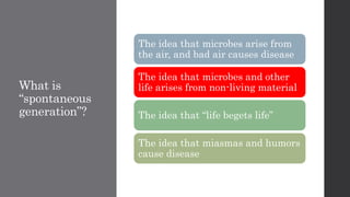
What is
“spontaneous
generation”?
The ideathat microbes arise from
the air, and bad air causes disease
The idea that microbes and other
life arises from non-living material
The idea that “life begets life”
The idea that miasmas and humors
cause disease
28.
What is
“spontaneous
generation”?
The ideathat microbes arise from
the air, and bad air causes disease
The idea that microbes and other
life arises from non-living material
The idea that “life begets life”
The idea that miasmas and humors
cause disease
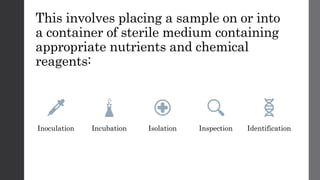
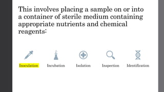
This involves placinga sample on or into
a container of sterile medium containing
appropriate nutrients and chemical
reagents:
Inoculation Incubation Isolation Inspection Identification
31.
This involves placinga sample on or into
a container of sterile medium containing
appropriate nutrients and chemical
reagents:
Inoculation Incubation Isolation Inspection Identification
32.
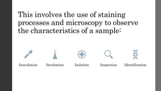
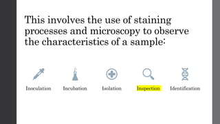
This involves theuse of staining
processes and microscopy to observe
the characteristics of a sample:
Inoculation Incubation Isolation Inspection Identification
33.
This involves theuse of staining
processes and microscopy to observe
the characteristics of a sample:
Inoculation Incubation Isolation Inspection Identification
34.
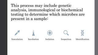
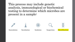
This process mayinclude genetic
analysis, immunological or biochemical
testing to determine which microbes are
present in a sample:
Inoculation Incubation Isolation Inspection Identification
35.
This process mayinclude genetic
analysis, immunological or biochemical
testing to determine which microbes are
present in a sample:
Inoculation Incubation Isolation Inspection Identification
36.
This process involvesseparating
specific colonies from a sample for
further study:
Inoculation Incubation Isolation Inspection Identification
37.
This process involvesseparating
specific colonies from a sample for
further study:
Inoculation Incubation Isolation Inspection Identification
38.
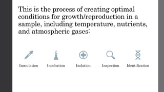
This is theprocess of creating optimal
conditions for growth/reproduction in a
sample, including temperature, nutrients,
and atmospheric gases:
Inoculation Incubation Isolation Inspection Identification
39.
This is theprocess of creating optimal
conditions for growth/reproduction in a
sample, including temperature, nutrients,
and atmospheric gases:
Inoculation Incubation Isolation Inspection Identification
40.
In which domain/swould you find
prokaryotes?
ARCHAEA EUKARYA BACTERIA
41.
In which domain/swould you find
prokaryotes?
ARCHAEA EUKARYA BACTERIA
In which domainwould you place
fungi?
ARCHAEA EUKARYA BACTERIA
55.
In which domainwould you place
fungi?
ARCHAEA EUKARYA BACTERIA
56.
In which domainwould you place
algae?
ARCHAEA EUKARYA BACTERIA
57.
In which domainwould you place
algae?
ARCHAEA EUKARYA BACTERIA
58.
In which domainwould you place
algae?
ARCHAEA EUKARYA BACTERIA
59.
In which domainwould you place
protozoans?
ARCHAEA EUKARYA BACTERIA
60.
In which domainwould you place
protozoans?
ARCHAEA EUKARYA BACTERIA
61.
Which of thefollowing statements is in
correct order from largest to smallest?
• Viruses, bacteria, eukaryotic cells, helminth
• Bacteria, viruses, helminth, eukaryotic cells
• Eukaryotic cells, helminth, viruses, bacteria
• Helminth, eukaryotic cells, bacteria, viruses
62.
Which of thefollowing statements is in
correct order from largest to smallest?
• Viruses, bacteria, eukaryotic cells, helminth
• Bacteria, viruses, helminth, eukaryotic cells
• Eukaryotic cells, helminth, viruses, bacteria
• Helminth, eukaryotic cells, bacteria, viruses
A ____ dyehas a _____ charge, causing it to
_____ charged particles in the cell envelope.
• Basic, positive, bind to
• Acidic, negative, bind to
• Basic, positive, repel
• Acidic, positive, repel
65.
A ____ dyehas a _____ charge, causing it to
_____ charged particles in the cell envelope.
• Basic, positive, bind to
• Acidic, negative, bind to
• Basic, positive, repel
• Acidic, positive, repel
66.
A ____ dyehas a ____ charge, which is ______ by
charged particles in the cell envelope.
• Basic, positive, repelled
• Basic, negative, bound
• Acidic, negative, repelled
• Acidic, negative, bound
67.
A ____ dyehas a ____ charge, which is ______ by
charged particles in the cell envelope.
• Basic, positive, repelled
• Basic, negative, bound
• Acidic, negative, repelled
• Acidic, negative, bound
68.
A positive staincolors the ____, and a
negative stain creates a _____.
• Background, cell
• Cell, silhouette
• Cell, contrast
• Envelope, colored sample
69.
A positive staincolors the ____, and a
negative stain creates a _____.
• Background, cell
• Cell, silhouette
• Cell, contrast
• Envelope, colored sample
70.
Which statement liststhe reagents of
the Gram stain in the correct order?
• Gram’s iodine, alcohol, safranin,
crystal violet
• Safranin, alcohol, crystal violet,
iodine
• Crystal violet, alcohol, Gram’s iodine,
safranin
• Crystal violet, Gram’s iodine, alcohol,
safranin
71.
Which statement liststhe reagents of
the Gram stain in the correct order?
• Gram’s iodine, alcohol, safranin,
crystal violet
• Safranin, alcohol, crystal violet,
iodine
• Crystal violet, alcohol, Gram’s iodine,
safranin
• Crystal violet, Gram’s iodine, alcohol,
safranin
72.
How does Gramstaining differentiate
between Gram positive and Gram
negative bacteria?
• Gram negative bacteria turns blue, as the crystal violet is trapped by the
thick peptidoglycan cell wall, but is washed out of the thin Gram-positive
cell wall, which is permeable to the red safranin added in the last step
• Gram positive bacteria turns blue, as the crystal violet is trapped by the
thick peptidoglycan cell wall, but is washed out of the thin Gram-negative
cell wall, which is permeable to the red safranin added in the last step
• Gram positive bacteria is colorized in the Gram stain, but Gram negative
bacteria is not colorized
• Gram negative bacteria cannot be identified by the Gram stain, because it
has a waxy, impervious cell wall
73.
How does Gramstaining differentiate
between Gram positive and Gram
negative bacteria?
• Gram negative bacteria turns blue, as the crystal violet is trapped by the
thick peptidoglycan cell wall, but is washed out of the thin Gram-positive
cell wall, which is permeable to the red safranin added in the last step
• Gram positive bacteria turns blue, as the crystal violet is trapped by the
thick peptidoglycan cell wall, but is washed out of the thin Gram-negative
cell wall, which is permeable to the red safranin added in the last step
• Gram positive bacteria is colorized in the Gram stain, but Gram negative
bacteria is not colorized
• Gram negative bacteria cannot be identified by the Gram stain, because it
has a waxy, impervious cell wall
74.
Which of thefollowing can lead to
erroneous or inconclusive results in a
Gram stain?
• Overlong application of decolorizing agent washing crystal violet out of
Gram-positive bacteria
• Old bacterial samples (greater than 24 hours old) that are unable to take up
enough dye to yield conclusive results
• Under-colorizing the sample, so the bacteria are unable to take up the
crystal violet or safranin
• More than one but not all of these
• All of these may lead to inconclusive or incorrect results
75.
Which of thefollowing can lead to
erroneous or inconclusive results in a
Gram stain?
• Overlong application of decolorizing agent washing crystal violet out of
Gram-positive bacteria
• Old bacterial samples (greater than 24 hours old) that are unable to take up
enough dye to yield conclusive results
• Under-colorizing the sample, so the bacteria are unable to take up the
crystal violet or safranin
• More than one but not all of these
• All of these may lead to inconclusive or incorrect results
76.
Why must Mycobacteriumbe
detected with an acid fast stain, and
not a simple stain or a Gram stain?
• Mycobacterium can be detected with a Gram stain using an augmented
procedure
• Mycobacterium does not have a peptidoglycan cell wall, so it will not bind
crystal violet or safranin
• Mycobacterium has high levels of mycolic acids in the cell wall, making it
impervious to most staining techniques
• Mycobacterium is a sporulating bacteria that can only be detected with an
endospore stain
77.
Why must Mycobacteriumbe
detected with an acid fast stain, and
not a simple stain or a Gram stain?
• Mycobacterium can be detected with a Gram stain using an augmented
procedure
• Mycobacterium does not have a peptidoglycan cell wall, so it will not bind
crystal violet or safranin
• Mycobacterium has high levels of mycolic acids in the cell wall, making it
impervious to most staining techniques
• Mycobacterium is a sporulating bacteria that can only be detected with an
endospore stain
78.
Which statement liststhe steps of the
acid-fast stain in the correct order?
• Methylene blue is applied to sample, excess stain is rinsed away, sample is
decolorized with acid-alcohol, carbol-fuchsin stain is applied to sample
• Carbol-fuchsin stain is applied to sample, excess stain is rinsed away,
sample is decolorized with acid-alcohol, methylene blue is applied to sample.
• Carbol-fuchsin stain is applied to sample, sample is decolorized with acid-
alcohol, methylene blue is applied to sample, excess stain is rinsed away
• Methylene blue is applied to sample, excess stain is rinsed away, carbol-
fuchsin stain is applied to sample, sample is decolorized with acid-alcohol
79.
Which statement liststhe steps of the
acid-fast stain in the correct order?
• Methylene blue is applied to sample, excess stain is rinsed away, sample is
decolorized with acid-alcohol, carbol-fuchsin stain is applied to sample
• Carbol-fuchsin stain is applied to sample, excess stain is rinsed away,
sample is decolorized with acid-alcohol, methylene blue is applied to sample.
• Carbol-fuchsin stain is applied to sample, sample is decolorized with acid-
alcohol, methylene blue is applied to sample, excess stain is rinsed away
• Methylene blue is applied to sample, excess stain is rinsed away, carbol-
fuchsin stain is applied to sample, sample is decolorized with acid-alcohol
80.
In the acid-faststain, acid-fast organisms
appear ____ and other cells appear ____.
Pink,
colorless
1
Blue,
colorless
2
Blue, pink
3
Pink, blue
4
81.
In the acid-faststain, acid-fast organisms
appear ____ and other cells appear ____.
Pink,
colorless
1
Blue,
colorless
2
Blue, pink
3
Pink, blue
4
This bacterium causesa lung disease that can
be dormant for years:
Mycobacterium tuberculosis
Bacillus anthracis
Clostridium tetani
Clostridium perfringens
Escheria coli
84.
This bacterium causesa lung disease that can
be dormant for years:
Mycobacterium tuberculosis
Bacillus anthracis
Clostridium tetani
Clostridium perfringens
Escheria coli
85.
This sporulating bacteriumcauses a disease that is
linked to livestock and leather production:
Mycobacterium tuberculosis
Bacillus anthracis
Clostridium tetani
Clostridium perfringens
Escheria coli
86.
This sporulating bacteriumcauses a disease that is
linked to livestock and leather production:
Mycobacterium tuberculosis
Bacillus anthracis
Clostridium tetani
Clostridium perfringens
Escheria coli
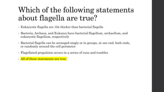
Which of thefollowing statements
about flagella are true?
• Eukaryotic flagella are 10x thicker than bacterial flagella
• Bacteria, Archaea, and Eukarya have bacterial flagellum, archaellum, and
eukaryotic flagellum, respectively
• Bacterial flagella can be arranged singly or in groups, at one end, both ends,
or randomly around the cell perimeter
• Flagellated propulsion occurs in a series of runs and tumbles
• All of these statements are true
95.
Which of thefollowing statements
about flagella are true?
• Eukaryotic flagella are 10x thicker than bacterial flagella
• Bacteria, Archaea, and Eukarya have bacterial flagellum, archaellum, and
eukaryotic flagellum, respectively
• Bacterial flagella can be arranged singly or in groups, at one end, both ends,
or randomly around the cell perimeter
• Flagellated propulsion occurs in a series of runs and tumbles
• All of these statements are true
96.
In this arrangement,flagella are clumped into
small bunches emerging from the same site:
Monotrichous
Lophotrichous
Amphitrichous
Peritrichous
97.
In this arrangement,flagella are clumped into
small bunches emerging from the same site:
Monotrichous
Lophotrichous
Amphitrichous
Peritrichous
98.
In this arrangement,a single flagellum
extends from one end of the cell:
Monotrichous
Lophotrichous
Amphitrichous
Peritrichous
99.
In this arrangement,a single flagellum
extends from one end of the cell:
Monotrichous
Lophotrichous
Amphitrichous
Peritrichous
100.
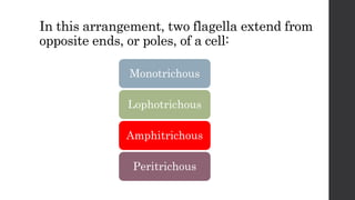
In this arrangement,two flagella extend from
opposite ends, or poles, of a cell:
Monotrichous
Lophotrichous
Amphitrichous
Peritrichous
101.
In this arrangement,two flagella extend from
opposite ends, or poles, of a cell:
Monotrichous
Lophotrichous
Amphitrichous
Peritrichous
102.
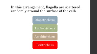
In this arrangement,flagella are scattered
randomly around the surface of the cell:
Monotrichous
Lophotrichous
Amphitrichous
Peritrichous
103.
In this arrangement,flagella are scattered
randomly around the surface of the cell:
Monotrichous
Lophotrichous
Amphitrichous
Peritrichous
104.
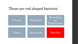
These are rodshaped bacteria:
Coccus Spirillum
Branching
or
filamentous
Vibrio Spirochete Bacillus
105.
These are rodshaped bacteria:
Coccus Spirillum
Branching
or
filamentous
Vibrio Spirochete Bacillus
106.
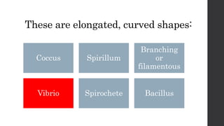
These are elongated,curved shapes:
Coccus Spirillum
Branching
or
filamentous
Vibrio Spirochete Bacillus
107.
These are elongated,curved shapes:
Coccus Spirillum
Branching
or
filamentous
Vibrio Spirochete Bacillus
108.
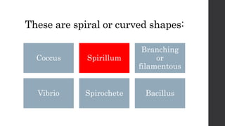
These are spiralor curved shapes:
Coccus Spirillum
Branching
or
filamentous
Vibrio Spirochete Bacillus
109.
These are spiralor curved shapes:
Coccus Spirillum
Branching
or
filamentous
Vibrio Spirochete Bacillus
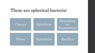
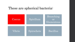
110.
These are sphericalbacteria:
Coccus Spirillum
Branching
or
filamentous
Vibrio Spirochete Bacillus
111.
These are sphericalbacteria:
Coccus Spirillum
Branching
or
filamentous
Vibrio Spirochete Bacillus
112.
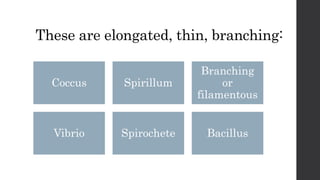
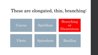
These are elongated,thin, branching:
Coccus Spirillum
Branching
or
filamentous
Vibrio Spirochete Bacillus
113.
These are elongated,thin, branching:
Coccus Spirillum
Branching
or
filamentous
Vibrio Spirochete Bacillus
114.
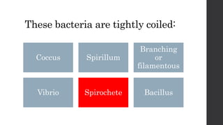
These bacteria aretightly coiled:
Coccus Spirillum
Branching
or
filamentous
Vibrio Spirochete Bacillus
115.
These bacteria aretightly coiled:
Coccus Spirillum
Branching
or
filamentous
Vibrio Spirochete Bacillus
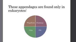
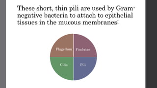
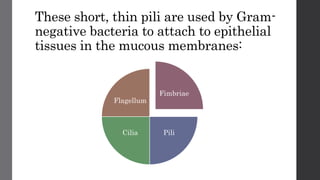
These short, thinpili are used by Gram-
negative bacteria to attach to epithelial
tissues in the mucous membranes:
Fimbriae
PiliCilia
Flagellum
119.
These short, thinpili are used by Gram-
negative bacteria to attach to epithelial
tissues in the mucous membranes:
Fimbriae
PiliCilia
Flagellum
120.
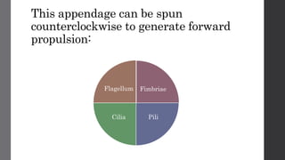
This appendage canbe spun
counterclockwise to generate forward
propulsion:
Fimbriae
PiliCilia
Flagellum
121.
This appendage canbe spun
counterclockwise to generate forward
propulsion:
Fimbriae
PiliCilia
Flagellum
122.
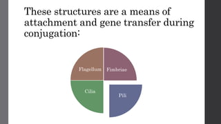
These structures area means of
attachment and gene transfer during
conjugation:
Fimbriae
PiliCilia
Flagellum
123.
These structures area means of
attachment and gene transfer during
conjugation:
Fimbriae
Pili
Cilia
Flagellum
124.
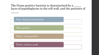
The Gram positivebacteria is characterized by a _____
layer of peptidoglycan in the cell wall, and the presence of
_____.
Thin, lipopolysaccharides
Thin, porins
Thick, tetrapeptides
Thick, teichoic acids
125.
The Gram positivebacteria is characterized by a _____
layer of peptidoglycan in the cell wall, and the presence of
_____.
Thin, lipopolysaccharides
Thin, porins
Thick, tetrapeptides
Thick, teichoic acids
126.
The Gram negativebacteria is characterized by a _____
layer of peptidoglycan in the cell wall, and the presence of
_____.
Thin, lipopolysaccharides
Thin, porins
Thick, tetrapeptides
Thick, teichoic acids
127.
The Gram negativebacteria is characterized by a _____
layer of peptidoglycan in the cell wall, and the presence of
_____.
Thin, lipopolysaccharides
Thin, porins
Thick, tetrapeptides
Thick, teichoic acids
128.
This structural featureof the Gram-
negative cell wall is an endotoxin,
making infections by these bacteria
severe:
• Periplasm
• Teichoic acids
• Peptidoglycan
• Lipopolysaccharide
129.
This structural featureof the Gram-
negative cell wall is an endotoxin,
making infections by these bacteria
severe:
• Periplasm
• Teichoic acids
• Peptidoglycan
• Lipopolysaccharide
130.
Which statement placesthe layers off the
Gram negative bacterial cell envelope in the
correct order from superficial to deep?
Which of thefollowing are industrial
uses for microbes?
• Fermenting yogurt, cheese, beer & wine
• Producing insulin for human use
• “Bioremediation” to break down pollutants like PCBs and oil spills
• Synthesis of plastics, cellulose, biofuels
• Synthesis of antibiotics and amino acids
• All of these
135.
Which of thefollowing are industrial
uses for microbes?
• Fermenting yogurt, cheese, beer & wine
• Producing insulin for human use
• “Bioremediation” to break down pollutants like PCBs and oil spills
• Synthesis of plastics, cellulose, biofuels
• Synthesis of antibiotics and amino acids
• All of these
136.
What qualifies anillness as an
emerging infectious disease?
• It is a disease that has only started to infect humans in the last 35 years
• It is a disease that has become more common in the last 35 years
• It is a disease that has spread from a limited population to the global
population in the past 35 years
• It is a disease that has emerged in animals in the past 35 years and has the
potential to infect humans
137.
What qualifies anillness as an
emerging infectious disease?
• It is a disease that has only started to infect humans in the last 35 years
• It is a disease that has become more common in the last 35 years
• It is a disease that has spread from a limited population to the global
population in the past 35 years
• It is a disease that has emerged in animals in the past 35 years and has the
potential to infect humans
138.
What is acontributing factor to
emerging infectious diseases?
• An organism may become resistant to antibiotics and be harder to treat
• A pathogen may develop the ability to infect human hosts
• A pathogen may evolve to become more toxic or virulent
• Global warming may increase the range of warm climate pathogens
• Population spread may bring humans into new or increased contact with
animal populations
• All of these
139.
What is acontributing factor to
emerging infectious diseases?
• An organism may become resistant to antibiotics and be harder to treat
• A pathogen may develop the ability to infect human hosts
• A pathogen may evolve to become more toxic or virulent
• Global warming may increase the range of warm climate pathogens
• Population spread may bring humans into new or increased contact with
animal populations
• All of these
140.
Which of thefollowing are acellular
infectious agents?
Prions Bacteria Prokaryotes
Viruses Viroids Protists
141.
Which of thefollowing are acellular
infectious agents?
Prions Bacteria Prokaryotes
Viruses Viroids Protists