SlideShare a Scribd company logo
History of Microbiology
Early Studies
Before 17th century, study of microbiology was hampered
by the lack of appropriate tools to observe microbes.
Robert Hooke: In 1665 built a compound light microscope
and used it to observe thin slices of cork. Coined the word cell.
Anton van Leeuwenhoeck: In 1673 was the first person to
observe live microorganisms which he called “animalcules”
(bacteria, protozoa), using single-lens microscopes that he
designed.
Before 1860s many scientists believed in Spontaneous
generation, i.e.: That living organisms could arise
spontaneously from nonliving matter:
 Mice come from rags in a basket.
 Maggots come from rotting meat.
 Ants come from honey.
 Microbes come from spoiled broth.
Spontaneous Generation vs Biogenesis
Francesco Redi: In 1668 proved that maggots do not arise
spontaneously from decaying meat.
Lazaro Spallanzani: In 1765 found that nutrient broth that
had been heated in a sealed flask would not become
contaminated with microbes.
 Some proponents of spontaneous generation argued that
boiling had destroyed the “life force” of air in flask.
 Others argued that microbes were different from other life
forms.
Redi's Experiment
In 1668, Francesco Redi, an Italian scientist, designed a scientific
experiment to test the spontaneous creation of maggots.
 He placed fresh meat in each of two different jars.
 One jar was left open; the other was covered with a cloth.
Days later, the open jar contained maggots, whereas the covered jar
contained no maggots.
He did note that maggots were found on the exterior surface of the cloth
that covered the jar.
Redi successfully demonstrated that the maggots came from fly eggs and
thereby helped to disprove spontaneous generation
Redi's Experiment and Needham's Rebuttal
In England, John Needham challenged Redi's
findings by conducting an experiment in which he placed
a broth, or “gravy,” into a bottle, heated the bottle to kill
anything inside, then sealed it. Days later, he reported the
presence of life in the broth and announced that life had
been created from nonlife.
In actuality, he did not heat it long enough to kill all
the microbes.
Needham's Rebuttal
Lazzaro Spallanzani, also an Italian scientist, reviewed both
Redi's and Needham's data and experimental design and concluded
that perhaps Needham's heating of the bottle did not kill everything
inside.
 He constructed his own experiment by placing broth in each of
two separate bottles, boiling the broth in both bottles, then sealing
one bottle and leaving the other open.
Spallanzani's Experiment
Days later, the unsealed bottle was teeming with small living
things that he could observe more clearly with the newly invented
microscope.
The sealed bottle showed no signs of life.
 This certainly excluded spontaneous generation as a viable
theory.
Scientists of that day deprived Spallanzani that the sealed bottle
was deprived of air.
So although his experiment was successful, a strong rebuttal
blunted his claims.
Louis Pasteur, the notable French scientist, accepted the
challenge to re-create the experiment of Lazzaro Spallanzani.
He designed two types of flasks, one with long S shaped necks
and another with straight necks.
He placed a nutrient-enriched broth in both the jars, boiled the
broth inside the jars and left them open.
Kept both the jars open for long time.
Straight necked flasks showed the presence of life whereas S
necked flasks showed no life up to one year.
Pasteur's Swan necked flask Experiment
He then broke off the top of the bottle, exposing it more directly
to the air, and noted life-forms in the broth within days
The reason behind it was that the air paricles carrying minute
life forms were trapped at the deepest portion of the neck because
of gravity.
Pasteur's Swan necked flask Experiment
Pasteur’s Contributions:
Pasteurization: Developed a process in which liquids are
heated (at 65oC) to kill most bacteria responsible for spoilage.
Disease Causes: Identified three different microbes that caused
silkworm diseases.
Vaccine: Developed a vaccine for rabies from dried spinal
cords of infected rabbits.
 Directed Pasteur Institute until his death in 1895.
History of Microbiology
Golden Age: 1857-1914
History of Microbiology
Golden Age: 1857-1914
Germ Theory of Disease: Belief that microbes cause diseases.
Before, most people believed diseases were caused by divine
punishment, poisonous vapors, curses, witchcraft, etc.
Agostino Bassi (1835): Found that a fungus was responsible for
a silkworm disease.
Ignaz Semmelweis (1840s): Demonstrated that childbirth fever
was transmitted from one patient to another, by physicians who
didn’t disinfect their hands. He was ostracized by colleagues.
Before Pasteur disproved spontaneous generation, he decided to
determine why some bottles of wine soured over time. He observed wine that
had soured and compared it to wine that had not. He determined that all
soured wines contained a large number of cells. Yeast cells are required for
wine fermentation, so even good wine would have yeast cells. But, sour wine
was full of many smaller cells that were not yeast. Pasteur reasoned that sour
wines had been contaminated with microbes, and these contaminating
microbes were causing the poor quality.
Based on this, Pasteur postulated the germ theory of disease, which
states that microorganisms are the causes of infectious disease.
The Germ Theory of Disease
Pasteur's attempts to prove the germ theory were
unsuccessful. However, the German scientist Robert Koch
provided the proof by cultivating anthrax bacteria apart from
any other type of organism. He then injected pure cultures of
the bacilli into mice and showed that the bacilli invariably
caused anthrax. The procedures used by Koch came to be
known as Koch's postulates.
Koch's postulates
Four criteria that were established by Robert Koch to
identify the causative agent of a particular disease, these
include:
•the microorganism or other pathogen must be present in all
cases of the disease.
•the pathogen can be isolated from the diseased host and grown
in pure culture.
•the pathogen from the pure culture must cause the disease
when inoculated into a healthy, susceptible laboratory animal.
•the pathogen must be reisolated from the new host and shown
to be the same as the originally inoculated pathogen.
Modern Taxonomy for Microbial Diversity
Microorganisms are actually composed of very different
and taxonomically diverse groups of communities: archaea,
bacteria, fungi and viruses. The members of these groups or taxa
are distinct in terms of their morphology, physiology and
phylogeny and fall into both prokaryotic and eukaryotic domains.
They constitute a broad group of life system inhabiting the
known ecosystems on earth: terrestrial and marine; including
geographical locations considered to be extreme or inimical to
life.
Phenotypic techniques
The phenotypic methods are all those that do not include the
DNA/RNA sequencing or their typing methods. Study of
morphological characteristics and chemotaxonomic profiles is broadly
associated with phenotypic characterization.
Classical: Colony characteristics, biochemical and physiological analyses
The phenotypic features are the foundation for description of taxa. The
morphological, biochemical and physiological characteristics provide in-depth
information on a taxon. The morphology can include the colony characteristics
(colour, shape, pigmentation, production of slime etc.). Further, the features of
the cell are described as to shape, size, Gram reaction, extracellular material like
capsule, presence of endospores, flagella presence and location, motility and
inclusion bodies. Light microscopy is generally used to describe the broad cell
features; however electron microscopy is recommended for high resolution
images.
Numerical taxonomy
Analysis of huge volumes of phenotypic data to derive meaningful
relationships amongst a large number of microorganisms can be carried out
using computer programs . This system of analysis is called numerical
taxonomy. Giving numerical weightage to each trait is followed by analysis
of the data by the computer programs generating data matrices between each
pair of isolates according to the degree of similarity. Based on the similarity
data, cluster analysis are carried out (based on different algorithms) and
dendrograms (‘trees’) are generated showing the overall pattern of
similarity/dissimilarity amongst the various organisms being studied.
Cell wall composition
The peptidoglycan component of cell walls of bacteria does not
provide much information except for classifying into Gram-positive,
Gram-negative and acid-fast bacterial types. However, those in Gram-
positive cells contain different types of peptidoglycan depending on the
genus or species. The peptidoglycan structure can be analysed by
determining its type (A or B), mode of cross-linking (whether it is directly
linked or via interpeptide bridge and with amino acids in the bridge), and
the composition of amino acids (especially the diaminoacid) of the side
chain.
Fatty acid analyses
Different types of lipids are present in bacterial cells. Polar lipids are
present in the lipid bilayer of the cytoplasmic membrane. The diversity of polar
lipids is known to be large and many are yet to be structurally elucidated. While
in archaea, polar lipids are of types phospholipids, aminophospholipids,
glycolipids and phosphoglycolipids, in bacteria, apart from the ones seen in
archaea, there are also lipids derived from amino acids, capnines, sphingolipids
(glycol or phosphosphingolipids) and hopanoids . In Gram-negative bacteria,
lipopolysaccharides are present in the outer membranes. The type of sugar
present and the fatty acid type, the linkage of the fatty acid to the sugar (amide or
ester linkage) provide information on characteristic of the cell.
Genotypic techniques
Modern taxonomy has been influenced by genetic methods
and indeed, much of the classification and identification is predicated
on specific gene sequences. All the techniques involving DNA or RNA
fall under genotypic methods.
16S rDNA-based analyses
The technique, which is very nearly a gold standard for taxonomic
purposes today, is sequencing of the 16S rRNA gene of bacteria. The 23S
rRNA gene sequence is also considered in many studies but lack of
comprehensive databases for comparison is a drawback. Since the 16S
rRNA is present in all bacteria, is functionally constant and is composed
of conserved and variable regions, it has consistently served as a good
taxonomic marker for deriving taxonomic relationships
DNA base content
Determination of moles percent guanosine and cytosine
constitutes a classical method of establishment of genomic content. This
is now being used along with other genotyping methods to establish
taxonomic position of an organism. Within species, the G+C content
ranges within 3% and within genera 10%. Overall, the G+C content
ranges from 24-76% in bacteria.
DNA-DNA hybridization
This method is an indirect measurement of sequence similarity between
genomes. A cut-off value of 70% similarity is considered for establishment
of species. However, the method has to be reproducible between
laboratories and performed under standardized conditions, which is often a
drawback. Hence it is applied only where 16S rRNA gene sequences show
similarity values above 98%. There have been reports where 16S rRNA
gene sequence has shown 99% similarity and yet DNA-DNA hybridization
values have been 60% or less. Hence, this method has to be used with
caution and performed under highly standardized conditions.
Other genotyping methods
Earlier, sub-typing was done on the basis of biochemical profile
(biotyping), serological profile (serotyping), phase susceptibility (phage
typing) or antibiotic susceptibility. But currently DNA-typing methods are
preferred due to their reproducibility, ease of performance and high level of
discrimination between strains [1]. Genotyping methods such as Restriction
Fragment Length Polymorphism (RFLP), Randomly Amplified Polymorphic
DNA (RAPD), Amplified Fragment Length Polymorphism (AFLP),
Amplified Ribosomal DNA Restriction Analysis (ARDRA), Repetitive
Element-Polymerase Chain Reaction (REP-PCR), Ribotyping and Multi
Locus Sequence Analyses (MLSA) are some of the newer methods to
characterize a taxon.
Taxonomy of viruses
The definition of a virus ‘species’ is: "A virus species is a polythetic
class of viruses that constitutes a replicating lineage and occupies a particular
ecological niche". A virus isolate can refer to any virus as long as the virus has
existed for some time. Viruses are not considered to be either prokaryotes or
eukaryotes but have implication from health point of view; hence
characterization of viruses has increased considerably. Where earlier, only
electron microscopy was used, today sequencing of viral genomes constitutes
advancements and the database is increasing. According to International
Committee on Taxonomy of Viruses (ICTV), proposals are afoot to accept
online descriptions of viral taxa based on taxonomical details such as : dsDNA,
ssDNA, rtDNA, rtRNA, dsRNA, ssNRNA, ssPRNA, SAT (Satellites), VIR
(Viroids), UN (unassigned).
MICROBIAL NUTRITION
The microbial cells are extremely complex and in
addition to oxygen and hydrogen they contain four other
major elements such as carbon, nitrogen, phosphorus and
sulphur. The microorganisms in general do not need only
these six elements but also others, which are found in
lesser -quantity. Such elements are potassium, magnesium,
calcium, sodium, iron, manganese, cobalt copper,
molybdenum and zinc.
Most of the microorganisms need molecular oxygen for respiration. In
these, the oxygen serves as terminal electron acceptor and, such organisms
are referred to as ‘obligate aerobes’. As opposed to this there are a few
organisms, which do not use molecular oxygen as terminal electron acceptor.
These microbes are called ‘obligate anaerobes’. In fact, molecular oxygen is
toxic to these organisms. Aerobes, which can grow in the absence of oxygen,
are called ‘facultative anaerobes’ and the anaerobes which can grow in the
presence of oxygen are referred to as ‘facultative aerobes’. In addition to
these major classes, there are organisms, which grow best at reduced oxygen
pressure but are obligate aerobes and these are called ‘Microaerophilic’.
Some microorganisms manufacture their foods from inorganic
supplies to them and thus are able to subsist in an exclusively
inorganic environment: They are collectively called autotrophs.
Other micro organic metabolites; they must absorb from the
environment in certain minimum amounts and kinds of prefabricated
organic metabolites (the foods). Such microorganisms are collectively
called heterotrophs.
Autotrophic microorganisms, which manufacture foods from
inorganic sources, require not only external source of appropriate nutrient
raw materials but also external sources of energy. In some cases, external
energy for food manufacture is obtained from light and such
microorganisms are collectively called photosynthesizers. In other cases,
some inorganic nutrients serve as raw materials for food manufacture and
other inorganic nutrients, i.e., chemicals, serve as external energy sources.
Such microorganisms are collectively called chemosynthesizers.
Major nutritional types of microbes
Microbes can be categorized under four nutritional types depending
upon the source of carbon, electron and energy-
Light as a source of energy
Photolithoautotrophs: these microbes use light as the source of
energy, inorganic compounds as electron source and CO2 as carbon
source. Example: Green and Purple sulphur bacteria, cyanobacteria.
Photoorganoheterotrophs: these microbes use light as energy source
and organic compounds as electron and carbon source. Example: Green
and Purple non sulphur bacteria.
Inorganic or organic compounds as source of energy
Chemolithoautotrophs: These microbes use inorganic
compounds as source of energy as well as electrons. They also
use carbon-dioxide as a source of carbon. Example: Sulphur
oxidizing bacteria, hydrogen bacteria.
Chemoorganoheterotrophs: They use organic compounds as
the source of carbon, electrons and energy. Example: Fungi,
nonphotosynthetic bacteria, nitrifying bacteria.
CULTURE MEDIA
Substrates or mixtures of nutrients that provide proper growth of
microorganisms in laboratory are referred as culture media. The use of diverse kinds of
culture media made the cultivation of microbes easy and these culture media may be
divided into following tyoes
Natural media: These types media contain simple ingredients having
nutrients of unknown composition like peptone, beef extract, yeast extract, potato etc.
which provide a wide range of nutrients Ilike amino acids, peptides, nucleotides,
itamins, minerals, carbon source etc.) for better growth of different kinds of microbes.
Eg. Nutrient agar, potato dextrose agar.
Synthetic media: These media are constructed using specific chemicals in
their exact proportions. It is commonly used for growth of microbes haing simple
nutritional requirements. Eg. Czapek dox agar.
Basal media or minimal : It supplies only the minimal nutritional
requirements of a particular microorganism. These types of media support the
growth of most non-fastidious organisms. Eg. Nutrient agar and nutrient broth.
Enriched media: When extra nutrients like blood, serum, egg yolk etc are
added to a minimal medium it becomes enriched medium. It supports the
growth of nutritionally fastidious bacteria. Eg. Blood agar, chocolate agar.
All purpose media: This type of media is rich with wide varieties of nutrients
including growth factors and therefore supports the growth of wide number of
bacteria. Eg. Plate count agar, Heart infusion agar.
Selective media: The media that encourage the growth of only some specic
microbes and inhibit the growth of others. Eg. Pseudomonas agar
Differential media: The media used to identify different groups of microbes.
Eg. Blood agar, skim milk agar.
MICROBIAL GROWTH
As most of the microorganisms are unicellular in nature, microbial
growth does not refer to the growth of the cell size, but it denotes the
growth of cell number in a specific period.
Microbial growth curve:
The growth curve has four distinct phases as mentioned in the figure.
Fig: Microbial growth curve
Fig: Calculation of generation time

More Related Content

Similar to Microbio.pptx

Histroy and scope of microbiology
Histroy and scope of microbiologyHistroy and scope of microbiology
Histroy and scope of microbiology
SuganyaPaulraj
 
Introduction to Microbiology.pptx
Introduction to Microbiology.pptxIntroduction to Microbiology.pptx
Introduction to Microbiology.pptx
BinteHawah1
 
Introduction and History of Pharmaceutical microbiology.pptx
Introduction and History of Pharmaceutical microbiology.pptxIntroduction and History of Pharmaceutical microbiology.pptx
Introduction and History of Pharmaceutical microbiology.pptx
Dr. Kirankumar Hullatti
 
Micro bio dentistry.ppt
Micro bio dentistry.pptMicro bio dentistry.ppt
Micro bio dentistry.ppt
bhusalbhuban
 
History and Scope of Microbiology
History and Scope of MicrobiologyHistory and Scope of Microbiology
History and Scope of Microbiology
Pharmacy Universe
 
Introduction of microbiology kbk
Introduction of microbiology kbkIntroduction of microbiology kbk
Introduction of microbiology kbk
Uchaaaa Dhee
 
1 bio265 introduction to microbiology_dr di bonaventura_instructor
1 bio265  introduction to microbiology_dr di bonaventura_instructor1 bio265  introduction to microbiology_dr di bonaventura_instructor
1 bio265 introduction to microbiology_dr di bonaventura_instructor
Shabab Ali
 
1. History and Scope of microbiology (1).pptx
1. History and Scope of microbiology (1).pptx1. History and Scope of microbiology (1).pptx
1. History and Scope of microbiology (1).pptx
Shaikh Ayesha
 
History of microbiology
History of microbiologyHistory of microbiology
History of microbiology
Saajida Sultaana
 
Contributions of renowned scientists in Microbiology
Contributions of renowned scientists in MicrobiologyContributions of renowned scientists in Microbiology
Contributions of renowned scientists in Microbiology
Saajida Sultaana
 
Bio presentation
Bio presentationBio presentation
Bio presentation
chuckiecalsado
 
Distinguish between cellular and acellular. Give examples of microorg.pdf
Distinguish between cellular and acellular. Give examples of microorg.pdfDistinguish between cellular and acellular. Give examples of microorg.pdf
Distinguish between cellular and acellular. Give examples of microorg.pdf
arjuntiwari586
 
Introduction to microbiology
Introduction to microbiologyIntroduction to microbiology
Introduction to microbiology
Parthasarathy Seethapathy
 
Milestones in microbiology
Milestones in microbiologyMilestones in microbiology
Milestones in microbiology
Rebecca Thombre
 
Microbiology 1st
Microbiology 1stMicrobiology 1st
Microbiology 1st
Junaid Afzal
 
C:\Fakepath\ Start Here Ch01 Lecture
C:\Fakepath\ Start Here Ch01 LectureC:\Fakepath\ Start Here Ch01 Lecture
C:\Fakepath\ Start Here Ch01 Lecture
Debra Costa-Nino
 
Introduction microbiology
Introduction microbiologyIntroduction microbiology
Introduction microbiology
deepak deshkar
 
Pharmaceutical microbiology (unit 1)
Pharmaceutical microbiology (unit 1)Pharmaceutical microbiology (unit 1)
Pharmaceutical microbiology (unit 1)
himanshu kamboj
 
Microbiology
MicrobiologyMicrobiology
Microbiology
Nandy 91
 
Mbp 01 history microbiology
Mbp 01 history microbiologyMbp 01 history microbiology
Mbp 01 history microbiology
Subashkumar Rathinasamy
 

Similar to Microbio.pptx (20)

Histroy and scope of microbiology
Histroy and scope of microbiologyHistroy and scope of microbiology
Histroy and scope of microbiology
 
Introduction to Microbiology.pptx
Introduction to Microbiology.pptxIntroduction to Microbiology.pptx
Introduction to Microbiology.pptx
 
Introduction and History of Pharmaceutical microbiology.pptx
Introduction and History of Pharmaceutical microbiology.pptxIntroduction and History of Pharmaceutical microbiology.pptx
Introduction and History of Pharmaceutical microbiology.pptx
 
Micro bio dentistry.ppt
Micro bio dentistry.pptMicro bio dentistry.ppt
Micro bio dentistry.ppt
 
History and Scope of Microbiology
History and Scope of MicrobiologyHistory and Scope of Microbiology
History and Scope of Microbiology
 
Introduction of microbiology kbk
Introduction of microbiology kbkIntroduction of microbiology kbk
Introduction of microbiology kbk
 
1 bio265 introduction to microbiology_dr di bonaventura_instructor
1 bio265  introduction to microbiology_dr di bonaventura_instructor1 bio265  introduction to microbiology_dr di bonaventura_instructor
1 bio265 introduction to microbiology_dr di bonaventura_instructor
 
1. History and Scope of microbiology (1).pptx
1. History and Scope of microbiology (1).pptx1. History and Scope of microbiology (1).pptx
1. History and Scope of microbiology (1).pptx
 
History of microbiology
History of microbiologyHistory of microbiology
History of microbiology
 
Contributions of renowned scientists in Microbiology
Contributions of renowned scientists in MicrobiologyContributions of renowned scientists in Microbiology
Contributions of renowned scientists in Microbiology
 
Bio presentation
Bio presentationBio presentation
Bio presentation
 
Distinguish between cellular and acellular. Give examples of microorg.pdf
Distinguish between cellular and acellular. Give examples of microorg.pdfDistinguish between cellular and acellular. Give examples of microorg.pdf
Distinguish between cellular and acellular. Give examples of microorg.pdf
 
Introduction to microbiology
Introduction to microbiologyIntroduction to microbiology
Introduction to microbiology
 
Milestones in microbiology
Milestones in microbiologyMilestones in microbiology
Milestones in microbiology
 
Microbiology 1st
Microbiology 1stMicrobiology 1st
Microbiology 1st
 
C:\Fakepath\ Start Here Ch01 Lecture
C:\Fakepath\ Start Here Ch01 LectureC:\Fakepath\ Start Here Ch01 Lecture
C:\Fakepath\ Start Here Ch01 Lecture
 
Introduction microbiology
Introduction microbiologyIntroduction microbiology
Introduction microbiology
 
Pharmaceutical microbiology (unit 1)
Pharmaceutical microbiology (unit 1)Pharmaceutical microbiology (unit 1)
Pharmaceutical microbiology (unit 1)
 
Microbiology
MicrobiologyMicrobiology
Microbiology
 
Mbp 01 history microbiology
Mbp 01 history microbiologyMbp 01 history microbiology
Mbp 01 history microbiology
 

More from rnath286

Phaeophyceae.ppt
Phaeophyceae.pptPhaeophyceae.ppt
Phaeophyceae.ppt
rnath286
 
control of microbes.pptx
control of microbes.pptxcontrol of microbes.pptx
control of microbes.pptx
rnath286
 
BIOINFO unit 1.pptx
BIOINFO unit 1.pptxBIOINFO unit 1.pptx
BIOINFO unit 1.pptx
rnath286
 
cell membrane.pptx
cell membrane.pptxcell membrane.pptx
cell membrane.pptx
rnath286
 
coleochaetea.pptx
coleochaetea.pptxcoleochaetea.pptx
coleochaetea.pptx
rnath286
 
Volvox.pptx
Volvox.pptxVolvox.pptx
Volvox.pptx
rnath286
 
chara.pptx
chara.pptxchara.pptx
chara.pptx
rnath286
 
algae classification.pptx
algae classification.pptxalgae classification.pptx
algae classification.pptx
rnath286
 
virus.pptx
virus.pptxvirus.pptx
virus.pptx
rnath286
 

More from rnath286 (9)

Phaeophyceae.ppt
Phaeophyceae.pptPhaeophyceae.ppt
Phaeophyceae.ppt
 
control of microbes.pptx
control of microbes.pptxcontrol of microbes.pptx
control of microbes.pptx
 
BIOINFO unit 1.pptx
BIOINFO unit 1.pptxBIOINFO unit 1.pptx
BIOINFO unit 1.pptx
 
cell membrane.pptx
cell membrane.pptxcell membrane.pptx
cell membrane.pptx
 
coleochaetea.pptx
coleochaetea.pptxcoleochaetea.pptx
coleochaetea.pptx
 
Volvox.pptx
Volvox.pptxVolvox.pptx
Volvox.pptx
 
chara.pptx
chara.pptxchara.pptx
chara.pptx
 
algae classification.pptx
algae classification.pptxalgae classification.pptx
algae classification.pptx
 
virus.pptx
virus.pptxvirus.pptx
virus.pptx
 

Recently uploaded

The use of Nauplii and metanauplii artemia in aquaculture (brine shrimp).pptx
The use of Nauplii and metanauplii artemia in aquaculture (brine shrimp).pptxThe use of Nauplii and metanauplii artemia in aquaculture (brine shrimp).pptx
The use of Nauplii and metanauplii artemia in aquaculture (brine shrimp).pptx
MAGOTI ERNEST
 
Eukaryotic Transcription Presentation.pptx
Eukaryotic Transcription Presentation.pptxEukaryotic Transcription Presentation.pptx
Eukaryotic Transcription Presentation.pptx
RitabrataSarkar3
 
waterlessdyeingtechnolgyusing carbon dioxide chemicalspdf
waterlessdyeingtechnolgyusing carbon dioxide chemicalspdfwaterlessdyeingtechnolgyusing carbon dioxide chemicalspdf
waterlessdyeingtechnolgyusing carbon dioxide chemicalspdf
LengamoLAppostilic
 
The binding of cosmological structures by massless topological defects
The binding of cosmological structures by massless topological defectsThe binding of cosmological structures by massless topological defects
The binding of cosmological structures by massless topological defects
Sérgio Sacani
 
Cytokines and their role in immune regulation.pptx
Cytokines and their role in immune regulation.pptxCytokines and their role in immune regulation.pptx
Cytokines and their role in immune regulation.pptx
Hitesh Sikarwar
 
The debris of the ‘last major merger’ is dynamically young
The debris of the ‘last major merger’ is dynamically youngThe debris of the ‘last major merger’ is dynamically young
The debris of the ‘last major merger’ is dynamically young
Sérgio Sacani
 
Basics of crystallography, crystal systems, classes and different forms
Basics of crystallography, crystal systems, classes and different formsBasics of crystallography, crystal systems, classes and different forms
Basics of crystallography, crystal systems, classes and different forms
MaheshaNanjegowda
 
Travis Hills' Endeavors in Minnesota: Fostering Environmental and Economic Pr...
Travis Hills' Endeavors in Minnesota: Fostering Environmental and Economic Pr...Travis Hills' Endeavors in Minnesota: Fostering Environmental and Economic Pr...
Travis Hills' Endeavors in Minnesota: Fostering Environmental and Economic Pr...
Travis Hills MN
 
Oedema_types_causes_pathophysiology.pptx
Oedema_types_causes_pathophysiology.pptxOedema_types_causes_pathophysiology.pptx
Oedema_types_causes_pathophysiology.pptx
muralinath2
 
Thornton ESPP slides UK WW Network 4_6_24.pdf
Thornton ESPP slides UK WW Network 4_6_24.pdfThornton ESPP slides UK WW Network 4_6_24.pdf
Thornton ESPP slides UK WW Network 4_6_24.pdf
European Sustainable Phosphorus Platform
 
Medical Orthopedic PowerPoint Templates.pptx
Medical Orthopedic PowerPoint Templates.pptxMedical Orthopedic PowerPoint Templates.pptx
Medical Orthopedic PowerPoint Templates.pptx
terusbelajar5
 
EWOCS-I: The catalog of X-ray sources in Westerlund 1 from the Extended Weste...
EWOCS-I: The catalog of X-ray sources in Westerlund 1 from the Extended Weste...EWOCS-I: The catalog of X-ray sources in Westerlund 1 from the Extended Weste...
EWOCS-I: The catalog of X-ray sources in Westerlund 1 from the Extended Weste...
Sérgio Sacani
 
20240520 Planning a Circuit Simulator in JavaScript.pptx
20240520 Planning a Circuit Simulator in JavaScript.pptx20240520 Planning a Circuit Simulator in JavaScript.pptx
20240520 Planning a Circuit Simulator in JavaScript.pptx
Sharon Liu
 
bordetella pertussis.................................ppt
bordetella pertussis.................................pptbordetella pertussis.................................ppt
bordetella pertussis.................................ppt
kejapriya1
 
8.Isolation of pure cultures and preservation of cultures.pdf
8.Isolation of pure cultures and preservation of cultures.pdf8.Isolation of pure cultures and preservation of cultures.pdf
8.Isolation of pure cultures and preservation of cultures.pdf
by6843629
 
在线办理(salfor毕业证书)索尔福德大学毕业证毕业完成信一模一样
在线办理(salfor毕业证书)索尔福德大学毕业证毕业完成信一模一样在线办理(salfor毕业证书)索尔福德大学毕业证毕业完成信一模一样
在线办理(salfor毕业证书)索尔福德大学毕业证毕业完成信一模一样
vluwdy49
 
原版制作(carleton毕业证书)卡尔顿大学毕业证硕士文凭原版一模一样
原版制作(carleton毕业证书)卡尔顿大学毕业证硕士文凭原版一模一样原版制作(carleton毕业证书)卡尔顿大学毕业证硕士文凭原版一模一样
原版制作(carleton毕业证书)卡尔顿大学毕业证硕士文凭原版一模一样
yqqaatn0
 
Describing and Interpreting an Immersive Learning Case with the Immersion Cub...
Describing and Interpreting an Immersive Learning Case with the Immersion Cub...Describing and Interpreting an Immersive Learning Case with the Immersion Cub...
Describing and Interpreting an Immersive Learning Case with the Immersion Cub...
Leonel Morgado
 
Immersive Learning That Works: Research Grounding and Paths Forward
Immersive Learning That Works: Research Grounding and Paths ForwardImmersive Learning That Works: Research Grounding and Paths Forward
Immersive Learning That Works: Research Grounding and Paths Forward
Leonel Morgado
 
如何办理(uvic毕业证书)维多利亚大学毕业证本科学位证书原版一模一样
如何办理(uvic毕业证书)维多利亚大学毕业证本科学位证书原版一模一样如何办理(uvic毕业证书)维多利亚大学毕业证本科学位证书原版一模一样
如何办理(uvic毕业证书)维多利亚大学毕业证本科学位证书原版一模一样
yqqaatn0
 

Recently uploaded (20)

The use of Nauplii and metanauplii artemia in aquaculture (brine shrimp).pptx
The use of Nauplii and metanauplii artemia in aquaculture (brine shrimp).pptxThe use of Nauplii and metanauplii artemia in aquaculture (brine shrimp).pptx
The use of Nauplii and metanauplii artemia in aquaculture (brine shrimp).pptx
 
Eukaryotic Transcription Presentation.pptx
Eukaryotic Transcription Presentation.pptxEukaryotic Transcription Presentation.pptx
Eukaryotic Transcription Presentation.pptx
 
waterlessdyeingtechnolgyusing carbon dioxide chemicalspdf
waterlessdyeingtechnolgyusing carbon dioxide chemicalspdfwaterlessdyeingtechnolgyusing carbon dioxide chemicalspdf
waterlessdyeingtechnolgyusing carbon dioxide chemicalspdf
 
The binding of cosmological structures by massless topological defects
The binding of cosmological structures by massless topological defectsThe binding of cosmological structures by massless topological defects
The binding of cosmological structures by massless topological defects
 
Cytokines and their role in immune regulation.pptx
Cytokines and their role in immune regulation.pptxCytokines and their role in immune regulation.pptx
Cytokines and their role in immune regulation.pptx
 
The debris of the ‘last major merger’ is dynamically young
The debris of the ‘last major merger’ is dynamically youngThe debris of the ‘last major merger’ is dynamically young
The debris of the ‘last major merger’ is dynamically young
 
Basics of crystallography, crystal systems, classes and different forms
Basics of crystallography, crystal systems, classes and different formsBasics of crystallography, crystal systems, classes and different forms
Basics of crystallography, crystal systems, classes and different forms
 
Travis Hills' Endeavors in Minnesota: Fostering Environmental and Economic Pr...
Travis Hills' Endeavors in Minnesota: Fostering Environmental and Economic Pr...Travis Hills' Endeavors in Minnesota: Fostering Environmental and Economic Pr...
Travis Hills' Endeavors in Minnesota: Fostering Environmental and Economic Pr...
 
Oedema_types_causes_pathophysiology.pptx
Oedema_types_causes_pathophysiology.pptxOedema_types_causes_pathophysiology.pptx
Oedema_types_causes_pathophysiology.pptx
 
Thornton ESPP slides UK WW Network 4_6_24.pdf
Thornton ESPP slides UK WW Network 4_6_24.pdfThornton ESPP slides UK WW Network 4_6_24.pdf
Thornton ESPP slides UK WW Network 4_6_24.pdf
 
Medical Orthopedic PowerPoint Templates.pptx
Medical Orthopedic PowerPoint Templates.pptxMedical Orthopedic PowerPoint Templates.pptx
Medical Orthopedic PowerPoint Templates.pptx
 
EWOCS-I: The catalog of X-ray sources in Westerlund 1 from the Extended Weste...
EWOCS-I: The catalog of X-ray sources in Westerlund 1 from the Extended Weste...EWOCS-I: The catalog of X-ray sources in Westerlund 1 from the Extended Weste...
EWOCS-I: The catalog of X-ray sources in Westerlund 1 from the Extended Weste...
 
20240520 Planning a Circuit Simulator in JavaScript.pptx
20240520 Planning a Circuit Simulator in JavaScript.pptx20240520 Planning a Circuit Simulator in JavaScript.pptx
20240520 Planning a Circuit Simulator in JavaScript.pptx
 
bordetella pertussis.................................ppt
bordetella pertussis.................................pptbordetella pertussis.................................ppt
bordetella pertussis.................................ppt
 
8.Isolation of pure cultures and preservation of cultures.pdf
8.Isolation of pure cultures and preservation of cultures.pdf8.Isolation of pure cultures and preservation of cultures.pdf
8.Isolation of pure cultures and preservation of cultures.pdf
 
在线办理(salfor毕业证书)索尔福德大学毕业证毕业完成信一模一样
在线办理(salfor毕业证书)索尔福德大学毕业证毕业完成信一模一样在线办理(salfor毕业证书)索尔福德大学毕业证毕业完成信一模一样
在线办理(salfor毕业证书)索尔福德大学毕业证毕业完成信一模一样
 
原版制作(carleton毕业证书)卡尔顿大学毕业证硕士文凭原版一模一样
原版制作(carleton毕业证书)卡尔顿大学毕业证硕士文凭原版一模一样原版制作(carleton毕业证书)卡尔顿大学毕业证硕士文凭原版一模一样
原版制作(carleton毕业证书)卡尔顿大学毕业证硕士文凭原版一模一样
 
Describing and Interpreting an Immersive Learning Case with the Immersion Cub...
Describing and Interpreting an Immersive Learning Case with the Immersion Cub...Describing and Interpreting an Immersive Learning Case with the Immersion Cub...
Describing and Interpreting an Immersive Learning Case with the Immersion Cub...
 
Immersive Learning That Works: Research Grounding and Paths Forward
Immersive Learning That Works: Research Grounding and Paths ForwardImmersive Learning That Works: Research Grounding and Paths Forward
Immersive Learning That Works: Research Grounding and Paths Forward
 
如何办理(uvic毕业证书)维多利亚大学毕业证本科学位证书原版一模一样
如何办理(uvic毕业证书)维多利亚大学毕业证本科学位证书原版一模一样如何办理(uvic毕业证书)维多利亚大学毕业证本科学位证书原版一模一样
如何办理(uvic毕业证书)维多利亚大学毕业证本科学位证书原版一模一样
 

Microbio.pptx

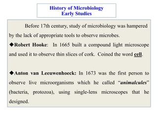
  • 1. History of Microbiology Early Studies Before 17th century, study of microbiology was hampered by the lack of appropriate tools to observe microbes. Robert Hooke: In 1665 built a compound light microscope and used it to observe thin slices of cork. Coined the word cell. Anton van Leeuwenhoeck: In 1673 was the first person to observe live microorganisms which he called “animalcules” (bacteria, protozoa), using single-lens microscopes that he designed.
  • 2. Before 1860s many scientists believed in Spontaneous generation, i.e.: That living organisms could arise spontaneously from nonliving matter:  Mice come from rags in a basket.  Maggots come from rotting meat.  Ants come from honey.  Microbes come from spoiled broth. Spontaneous Generation vs Biogenesis
  • 3. Francesco Redi: In 1668 proved that maggots do not arise spontaneously from decaying meat. Lazaro Spallanzani: In 1765 found that nutrient broth that had been heated in a sealed flask would not become contaminated with microbes.  Some proponents of spontaneous generation argued that boiling had destroyed the “life force” of air in flask.  Others argued that microbes were different from other life forms.
  • 4. Redi's Experiment In 1668, Francesco Redi, an Italian scientist, designed a scientific experiment to test the spontaneous creation of maggots.  He placed fresh meat in each of two different jars.  One jar was left open; the other was covered with a cloth. Days later, the open jar contained maggots, whereas the covered jar contained no maggots. He did note that maggots were found on the exterior surface of the cloth that covered the jar. Redi successfully demonstrated that the maggots came from fly eggs and thereby helped to disprove spontaneous generation Redi's Experiment and Needham's Rebuttal
  • 5. In England, John Needham challenged Redi's findings by conducting an experiment in which he placed a broth, or “gravy,” into a bottle, heated the bottle to kill anything inside, then sealed it. Days later, he reported the presence of life in the broth and announced that life had been created from nonlife. In actuality, he did not heat it long enough to kill all the microbes. Needham's Rebuttal
  • 6. Lazzaro Spallanzani, also an Italian scientist, reviewed both Redi's and Needham's data and experimental design and concluded that perhaps Needham's heating of the bottle did not kill everything inside.  He constructed his own experiment by placing broth in each of two separate bottles, boiling the broth in both bottles, then sealing one bottle and leaving the other open. Spallanzani's Experiment
  • 7. Days later, the unsealed bottle was teeming with small living things that he could observe more clearly with the newly invented microscope. The sealed bottle showed no signs of life.  This certainly excluded spontaneous generation as a viable theory. Scientists of that day deprived Spallanzani that the sealed bottle was deprived of air. So although his experiment was successful, a strong rebuttal blunted his claims.
  • 8. Louis Pasteur, the notable French scientist, accepted the challenge to re-create the experiment of Lazzaro Spallanzani. He designed two types of flasks, one with long S shaped necks and another with straight necks. He placed a nutrient-enriched broth in both the jars, boiled the broth inside the jars and left them open. Kept both the jars open for long time. Straight necked flasks showed the presence of life whereas S necked flasks showed no life up to one year. Pasteur's Swan necked flask Experiment
  • 9. He then broke off the top of the bottle, exposing it more directly to the air, and noted life-forms in the broth within days The reason behind it was that the air paricles carrying minute life forms were trapped at the deepest portion of the neck because of gravity.
  • 10. Pasteur's Swan necked flask Experiment
  • 11. Pasteur’s Contributions: Pasteurization: Developed a process in which liquids are heated (at 65oC) to kill most bacteria responsible for spoilage. Disease Causes: Identified three different microbes that caused silkworm diseases. Vaccine: Developed a vaccine for rabies from dried spinal cords of infected rabbits.  Directed Pasteur Institute until his death in 1895. History of Microbiology Golden Age: 1857-1914
  • 12. History of Microbiology Golden Age: 1857-1914 Germ Theory of Disease: Belief that microbes cause diseases. Before, most people believed diseases were caused by divine punishment, poisonous vapors, curses, witchcraft, etc. Agostino Bassi (1835): Found that a fungus was responsible for a silkworm disease. Ignaz Semmelweis (1840s): Demonstrated that childbirth fever was transmitted from one patient to another, by physicians who didn’t disinfect their hands. He was ostracized by colleagues.
  • 13. Before Pasteur disproved spontaneous generation, he decided to determine why some bottles of wine soured over time. He observed wine that had soured and compared it to wine that had not. He determined that all soured wines contained a large number of cells. Yeast cells are required for wine fermentation, so even good wine would have yeast cells. But, sour wine was full of many smaller cells that were not yeast. Pasteur reasoned that sour wines had been contaminated with microbes, and these contaminating microbes were causing the poor quality. Based on this, Pasteur postulated the germ theory of disease, which states that microorganisms are the causes of infectious disease. The Germ Theory of Disease
  • 14. Pasteur's attempts to prove the germ theory were unsuccessful. However, the German scientist Robert Koch provided the proof by cultivating anthrax bacteria apart from any other type of organism. He then injected pure cultures of the bacilli into mice and showed that the bacilli invariably caused anthrax. The procedures used by Koch came to be known as Koch's postulates. Koch's postulates
  • 15.
  • 16. Four criteria that were established by Robert Koch to identify the causative agent of a particular disease, these include: •the microorganism or other pathogen must be present in all cases of the disease. •the pathogen can be isolated from the diseased host and grown in pure culture. •the pathogen from the pure culture must cause the disease when inoculated into a healthy, susceptible laboratory animal. •the pathogen must be reisolated from the new host and shown to be the same as the originally inoculated pathogen.
  • 17. Modern Taxonomy for Microbial Diversity Microorganisms are actually composed of very different and taxonomically diverse groups of communities: archaea, bacteria, fungi and viruses. The members of these groups or taxa are distinct in terms of their morphology, physiology and phylogeny and fall into both prokaryotic and eukaryotic domains. They constitute a broad group of life system inhabiting the known ecosystems on earth: terrestrial and marine; including geographical locations considered to be extreme or inimical to life.
  • 18.
  • 19.
  • 20. Phenotypic techniques The phenotypic methods are all those that do not include the DNA/RNA sequencing or their typing methods. Study of morphological characteristics and chemotaxonomic profiles is broadly associated with phenotypic characterization.
  • 21. Classical: Colony characteristics, biochemical and physiological analyses The phenotypic features are the foundation for description of taxa. The morphological, biochemical and physiological characteristics provide in-depth information on a taxon. The morphology can include the colony characteristics (colour, shape, pigmentation, production of slime etc.). Further, the features of the cell are described as to shape, size, Gram reaction, extracellular material like capsule, presence of endospores, flagella presence and location, motility and inclusion bodies. Light microscopy is generally used to describe the broad cell features; however electron microscopy is recommended for high resolution images.
  • 22. Numerical taxonomy Analysis of huge volumes of phenotypic data to derive meaningful relationships amongst a large number of microorganisms can be carried out using computer programs . This system of analysis is called numerical taxonomy. Giving numerical weightage to each trait is followed by analysis of the data by the computer programs generating data matrices between each pair of isolates according to the degree of similarity. Based on the similarity data, cluster analysis are carried out (based on different algorithms) and dendrograms (‘trees’) are generated showing the overall pattern of similarity/dissimilarity amongst the various organisms being studied.
  • 23. Cell wall composition The peptidoglycan component of cell walls of bacteria does not provide much information except for classifying into Gram-positive, Gram-negative and acid-fast bacterial types. However, those in Gram- positive cells contain different types of peptidoglycan depending on the genus or species. The peptidoglycan structure can be analysed by determining its type (A or B), mode of cross-linking (whether it is directly linked or via interpeptide bridge and with amino acids in the bridge), and the composition of amino acids (especially the diaminoacid) of the side chain.
  • 24. Fatty acid analyses Different types of lipids are present in bacterial cells. Polar lipids are present in the lipid bilayer of the cytoplasmic membrane. The diversity of polar lipids is known to be large and many are yet to be structurally elucidated. While in archaea, polar lipids are of types phospholipids, aminophospholipids, glycolipids and phosphoglycolipids, in bacteria, apart from the ones seen in archaea, there are also lipids derived from amino acids, capnines, sphingolipids (glycol or phosphosphingolipids) and hopanoids . In Gram-negative bacteria, lipopolysaccharides are present in the outer membranes. The type of sugar present and the fatty acid type, the linkage of the fatty acid to the sugar (amide or ester linkage) provide information on characteristic of the cell.
  • 25. Genotypic techniques Modern taxonomy has been influenced by genetic methods and indeed, much of the classification and identification is predicated on specific gene sequences. All the techniques involving DNA or RNA fall under genotypic methods.
  • 26. 16S rDNA-based analyses The technique, which is very nearly a gold standard for taxonomic purposes today, is sequencing of the 16S rRNA gene of bacteria. The 23S rRNA gene sequence is also considered in many studies but lack of comprehensive databases for comparison is a drawback. Since the 16S rRNA is present in all bacteria, is functionally constant and is composed of conserved and variable regions, it has consistently served as a good taxonomic marker for deriving taxonomic relationships
  • 27. DNA base content Determination of moles percent guanosine and cytosine constitutes a classical method of establishment of genomic content. This is now being used along with other genotyping methods to establish taxonomic position of an organism. Within species, the G+C content ranges within 3% and within genera 10%. Overall, the G+C content ranges from 24-76% in bacteria.
  • 28. DNA-DNA hybridization This method is an indirect measurement of sequence similarity between genomes. A cut-off value of 70% similarity is considered for establishment of species. However, the method has to be reproducible between laboratories and performed under standardized conditions, which is often a drawback. Hence it is applied only where 16S rRNA gene sequences show similarity values above 98%. There have been reports where 16S rRNA gene sequence has shown 99% similarity and yet DNA-DNA hybridization values have been 60% or less. Hence, this method has to be used with caution and performed under highly standardized conditions.
  • 29. Other genotyping methods Earlier, sub-typing was done on the basis of biochemical profile (biotyping), serological profile (serotyping), phase susceptibility (phage typing) or antibiotic susceptibility. But currently DNA-typing methods are preferred due to their reproducibility, ease of performance and high level of discrimination between strains [1]. Genotyping methods such as Restriction Fragment Length Polymorphism (RFLP), Randomly Amplified Polymorphic DNA (RAPD), Amplified Fragment Length Polymorphism (AFLP), Amplified Ribosomal DNA Restriction Analysis (ARDRA), Repetitive Element-Polymerase Chain Reaction (REP-PCR), Ribotyping and Multi Locus Sequence Analyses (MLSA) are some of the newer methods to characterize a taxon.
  • 30. Taxonomy of viruses The definition of a virus ‘species’ is: "A virus species is a polythetic class of viruses that constitutes a replicating lineage and occupies a particular ecological niche". A virus isolate can refer to any virus as long as the virus has existed for some time. Viruses are not considered to be either prokaryotes or eukaryotes but have implication from health point of view; hence characterization of viruses has increased considerably. Where earlier, only electron microscopy was used, today sequencing of viral genomes constitutes advancements and the database is increasing. According to International Committee on Taxonomy of Viruses (ICTV), proposals are afoot to accept online descriptions of viral taxa based on taxonomical details such as : dsDNA, ssDNA, rtDNA, rtRNA, dsRNA, ssNRNA, ssPRNA, SAT (Satellites), VIR (Viroids), UN (unassigned).
  • 31. MICROBIAL NUTRITION The microbial cells are extremely complex and in addition to oxygen and hydrogen they contain four other major elements such as carbon, nitrogen, phosphorus and sulphur. The microorganisms in general do not need only these six elements but also others, which are found in lesser -quantity. Such elements are potassium, magnesium, calcium, sodium, iron, manganese, cobalt copper, molybdenum and zinc.
  • 32. Most of the microorganisms need molecular oxygen for respiration. In these, the oxygen serves as terminal electron acceptor and, such organisms are referred to as ‘obligate aerobes’. As opposed to this there are a few organisms, which do not use molecular oxygen as terminal electron acceptor. These microbes are called ‘obligate anaerobes’. In fact, molecular oxygen is toxic to these organisms. Aerobes, which can grow in the absence of oxygen, are called ‘facultative anaerobes’ and the anaerobes which can grow in the presence of oxygen are referred to as ‘facultative aerobes’. In addition to these major classes, there are organisms, which grow best at reduced oxygen pressure but are obligate aerobes and these are called ‘Microaerophilic’.
  • 33. Some microorganisms manufacture their foods from inorganic supplies to them and thus are able to subsist in an exclusively inorganic environment: They are collectively called autotrophs. Other micro organic metabolites; they must absorb from the environment in certain minimum amounts and kinds of prefabricated organic metabolites (the foods). Such microorganisms are collectively called heterotrophs.
  • 34. Autotrophic microorganisms, which manufacture foods from inorganic sources, require not only external source of appropriate nutrient raw materials but also external sources of energy. In some cases, external energy for food manufacture is obtained from light and such microorganisms are collectively called photosynthesizers. In other cases, some inorganic nutrients serve as raw materials for food manufacture and other inorganic nutrients, i.e., chemicals, serve as external energy sources. Such microorganisms are collectively called chemosynthesizers.
  • 35. Major nutritional types of microbes Microbes can be categorized under four nutritional types depending upon the source of carbon, electron and energy- Light as a source of energy Photolithoautotrophs: these microbes use light as the source of energy, inorganic compounds as electron source and CO2 as carbon source. Example: Green and Purple sulphur bacteria, cyanobacteria. Photoorganoheterotrophs: these microbes use light as energy source and organic compounds as electron and carbon source. Example: Green and Purple non sulphur bacteria.
  • 36. Inorganic or organic compounds as source of energy Chemolithoautotrophs: These microbes use inorganic compounds as source of energy as well as electrons. They also use carbon-dioxide as a source of carbon. Example: Sulphur oxidizing bacteria, hydrogen bacteria. Chemoorganoheterotrophs: They use organic compounds as the source of carbon, electrons and energy. Example: Fungi, nonphotosynthetic bacteria, nitrifying bacteria.
  • 37. CULTURE MEDIA Substrates or mixtures of nutrients that provide proper growth of microorganisms in laboratory are referred as culture media. The use of diverse kinds of culture media made the cultivation of microbes easy and these culture media may be divided into following tyoes Natural media: These types media contain simple ingredients having nutrients of unknown composition like peptone, beef extract, yeast extract, potato etc. which provide a wide range of nutrients Ilike amino acids, peptides, nucleotides, itamins, minerals, carbon source etc.) for better growth of different kinds of microbes. Eg. Nutrient agar, potato dextrose agar. Synthetic media: These media are constructed using specific chemicals in their exact proportions. It is commonly used for growth of microbes haing simple nutritional requirements. Eg. Czapek dox agar.
  • 38. Basal media or minimal : It supplies only the minimal nutritional requirements of a particular microorganism. These types of media support the growth of most non-fastidious organisms. Eg. Nutrient agar and nutrient broth. Enriched media: When extra nutrients like blood, serum, egg yolk etc are added to a minimal medium it becomes enriched medium. It supports the growth of nutritionally fastidious bacteria. Eg. Blood agar, chocolate agar. All purpose media: This type of media is rich with wide varieties of nutrients including growth factors and therefore supports the growth of wide number of bacteria. Eg. Plate count agar, Heart infusion agar. Selective media: The media that encourage the growth of only some specic microbes and inhibit the growth of others. Eg. Pseudomonas agar Differential media: The media used to identify different groups of microbes. Eg. Blood agar, skim milk agar.
  • 39. MICROBIAL GROWTH As most of the microorganisms are unicellular in nature, microbial growth does not refer to the growth of the cell size, but it denotes the growth of cell number in a specific period. Microbial growth curve: The growth curve has four distinct phases as mentioned in the figure.
  • 41. Fig: Calculation of generation time