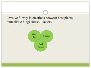
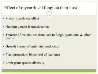
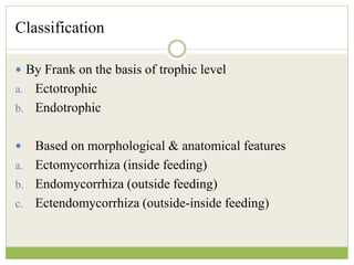
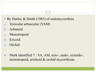
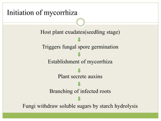
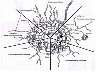
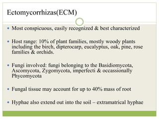
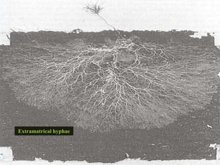
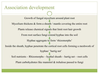
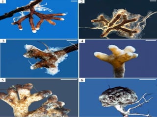
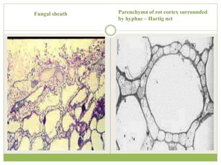
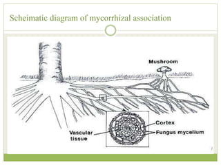
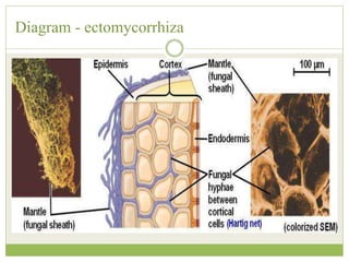
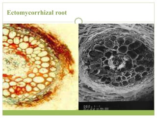
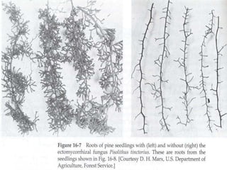
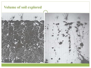
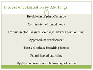
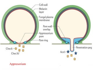
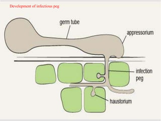
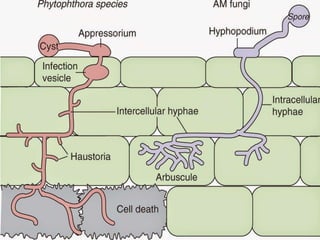
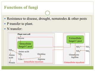
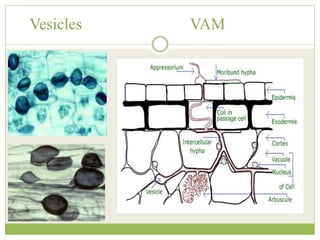
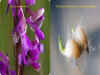
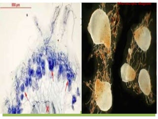
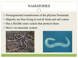
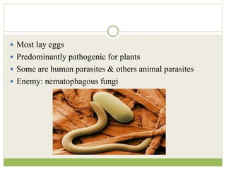
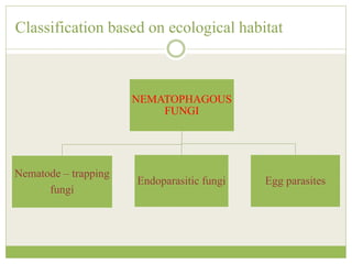
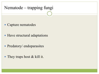
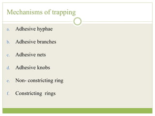
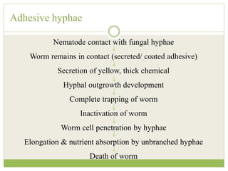
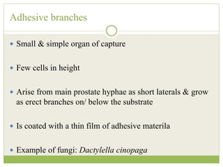
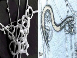
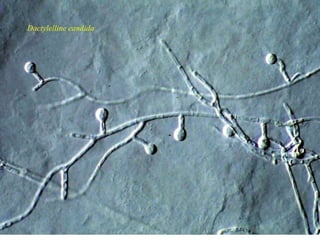
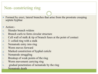
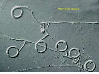
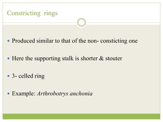
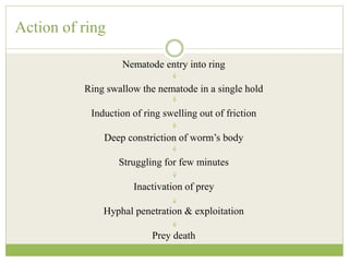
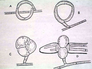
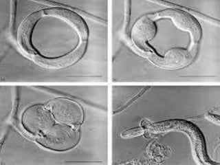
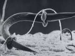
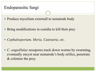
MYCORRHIZA &
NAMATOPHAGOUS
FUNGI
MICROBIAL ECOLOGY
INTRODUCTION
 Microbial interactions
 Animal- microbe
 Microbe- microbe
 Plant - microbe
 Plant - microbe interaction : mycorrhizae
 Destructive association : nematophagy
MYCORRHIZA
 Plural : mycorrhizas/ mycorrhizae
 Symbiotic association between a fungus and the roots of
a vascular plant
 Greek words : myker = fungus & rhiza = root
 Literally : fungus roots
 Abundant
 Both partners are depends on each other & thus have co-evolved
 The fungus role :-
a. plant’s root hairs
b. acts as an extension of the root systems
Involve 3- way interactions between host plants,
mutualistic fungi and soil factors
Host
plant
Fungus
Soil
factors
Effect of mycorrhizal fungi on their host
 Mycorrhizoshpere effect
 Nutrient uptake & translocation
 Transfer of metabolites from host to fungal symbionts & other
plants
 Growth hormone/ antibiotic production
 Plant protection/ biocontrol of pathogen
 Limit plant species diversity
Classification
 By Frank on the basis of trophic level
a. Ectotrophic
b. Endotrophic
 Based on morphological & anatomical features
a. Ectomycorrhiza (inside feeding)
b. Endomycorrhiza (outside feeding)
c. Ectendomycorrhiza (outside-inside feeding)
 By Harley & Smith (1983) of endomycorrhiza
a. Vesicular arbuscular (VAM)
b. Arbutoid
c. Monotropoid
d. Ericoid
e. Orchid
 Mark identified 7 : VA, AM, ecto-, endo-, ectendo-,
monotropoid, erichoid & orchid mycorrhizae.
Initiation of mycorrhiza
Host plant exudates(seedling stage)
Triggers fungal spore germination
Establishment of mycorrhiza
Plant secrete auxins
Branching of infected roots
Fungi withdraw soluble sugars by starch hydrolysis
Ectomycorrhizas(ECM)
 Most conspicuous, easily recognized & best characterized
 Host range: 10% of plant families, mostly woody plants
including the birch, dipterocarp, eucalyptus, oak, pine, rose
families & orchids.
 Fungi involved: fungi belonging to the Basidiomycota,
Ascomycota, Zygomycota, imperfecti & occassionally
Phycomycota
 Fungal tissue may account for up to 40% mass of root
 Hyphae also extend out into the soil – extramatrical hyphae
Extramatrical hyphae
Association development
Growth of fungal mycelium around plant root
Mycelium thickens & form a sheath / mantle covering the entire root
Plants release chemical signals that limit root hair growth
From root surface fungi extend hyphae into the soil
Hyphae aggregate to form ‘rhizomorphs’
Inside the sheath, hyphae penetrate the cortical root cells forming a meshwork of
hyphae/ ‘hartig net’
Soil nutrients – Rhizomorphs – hyphal sheath – hartig net – root cells
Plant carbohydrates like mannitol & trehalose passed to fungi
Fungal sheath Parenchyma of rot cortex surrounded
by hyphae – Hartig net
Scheimatic diagram of mycorrhizal association
Diagram - ectomycorrhiza
Ectomycorrhizal root
Benefits to trees
 Numerous studies have shown that tree growth is better when
mycorrhizae are present
 Fungi increase supply of inorganic nutrients to tree
 Plant hormones produced by fungus changes the physiological state
of roots – physiologically active root area for nutrient and water
absorption is increased
 Increases tolerance of plant to drought, high temperatures, pH
extremes, heavy metals
 Increases resistance to infection by root pathogens – provides a
physical barrier
Volume of soil explored
Ectomycorrhizal fungi
 Can also grow saprotrophically
 Many have been cultured
 Most do not have the capability to degrade complex plant
polymers (e.g. cellulose and lignin)
 Depend on soluble carbohydrates
 Many have organic growth factor requirements – vitamins, amino
acids
 Not decomposers but depend on plant
Benefits to fungus
 Provided with source of carbon (C) and energy (Plants
provided with CO2 demonstrated that C appears in
fungus)
 Sucrose from plant converted into trehalose & mannitol
by fungus
 Estimates that up to 10% (or more) of photosynthate
produced by trees is passed to mycorrhizae and other
rhizosphere organisms
Endo/arbuscule mycorrhiza (AM)
 Most common
 Fungi : Glomeromycetes
 Host : wild crop plants (90 %) & tropical trees
 Grows in between the cells of the plant.
 Mycorrhiza increase the surface area of the root.
 Absorb nutrients from soil especially phosphorus and
micronutrients through hyphae and mobilize into the host cell.
Features
 Mycorrhiza possess vesicles and arbuscules.
 Not as well characterized as ectomycorrhizae
 Root is not altered in morphology – difficult to determine
when roots are infected – must clear and stain followed by
microscopic examination
 Fungi are obligate biotrophs – cannot be grown in axenic
culture – so difficult to conduct experiments
Spores
•Form large spores that superficially resemble zygospores, but not formed from fusion of
gametangia
•Spore diameters range from 50 to 400 μm
Arbuscules
 Fungal hyphae- penetrate cortical cells- grow
intracellularly- form coils, swellings/ minute branches-
arbuscule (minute branches).
 Surrounded by plant cell membrane
 Typically disintegrate after 2 weeks in plant cell and
release nutrients
 Thought to be site of nutrient exchange
Arbuscule’s diagram
Process of colonization by AM fungi
Breakdown of plant C storage
Germination of fungal pores
External molecular signal exchange between plant & fungi
Appresorium development
Host cell release branching factors
Fungal hyphal branching
Hyphae colonize root cells forming arbuscule
Appresorium
Develepment of infectious peg
Functions of fungi
 Resistance to disease, drought, nematodes & other pests
 P transfer to plant.
 N transfer:
Intracellular
fungal C pool
Extracellular
fungal C pool
Hexose
Plant root cell
Amino acids
NH4 Ornithine
Urea Arginine
Intracellular mycelium
NH4
Amino
acids
Protein
NO3 NO3
NH4 NH4
Glutathione
Arginine
Extracellular mycelium
Types
a. Vesicular arbuscular (VAM)
b. Arbutoid
c. Monotropoid
d. Ericoid
e. Orchid
Vesicular Arbuscular mycorrhizae (VAM)
 Most common – 90% vascular plants
 Host : cultivated & wild plants and found in bryophyte,
pterodophytes, gymnosperms & angiosperms.
 Fungi: family Endogonacea of Zygomycotina.
 Hyphae: aseptate, intra- & inter- celullar in cortex
 Its development played a crucial role in the initial colonization of
land by plants and in the evolution of the vascular plants.
• VAM fungi capture nutrients like P, S, N and micronutrients from the soil.
• Form arbuscules and vesicles
• Vesicles :-
a. Spherical- oval intercalary, terminal, thickwalled structures, formed in the
intracellular spaces or occasionally in the cortex cells.
b. Radial & multinucleate
c. Have fat granules
d. Act as storage organ
e. Upon degeneration & eventual decomposition they provide nutrition to host.
Vesicles VAM
Arbutoid mycorrhizae
 Host : woody shrubs & tress - order Ericales
 Roots : herorhizic
 Mycosymbionts: Basidiomycetes
 Fungi penetrate cortical cells & form hartig net
 Sometimes show transition between ecto- & endo-
mycorrhizae – ectendomycorrhizae
 Fungal sheath or mantle (20 - 80 mm) covers the roots
 Nutrients scavenged by the mycelium and rhizomorphs
have to pass through the sheath and into the short roots.
 The sheath - an important store of nutrients, to be released
to the plant when nutrient levels are sufficiently depleted.
 Similar to ectomycorrhiza except that it’s hyphae
penetrate the outer cortical cells & form coils that the
latter can’t.
Mycorrhizal root
Monotropoid mycorrhizae
 Host:-
a. Achlorophyllus plants of the family Monotropacea
b. Completely depend on the fungus for C & energy
c. Roots form ball throughout which fungal mycelium ramifies enclosing the
roots of neighboring green plants.
 Fungi: Basidiomycetes
 Root ball:-
a. survival organ during winter
b. form flowering shoots during favorable condition
 Form sheat & hartig net
 Structure & function change with seasonal development of host.
Monotropoid mycorrhizael association
Ericoid mycorrhizae
Orchid mycorrhizae
 Orchidaceous mycorrhizae
 Orchid- Basidiomycete association
 Orchids show absolute dependency on ther fungal partner
 Orchid seeds won’t germinate unless fungal colonization since
they lack chorophyll.
 Fungi: saprophytic
 Orchids: parasitic
Anacamptis morio The germinated seed with fungi hyphae
Microscopic diagram
NAMATODES
 Nonsegmented roundworms of the phylum Nematode
 Majority are free-living in soil & fresh and salt waters
 Has a flexible outer cuticle that protects them
 Move via muscular system
 Most lay eggs
 Predominantly pathogenic for plants
 Some are human parasites & others animal parasites
 Enemy: nematophagous fungi
NAMATOPHAGY
 Eating of nematodes by fungi
 Fungi - predacious/ namatophagous/ nematode trapping
 Fungi mechanically attack & kill the worms resulting in
consumption of the worm
NEMATOPHAGOUS FUNGI
 Obligate parasites of nematodes
 Majority of them are facultative saprophytes
 Used for biological control of plant-parasitic nematodes
 Source: surface litter & decaying organic matter
Taxonomy and phylogeny
 Fungal taxa: Ascomycetes (and their hyphomycete
anamorphs), Basidiomycetes, Zygomycetes,
Chytridiomycetes and Oomycetes
 Nematophagous habit evolved from lignolytic
&cellulolytic fungi as an adaptation to overcome
competition for nutrients in soil.
Classification based on ecological habitat
NEMATOPHAGOUS
FUNGI
Nematode – trapping
fungi
Endoparasitic fungi Egg parasites
Nematode – trapping fungi
 Capture nematodes
 Have structural adaptations
 Predatory/ endoparasites
 They traps host & kill it.
Mechanisms of trapping
a. Adhesive hyphae
b. Adhesive branches
c. Adhesive nets
d. Adhesive knobs
e. Non- constricting ring
f. Constricting rings
Adhesive hyphae
Nematode contact with fungal hyphae
Worm remains in contact (secreted/ coated adhesive)
Secretion of yellow, thick chemical
Hyphal outgrowth development
Complete trapping of worm
Inactivation of worm
Worm cell penetration by hyphae
Elongation & nutrient absorption by unbranched hyphae
Death of worm
Stylopage hadra
Adhesive branches
 Small & simple organ of capture
 Few cells in height
 Arise from main prostate hyphae as short laterals & grow
as erect branches on/ below the substrate
 Is coated with a thin film of adhesive materila
 Example of fungi: Dactylella cinopaga
Dactylella cinopaga
Adhesive nets
 Formed by adhesive fungal hyphae
 Single loop like – complex multibranched network
 Example: Anthrobortrys musiformis
 Action similar to that of adhesive hyphae except that they
never secrete adhesive material but coated.
Adhesive knobs
 Distant adhesive globose- sub- globose cell
 Produced at the apex of a slender non- adhesive stalk
with 1- 3 cells
 Action:-
Production of a thin adhesive film over the knob surface
Nematode trapping by knob
Attack by several more knobs
Knob penetration
Nematode death
Dactylelline candida
Non- constricting ring
 Formed by erect, lateral branches that arise from the prostrate creeping
septate hyphae
 Action:-
a. Slender branch widens
b. Branch curls to form circular structure
c. Cell wall of stalk & tip of branch fuses at the point of contact
d. 3- celled ring with a stalk
e. Nematode entry into ring
f. Worm moves forward
g. Marked constriction of hyphal cuticle
h. Nematode struggling
i. Breakup of weak points of the ring
j. Worm movement carrying ring
k. gradual penetration of nematode by the ring
l. Nematode death
Dactylelline candida
Constricting rings
 Produced similar to that of the non- consticting one
 Here the supporting stalk is shorter & stouter
 3- celled ring
 Example: Arthrobotrys anchonia
Action of ring
Nematode entry into ring
Ring swallow the nematode in a single hold
Induction of ring swelling out of friction
Deep constriction of worm’s body
Struggling for few minutes
Inactivation of prey
Hyphal penetration & exploitation
Prey death
Endoparasitic fungi
 Produce mycelium externall to nematode body
 Bring modifications in conidia to kill their prey
 Cephalosporium, Meria, Catenaria, etc.
 C. anguillulae zoospores track down worms by swarming,
eventually encyst near nematode’s body orifice, penetrate
& colonize the prey.
Meria spp. Hirsutella rhossiliensis
Egg parasites
 Saprophytic
 Action:-
Contact between fungi & nematode egg
Hyphae develops a terminal swollen structure
Hyphal narrow infectious tube develops
Tube penetrates the egg shell
Hyphae swells up & forms a penetration bulk
From the bulk develops numerous absorption hyphae
The hyhae consume egg nutrients
Dactyllela oviparasitica
REFERENCES
1. Charles P. Gerba, Ian L.Pepper, Raina M. Maier- Environmental
Microbiology- second edition pg.no: 466
2. Ajith KR Banerjee, Nirmalya Banerjee- Fundamentals of
Microbiology & Immunology- 2006 edition- pg.no.: 153
3. Dubey R.C., Maheswari D.K.- A Textbook of Microbiolgy-
multicolor edition- pg. no.: 720- 724, 726- 727, 733- 735.
4. Dube H.C.- An Introduction to Fungi- second revised edition-
pg.no.: 455, 456, 458, 460, 461, 463.
5. Christopher I. Woolverton, Joanne M Willey, Linda M. Sherwood-
Presscott’s Microbiology- eight edition- pg. no: 699- 702
6. John Webster, Roland W.S. Weber- Introducing Fungi- third edition-
pg.no.: 218
7. Pelczar, Michael Joseph – Microbiology- edition 2010- pg.no: 362 -
363

MICROBIAL ECOLOGY

  • 1.
  • 2.
    INTRODUCTION  Microbial interactions Animal- microbe  Microbe- microbe  Plant - microbe  Plant - microbe interaction : mycorrhizae  Destructive association : nematophagy
  • 3.
    MYCORRHIZA  Plural :mycorrhizas/ mycorrhizae  Symbiotic association between a fungus and the roots of a vascular plant  Greek words : myker = fungus & rhiza = root  Literally : fungus roots
  • 4.
     Abundant  Bothpartners are depends on each other & thus have co-evolved  The fungus role :- a. plant’s root hairs b. acts as an extension of the root systems
  • 5.
    Involve 3- wayinteractions between host plants, mutualistic fungi and soil factors Host plant Fungus Soil factors
  • 6.
    Effect of mycorrhizalfungi on their host  Mycorrhizoshpere effect  Nutrient uptake & translocation  Transfer of metabolites from host to fungal symbionts & other plants  Growth hormone/ antibiotic production  Plant protection/ biocontrol of pathogen  Limit plant species diversity
  • 7.
    Classification  By Frankon the basis of trophic level a. Ectotrophic b. Endotrophic  Based on morphological & anatomical features a. Ectomycorrhiza (inside feeding) b. Endomycorrhiza (outside feeding) c. Ectendomycorrhiza (outside-inside feeding)
  • 8.
     By Harley& Smith (1983) of endomycorrhiza a. Vesicular arbuscular (VAM) b. Arbutoid c. Monotropoid d. Ericoid e. Orchid  Mark identified 7 : VA, AM, ecto-, endo-, ectendo-, monotropoid, erichoid & orchid mycorrhizae.
  • 9.
    Initiation of mycorrhiza Hostplant exudates(seedling stage) Triggers fungal spore germination Establishment of mycorrhiza Plant secrete auxins Branching of infected roots Fungi withdraw soluble sugars by starch hydrolysis
  • 11.
    Ectomycorrhizas(ECM)  Most conspicuous,easily recognized & best characterized  Host range: 10% of plant families, mostly woody plants including the birch, dipterocarp, eucalyptus, oak, pine, rose families & orchids.  Fungi involved: fungi belonging to the Basidiomycota, Ascomycota, Zygomycota, imperfecti & occassionally Phycomycota  Fungal tissue may account for up to 40% mass of root  Hyphae also extend out into the soil – extramatrical hyphae
  • 12.
  • 13.
    Association development Growth offungal mycelium around plant root Mycelium thickens & form a sheath / mantle covering the entire root Plants release chemical signals that limit root hair growth From root surface fungi extend hyphae into the soil Hyphae aggregate to form ‘rhizomorphs’ Inside the sheath, hyphae penetrate the cortical root cells forming a meshwork of hyphae/ ‘hartig net’ Soil nutrients – Rhizomorphs – hyphal sheath – hartig net – root cells Plant carbohydrates like mannitol & trehalose passed to fungi
  • 15.
    Fungal sheath Parenchymaof rot cortex surrounded by hyphae – Hartig net
  • 16.
    Scheimatic diagram ofmycorrhizal association
  • 17.
  • 18.
  • 20.
    Benefits to trees Numerous studies have shown that tree growth is better when mycorrhizae are present  Fungi increase supply of inorganic nutrients to tree  Plant hormones produced by fungus changes the physiological state of roots – physiologically active root area for nutrient and water absorption is increased  Increases tolerance of plant to drought, high temperatures, pH extremes, heavy metals  Increases resistance to infection by root pathogens – provides a physical barrier
  • 21.
  • 22.
    Ectomycorrhizal fungi  Canalso grow saprotrophically  Many have been cultured  Most do not have the capability to degrade complex plant polymers (e.g. cellulose and lignin)  Depend on soluble carbohydrates  Many have organic growth factor requirements – vitamins, amino acids  Not decomposers but depend on plant
  • 23.
    Benefits to fungus Provided with source of carbon (C) and energy (Plants provided with CO2 demonstrated that C appears in fungus)  Sucrose from plant converted into trehalose & mannitol by fungus  Estimates that up to 10% (or more) of photosynthate produced by trees is passed to mycorrhizae and other rhizosphere organisms
  • 24.
    Endo/arbuscule mycorrhiza (AM) Most common  Fungi : Glomeromycetes  Host : wild crop plants (90 %) & tropical trees  Grows in between the cells of the plant.  Mycorrhiza increase the surface area of the root.  Absorb nutrients from soil especially phosphorus and micronutrients through hyphae and mobilize into the host cell.
  • 25.
    Features  Mycorrhiza possessvesicles and arbuscules.  Not as well characterized as ectomycorrhizae  Root is not altered in morphology – difficult to determine when roots are infected – must clear and stain followed by microscopic examination  Fungi are obligate biotrophs – cannot be grown in axenic culture – so difficult to conduct experiments
  • 26.
    Spores •Form large sporesthat superficially resemble zygospores, but not formed from fusion of gametangia •Spore diameters range from 50 to 400 μm
  • 27.
    Arbuscules  Fungal hyphae-penetrate cortical cells- grow intracellularly- form coils, swellings/ minute branches- arbuscule (minute branches).  Surrounded by plant cell membrane  Typically disintegrate after 2 weeks in plant cell and release nutrients  Thought to be site of nutrient exchange
  • 28.
  • 29.
    Process of colonizationby AM fungi Breakdown of plant C storage Germination of fungal pores External molecular signal exchange between plant & fungi Appresorium development Host cell release branching factors Fungal hyphal branching Hyphae colonize root cells forming arbuscule
  • 30.
  • 31.
  • 33.
    Functions of fungi Resistance to disease, drought, nematodes & other pests  P transfer to plant.  N transfer: Intracellular fungal C pool Extracellular fungal C pool Hexose Plant root cell Amino acids NH4 Ornithine Urea Arginine Intracellular mycelium NH4 Amino acids Protein NO3 NO3 NH4 NH4 Glutathione Arginine Extracellular mycelium
  • 34.
    Types a. Vesicular arbuscular(VAM) b. Arbutoid c. Monotropoid d. Ericoid e. Orchid
  • 35.
    Vesicular Arbuscular mycorrhizae(VAM)  Most common – 90% vascular plants  Host : cultivated & wild plants and found in bryophyte, pterodophytes, gymnosperms & angiosperms.  Fungi: family Endogonacea of Zygomycotina.  Hyphae: aseptate, intra- & inter- celullar in cortex  Its development played a crucial role in the initial colonization of land by plants and in the evolution of the vascular plants.
  • 36.
    • VAM fungicapture nutrients like P, S, N and micronutrients from the soil. • Form arbuscules and vesicles • Vesicles :- a. Spherical- oval intercalary, terminal, thickwalled structures, formed in the intracellular spaces or occasionally in the cortex cells. b. Radial & multinucleate c. Have fat granules d. Act as storage organ e. Upon degeneration & eventual decomposition they provide nutrition to host.
  • 37.
  • 38.
    Arbutoid mycorrhizae  Host: woody shrubs & tress - order Ericales  Roots : herorhizic  Mycosymbionts: Basidiomycetes  Fungi penetrate cortical cells & form hartig net  Sometimes show transition between ecto- & endo- mycorrhizae – ectendomycorrhizae
  • 39.
     Fungal sheathor mantle (20 - 80 mm) covers the roots  Nutrients scavenged by the mycelium and rhizomorphs have to pass through the sheath and into the short roots.  The sheath - an important store of nutrients, to be released to the plant when nutrient levels are sufficiently depleted.  Similar to ectomycorrhiza except that it’s hyphae penetrate the outer cortical cells & form coils that the latter can’t.
  • 41.
  • 42.
    Monotropoid mycorrhizae  Host:- a.Achlorophyllus plants of the family Monotropacea b. Completely depend on the fungus for C & energy c. Roots form ball throughout which fungal mycelium ramifies enclosing the roots of neighboring green plants.  Fungi: Basidiomycetes  Root ball:- a. survival organ during winter b. form flowering shoots during favorable condition  Form sheat & hartig net  Structure & function change with seasonal development of host.
  • 43.
  • 44.
  • 45.
    Orchid mycorrhizae  Orchidaceousmycorrhizae  Orchid- Basidiomycete association  Orchids show absolute dependency on ther fungal partner  Orchid seeds won’t germinate unless fungal colonization since they lack chorophyll.  Fungi: saprophytic  Orchids: parasitic
  • 46.
    Anacamptis morio Thegerminated seed with fungi hyphae
  • 47.
  • 48.
    NAMATODES  Nonsegmented roundwormsof the phylum Nematode  Majority are free-living in soil & fresh and salt waters  Has a flexible outer cuticle that protects them  Move via muscular system
  • 49.
     Most layeggs  Predominantly pathogenic for plants  Some are human parasites & others animal parasites  Enemy: nematophagous fungi
  • 50.
    NAMATOPHAGY  Eating ofnematodes by fungi  Fungi - predacious/ namatophagous/ nematode trapping  Fungi mechanically attack & kill the worms resulting in consumption of the worm
  • 51.
    NEMATOPHAGOUS FUNGI  Obligateparasites of nematodes  Majority of them are facultative saprophytes  Used for biological control of plant-parasitic nematodes  Source: surface litter & decaying organic matter
  • 52.
    Taxonomy and phylogeny Fungal taxa: Ascomycetes (and their hyphomycete anamorphs), Basidiomycetes, Zygomycetes, Chytridiomycetes and Oomycetes  Nematophagous habit evolved from lignolytic &cellulolytic fungi as an adaptation to overcome competition for nutrients in soil.
  • 53.
    Classification based onecological habitat NEMATOPHAGOUS FUNGI Nematode – trapping fungi Endoparasitic fungi Egg parasites
  • 54.
    Nematode – trappingfungi  Capture nematodes  Have structural adaptations  Predatory/ endoparasites  They traps host & kill it.
  • 55.
    Mechanisms of trapping a.Adhesive hyphae b. Adhesive branches c. Adhesive nets d. Adhesive knobs e. Non- constricting ring f. Constricting rings
  • 56.
    Adhesive hyphae Nematode contactwith fungal hyphae Worm remains in contact (secreted/ coated adhesive) Secretion of yellow, thick chemical Hyphal outgrowth development Complete trapping of worm Inactivation of worm Worm cell penetration by hyphae Elongation & nutrient absorption by unbranched hyphae Death of worm
  • 57.
  • 59.
    Adhesive branches  Small& simple organ of capture  Few cells in height  Arise from main prostate hyphae as short laterals & grow as erect branches on/ below the substrate  Is coated with a thin film of adhesive materila  Example of fungi: Dactylella cinopaga
  • 60.
  • 61.
    Adhesive nets  Formedby adhesive fungal hyphae  Single loop like – complex multibranched network  Example: Anthrobortrys musiformis  Action similar to that of adhesive hyphae except that they never secrete adhesive material but coated.
  • 63.
    Adhesive knobs  Distantadhesive globose- sub- globose cell  Produced at the apex of a slender non- adhesive stalk with 1- 3 cells  Action:- Production of a thin adhesive film over the knob surface Nematode trapping by knob Attack by several more knobs Knob penetration Nematode death
  • 64.
  • 65.
    Non- constricting ring Formed by erect, lateral branches that arise from the prostrate creeping septate hyphae  Action:- a. Slender branch widens b. Branch curls to form circular structure c. Cell wall of stalk & tip of branch fuses at the point of contact d. 3- celled ring with a stalk e. Nematode entry into ring f. Worm moves forward g. Marked constriction of hyphal cuticle h. Nematode struggling i. Breakup of weak points of the ring j. Worm movement carrying ring k. gradual penetration of nematode by the ring l. Nematode death
  • 66.
  • 67.
    Constricting rings  Producedsimilar to that of the non- consticting one  Here the supporting stalk is shorter & stouter  3- celled ring  Example: Arthrobotrys anchonia
  • 68.
    Action of ring Nematodeentry into ring Ring swallow the nematode in a single hold Induction of ring swelling out of friction Deep constriction of worm’s body Struggling for few minutes Inactivation of prey Hyphal penetration & exploitation Prey death
  • 72.
    Endoparasitic fungi  Producemycelium externall to nematode body  Bring modifications in conidia to kill their prey  Cephalosporium, Meria, Catenaria, etc.  C. anguillulae zoospores track down worms by swarming, eventually encyst near nematode’s body orifice, penetrate & colonize the prey.
  • 73.
    Meria spp. Hirsutellarhossiliensis
  • 74.
    Egg parasites  Saprophytic Action:- Contact between fungi & nematode egg Hyphae develops a terminal swollen structure Hyphal narrow infectious tube develops Tube penetrates the egg shell Hyphae swells up & forms a penetration bulk From the bulk develops numerous absorption hyphae The hyhae consume egg nutrients
  • 75.
  • 76.
    REFERENCES 1. Charles P.Gerba, Ian L.Pepper, Raina M. Maier- Environmental Microbiology- second edition pg.no: 466 2. Ajith KR Banerjee, Nirmalya Banerjee- Fundamentals of Microbiology & Immunology- 2006 edition- pg.no.: 153 3. Dubey R.C., Maheswari D.K.- A Textbook of Microbiolgy- multicolor edition- pg. no.: 720- 724, 726- 727, 733- 735. 4. Dube H.C.- An Introduction to Fungi- second revised edition- pg.no.: 455, 456, 458, 460, 461, 463. 5. Christopher I. Woolverton, Joanne M Willey, Linda M. Sherwood- Presscott’s Microbiology- eight edition- pg. no: 699- 702 6. John Webster, Roland W.S. Weber- Introducing Fungi- third edition- pg.no.: 218 7. Pelczar, Michael Joseph – Microbiology- edition 2010- pg.no: 362 - 363