Downloaded 372 times

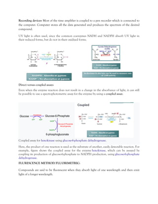
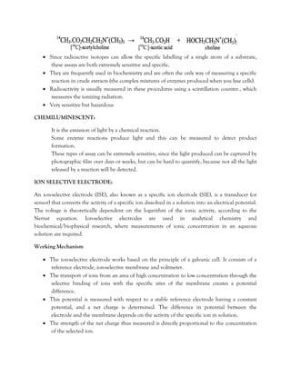
![At low concentrations, the intensity of fluorescence (fr) is related to the intensity of the incident
light (fo) of appropriate wavelength by the relationship:
Where is the molar absorption coefficient, c the molar concentration, l the length of the light-path
and q the quantum efficiency (i.e. the number of quanta fluoresced divided by the number of
quanta absorbed)
An example of these assays is again the use of the nucleotide coenzymes NADH and NADPH.
Here, the reduced forms are fluorescent and the oxidised forms nonfluorescent.
Oxidation reactions can therefore be followed by a decrease in fluorescence and reduction
reactions by an increase.
More sensitive than spectrophotometric assays, but can suffer from interference caused by
impurities and the instability of many fluorescent compounds when exposed to light.
Detection in small quantities
Non dangerous
Other example:-
RADIOISOTOPIC METHOD:
• The use of a radioactively-labelled substrate can be valuable in enzymatic analysis. The
isotopes most commonly used for labelling purposes are 3H (tritium), 14C(carbon),
32P(phosphorous), 35S(sulphur) and 131I(iodine). All of these isotopes emit beta-radiation
(electrons) as they decay.
• After the enzyme-catalysed reaction has progressed for a specified period, it is terminated.
The substrate is then separated from the product, usually by chromatography or
electrophoresis, and the product concentration is determined indirectly by measuring the
radioactivity of the product fraction.
• A typical example of enzymatic analysis by a radiochemical procedure is that involving the
cholinesterase-catalysed hydrolysis of [ 14C]-acetylcholine.](https://image.slidesharecdn.com/enzymeassay1-180329151646/85/methods-of-enzyme-assay-5-320.jpg)
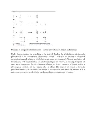


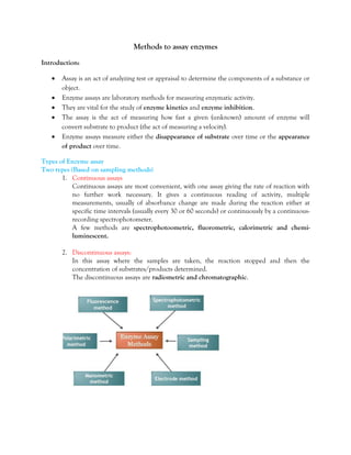
The document outlines various methods for assaying enzyme activity, emphasizing the significance of enzyme assays in fields such as enzyme kinetics and inhibition. It details continuous and discontinuous assay types, various detection methods (spectrophotometric, fluorescence, radioisotopic, chemiluminescent, etc.), and immunochemical techniques such as enzyme immunoassays. Ultimately, these assays enable the measurement of enzyme activity and have broad applications in biochemistry and diagnostics.


![ANIMAL_CELL_,_TISSUE_AND_ORGAN_CULTURE[1].pptx](https://cdn.slidesharecdn.com/ss_thumbnails/animalcelltissueandorganculture1-260204172026-4462b440-thumbnail.jpg?width=640&height=640&fit=bounds)


![Polymer [ बहुलक ] Chemistry Notes PDF - Irfanullah Mehar - JJ Sir Chemistry.pdf](https://cdn.slidesharecdn.com/ss_thumbnails/polymerchemistrynotespdf-irfanullahmehar-jjsirchemistry-260210172118-3f9b37f7-thumbnail.jpg?width=640&height=640&fit=bounds)

