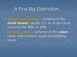
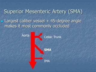
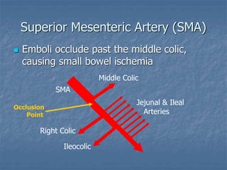
Acute Mesenteric
Ischemia and Infarction
Dr. Muhammad Noman Rashid
Senior Registrar
Department of Interventional Cardiology
Ziauddin Medical University Hospital
A First Big Distinction…
 Mesenteric Ischemia – ischemia of the
small bowel, usually 2/2 an acute cause
involving the SMA or SMV.
 Ischemic colitis – ischemia of the colon,
rarely with a known acute precipitating
cause.
Superior Mesenteric Artery (SMA)
 Largest caliber vessel + 45-degree angle
makes it most commonly occluded
Celiac Trunk
IMA
SMA
Aorta
Superior Mesenteric Artery (SMA)
 Emboli occlude past the middle colic,
causing small bowel ischemia
SMA
Middle Colic
Right Colic
Ileocolic
Jejunal & Ileal
Arteries
Occlusion
Point
Etiologies of Acute Mesenteric
Ischemia (AMI)
 SMA Occlusion (at least 60% of cases)
 Embolism: MI, Afib, Endocarditis, Valve d/o
 Thrombosis: Atherosclerosis – plaque rupture
 Nonocclusive Mesenteric Ischemia (NOMI)
 Atherosclerosis + shock + vasopressors
 Mesenteric Venous Thrombosis (MVT)
 Primary clotting disorder
Etiologies of Acute Mesenteric
Ischemia (AMI)
 Focal small bowel ischemia - rare
 Partial malrotation, volvulus, mesenteric
hematoma, strangulated hernia
 Unknown
 ?Mesenteric small vessel disease
History & Physical
Classic Presentation:
 Rapid onset of severe, unrelenting
periumbilical pain
 Pain out of proportion to findings on
physical examination.
 Nausea and vomiting
 Forceful/urgent bowel evacuation
 Risk factors for acute mesenteric ischemia
History & Physical
SMA Thrombosis:
 Prodrome of postprandial pain/nausea and
weight loss
 Presentation with classic symptoms
Non-occlusive Mesenteric Ischemia:
 Unexplained decline in clinical status or
failure to follow expected recovery
History & Physical
Mesenteric Venous Thrombosis:
 Fever
 Abdominal distension
 Hemoccult positive stool
Laboratory Findings
 Anion gap metabolic acidosis
 Elevated arterial/venous lactate
 Leukocytosis
 Hemoconcentration
 Elevated LDH, amylase, AST, and CPK
 Elevated K and Phos are late signs
Radiology
 Plain films – thumbprinting, thickened
bowel (<40% sensitivity)
 CT – thickened/dilated bowel, intramural
hematoma, pneumatosis (64% sensitivity)
 MRI – promising but untested to date
 Mesenteric angiography – test of choice;
can identify the type of AMI
Differential Diagnosis
Other serious conditions to consider:
 Pancreatitis
 Acute Diverticulitis
 Acute Cholecystitis
 Small bowel obstruction
 Perforation of a viscous
 Ruptured aneurysm
Treatment
 Resuscitation with fluids/blood products
 Anticoagulation
 Infusion of a vasodilator
 Glucagon systemically OR
 Papaverine through a catheter
From Ischemia to Infarction
 Marked by peritoneal signs, fever
 Emergent laporatomy
 Restoration of interrupted blood flow with
arteriotomy or bypass graft
 Resection of infarcted bowel
 Second-look in 24-48 hours
 Vasodilators and careful pressor use
A Word on Ischemic Colitis
 Presentation: less & more focal pain
(usually left-sided), more bloody diarrhea,
>90% are over 60 years old.
 Etiology rarely identified: ?small vessel
disease +/- hypoperfusion
 Episodes usually self limited except when
stricture or gangrene develops
 Colonoscopy is initial evaluation of choice
References
 Netter FH, Atlas of Human Anatomy
 Oldenburg et al. Arch Intern Med 164:1054 2004
 Scott JR et al. AJR 113:2 “Acute Mesenteric
Infarction” 1971
 UptoDate Online: Article on “Acute Mesenteric
Ischemia”
 UptoDate Online: Article on “Ischemic colitis”

Medical Information Mesenteric Ischemia.ppt

  • 1.
    Acute Mesenteric Ischemia andInfarction Dr. Muhammad Noman Rashid Senior Registrar Department of Interventional Cardiology Ziauddin Medical University Hospital
  • 2.
    A First BigDistinction…  Mesenteric Ischemia – ischemia of the small bowel, usually 2/2 an acute cause involving the SMA or SMV.  Ischemic colitis – ischemia of the colon, rarely with a known acute precipitating cause.
  • 3.
    Superior Mesenteric Artery(SMA)  Largest caliber vessel + 45-degree angle makes it most commonly occluded Celiac Trunk IMA SMA Aorta
  • 4.
    Superior Mesenteric Artery(SMA)  Emboli occlude past the middle colic, causing small bowel ischemia SMA Middle Colic Right Colic Ileocolic Jejunal & Ileal Arteries Occlusion Point
  • 5.
    Etiologies of AcuteMesenteric Ischemia (AMI)  SMA Occlusion (at least 60% of cases)  Embolism: MI, Afib, Endocarditis, Valve d/o  Thrombosis: Atherosclerosis – plaque rupture  Nonocclusive Mesenteric Ischemia (NOMI)  Atherosclerosis + shock + vasopressors  Mesenteric Venous Thrombosis (MVT)  Primary clotting disorder
  • 6.
    Etiologies of AcuteMesenteric Ischemia (AMI)  Focal small bowel ischemia - rare  Partial malrotation, volvulus, mesenteric hematoma, strangulated hernia  Unknown  ?Mesenteric small vessel disease
  • 7.
    History & Physical ClassicPresentation:  Rapid onset of severe, unrelenting periumbilical pain  Pain out of proportion to findings on physical examination.  Nausea and vomiting  Forceful/urgent bowel evacuation  Risk factors for acute mesenteric ischemia
  • 8.
    History & Physical SMAThrombosis:  Prodrome of postprandial pain/nausea and weight loss  Presentation with classic symptoms Non-occlusive Mesenteric Ischemia:  Unexplained decline in clinical status or failure to follow expected recovery
  • 9.
    History & Physical MesentericVenous Thrombosis:  Fever  Abdominal distension  Hemoccult positive stool
  • 10.
    Laboratory Findings  Aniongap metabolic acidosis  Elevated arterial/venous lactate  Leukocytosis  Hemoconcentration  Elevated LDH, amylase, AST, and CPK  Elevated K and Phos are late signs
  • 11.
    Radiology  Plain films– thumbprinting, thickened bowel (<40% sensitivity)  CT – thickened/dilated bowel, intramural hematoma, pneumatosis (64% sensitivity)  MRI – promising but untested to date  Mesenteric angiography – test of choice; can identify the type of AMI
  • 12.
    Differential Diagnosis Other seriousconditions to consider:  Pancreatitis  Acute Diverticulitis  Acute Cholecystitis  Small bowel obstruction  Perforation of a viscous  Ruptured aneurysm
  • 13.
    Treatment  Resuscitation withfluids/blood products  Anticoagulation  Infusion of a vasodilator  Glucagon systemically OR  Papaverine through a catheter
  • 14.
    From Ischemia toInfarction  Marked by peritoneal signs, fever  Emergent laporatomy  Restoration of interrupted blood flow with arteriotomy or bypass graft  Resection of infarcted bowel  Second-look in 24-48 hours  Vasodilators and careful pressor use
  • 15.
    A Word onIschemic Colitis  Presentation: less & more focal pain (usually left-sided), more bloody diarrhea, >90% are over 60 years old.  Etiology rarely identified: ?small vessel disease +/- hypoperfusion  Episodes usually self limited except when stricture or gangrene develops  Colonoscopy is initial evaluation of choice
  • 16.
    References  Netter FH,Atlas of Human Anatomy  Oldenburg et al. Arch Intern Med 164:1054 2004  Scott JR et al. AJR 113:2 “Acute Mesenteric Infarction” 1971  UptoDate Online: Article on “Acute Mesenteric Ischemia”  UptoDate Online: Article on “Ischemic colitis”