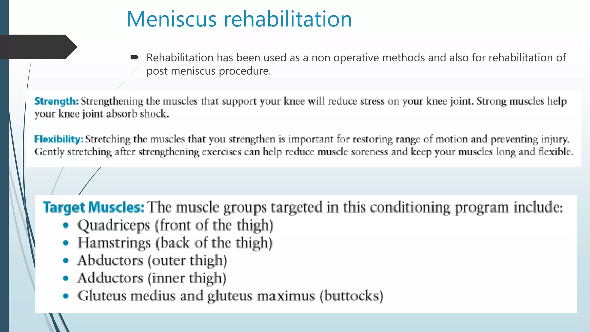
meniscus injury explained with treatment and videos to help understand the use of MRI to help understand injury to meniscus and help diagnose meniscal tear
Tendoachilles rupture and its managementRohan Vakta
Achilles tendon is the strongest tendon of body. There are many causes of its rupture. It can be acute or chronic rupture. Management of chronic rupture by semitendinosus tendon is mentioned here.
Tendoachilles rupture and its managementRohan Vakta
Achilles tendon is the strongest tendon of body. There are many causes of its rupture. It can be acute or chronic rupture. Management of chronic rupture by semitendinosus tendon is mentioned here.
Safe surgical dislocation for femoral head fractures.dr mohamed ashraf,dr rah...drashraf369
femoral head fractures are very complex fractures that need immediate and prompt surgical intervention.conventional surgical appproaches to hip may lead to short and long term complications.dr mohamed ashraf ,dr rahul thampi et al are presenting their experience with gantz safe surgical dislocation approach to surgical management of femoral head fractures
MCL. LCL.ALL injuries
To understand the relevant anatomy of the side ligaments of the knee
To study the mechanism of injury of each ligament and how to diagnose such injury
To highlight the different treatment options in acute or chronic situations
The presentation investigates the following characteristics of the meniscus;
Role of the Meniscus
Material Properties
Structural Limitations / Failure Limits
Mechanism & Treatment of Injuries
MENISCUS REPAIR I Dr.RAJAT JANGIR JAIPUR
#aclsurgeryjaipur #aclsurgeryhindia #aclsurgerytaekwondo
Acl reconstruction in jaipur | Acl reconstruction in taekwondo | Acl injury in football player surgery | Acl reconstruction surgery in football | acl surgery | Acl surgery ke baad physiotherapy | Acl surgery in jaipur | acl surgery recovery | Best acl surgeon in jaipur | Best ligament doctor in hindi | Best acl surgeon in india | Meniscus repair surgery in jaipur | Sports injury doctor | Acl injury in football players | Acl injury in taekwondo | acl tear | Best knee surgeon in jaipur
#allinsideacl #internalbrace #drrajatjangir #bestaclsurgeon #aclexpert #bestkneesurgeon
To Know more about ACL Injury, Click the links below:
1. ACL surgery 7 different Techniques we do at our center - "Not single technique best for all"
https://youtu.be/oWkIr8IXvr8
2. Everything about ACL Injury tear surgery in Hindi I
https://youtu.be/bqpjkAkwZ14
3. Best Screw for ACL tear surgery in Hindi
https://youtu.be/1LGpU1NHiIs
4. ACL Injury Tear Surgery Recovery : All your questions & queries solved by Dr.Rajat Jangir
https://youtu.be/SIAPWiMbOqs
5. Partial ACL Tear Surgery or not ! ACL आधा टूटा हो तो क्या करें ?
https://youtu.be/NEJRPKskJTI
6. 5 Symptoms of ACL Injury tear इंजरी के पांच लक्षण ?
https://youtu.be/EXpgy19Jxzw
7. PRP injection therapy in Partial ACL TEARs
https://youtu.be/qyG1EYgS87E
Dr.RAJAT JANGIR(Asso Prof.)
Senior Consultant Arthroscopy and Joint Replacement
(Specialist in Shoulder Knee Hip Surgery)
Ligament and Joints Clinic
67/34 Mansarovar Jaipur
Whatsapp: shorturl.at/gnAEP
Appointment: +91 8104855900
Email: ligamentsurgeon@gmail.com
Google Page: https://g.page/KNEE-Shoulder-SURGERY?...
Facebook: https://www.facebook.com/Ligamentandj...
* Vast experience and specialisation in the field of Arthroscopy and sports surgery.
* M.S. orthopaedics from BJ Medical College, Civil hospital, Ahmedabad
* Fellowship in Arthroscopy and Sports injury with Prof Joon Ho Wang at Samsung Medical Center, South Korea
* Diploma in Sports Medicine from InternationaI Olympic Committee
* Invited as Athlete Medical Doctor at Rio Olympic 2016
* Done Rajasthan's first "All Inside Physeal Preserving ACL reconstruction" in 13 year old Athlete
Dr.Rajat is rated as one of the best orthopedic surgeon with with excellence in Knee Shoulder Arthroscopy surgeries as replacements'
shoulder instability is quite common because shoulder have highest range of motion but at the cost of stability. here discussing anatomy around the shoulder & treatment of shoulder instability
The upper limb consists of various joints that enable movement and provide flexibility. These joints can be classified into different types based on their structure and function.
Understanding the anatomy and function of these joints is crucial for assessing and managing conditions related to the upper limb, as well as for rehabilitation and therapeutic interventions. Joint injuries, arthritis, and other disorders may affect the functionality of these joints, and appropriate medical care may be necessary for optimal outcomes.
Approach to acute knee injuries (knee injury)mahadev deuja
approach to acute knee injuries include detail history, focused knee exam and imaging/invasive procedure,Diagnosis is made at history most of the times.History should include mechanism of Injury,location of pain, mechanical symptoms like swelling/ effusion...
The Stone Clinic is a sports medicine clinic in San Francisco, California, offering orthopaedic surgery and medical care, physical therapy and rehabilitation, and radiology imaging services. The Stone Clinic was founded by Kevin R. Stone, M.D., an orthopaedic surgeon, combining himself with a team of nurses, physical therapists, imaging specialists, and patient coordinators, in 1988 to focus on caring for injured athletes and people experiencing arthritis pain.
The Stone Clinic is founded on the goal of rehabilitating all patients to an operating level higher than before they were injured. The Stone Clinic specializes in sports medicine and injury treatment of knee, shoulder, and ankle joints. Stone has lectured and is recognized internationally as an authority on cartilage and meniscal growth, replacement, and repair. Stone and the Stone Clinic are known for the development of the paste grafting surgical technique in 1991, combined with meniscus replacement, which are biologic joint replacement procedures for the regeneration of the knee joint. Surgical procedures were subjected to rigorous outcomes analysis with the results reported in peer reviewed journals. The surgical techniques have been taught to surgeons in the US and worldwide, through lectures and videos.
Nursing students, medical students, residents, fellows, and other physicians from various institutions around the world, rotate through The Stone Clinic and mentor with Stone. The Stone Clinic hosts the annual Meniscus Transplantation Study Group Meeting as well as the annual Professional Women Athlete's Career Conference.
Pulmonary Thromboembolism - etilogy, types, medical- Surgical and nursing man...VarunMahajani
Disruption of blood supply to lung alveoli due to blockage of one or more pulmonary blood vessels is called as Pulmonary thromboembolism. In this presentation we will discuss its causes, types and its management in depth.
These simplified slides by Dr. Sidra Arshad present an overview of the non-respiratory functions of the respiratory tract.
Learning objectives:
1. Enlist the non-respiratory functions of the respiratory tract
2. Briefly explain how these functions are carried out
3. Discuss the significance of dead space
4. Differentiate between minute ventilation and alveolar ventilation
5. Describe the cough and sneeze reflexes
Study Resources:
1. Chapter 39, Guyton and Hall Textbook of Medical Physiology, 14th edition
2. Chapter 34, Ganong’s Review of Medical Physiology, 26th edition
3. Chapter 17, Human Physiology by Lauralee Sherwood, 9th edition
4. Non-respiratory functions of the lungs https://academic.oup.com/bjaed/article/13/3/98/278874
ARTIFICIAL INTELLIGENCE IN HEALTHCARE.pdfAnujkumaranit
Artificial intelligence (AI) refers to the simulation of human intelligence processes by machines, especially computer systems. It encompasses tasks such as learning, reasoning, problem-solving, perception, and language understanding. AI technologies are revolutionizing various fields, from healthcare to finance, by enabling machines to perform tasks that typically require human intelligence.
Title: Sense of Smell
Presenter: Dr. Faiza, Assistant Professor of Physiology
Qualifications:
MBBS (Best Graduate, AIMC Lahore)
FCPS Physiology
ICMT, CHPE, DHPE (STMU)
MPH (GC University, Faisalabad)
MBA (Virtual University of Pakistan)
Learning Objectives:
Describe the primary categories of smells and the concept of odor blindness.
Explain the structure and location of the olfactory membrane and mucosa, including the types and roles of cells involved in olfaction.
Describe the pathway and mechanisms of olfactory signal transmission from the olfactory receptors to the brain.
Illustrate the biochemical cascade triggered by odorant binding to olfactory receptors, including the role of G-proteins and second messengers in generating an action potential.
Identify different types of olfactory disorders such as anosmia, hyposmia, hyperosmia, and dysosmia, including their potential causes.
Key Topics:
Olfactory Genes:
3% of the human genome accounts for olfactory genes.
400 genes for odorant receptors.
Olfactory Membrane:
Located in the superior part of the nasal cavity.
Medially: Folds downward along the superior septum.
Laterally: Folds over the superior turbinate and upper surface of the middle turbinate.
Total surface area: 5-10 square centimeters.
Olfactory Mucosa:
Olfactory Cells: Bipolar nerve cells derived from the CNS (100 million), with 4-25 olfactory cilia per cell.
Sustentacular Cells: Produce mucus and maintain ionic and molecular environment.
Basal Cells: Replace worn-out olfactory cells with an average lifespan of 1-2 months.
Bowman’s Gland: Secretes mucus.
Stimulation of Olfactory Cells:
Odorant dissolves in mucus and attaches to receptors on olfactory cilia.
Involves a cascade effect through G-proteins and second messengers, leading to depolarization and action potential generation in the olfactory nerve.
Quality of a Good Odorant:
Small (3-20 Carbon atoms), volatile, water-soluble, and lipid-soluble.
Facilitated by odorant-binding proteins in mucus.
Membrane Potential and Action Potential:
Resting membrane potential: -55mV.
Action potential frequency in the olfactory nerve increases with odorant strength.
Adaptation Towards the Sense of Smell:
Rapid adaptation within the first second, with further slow adaptation.
Psychological adaptation greater than receptor adaptation, involving feedback inhibition from the central nervous system.
Primary Sensations of Smell:
Camphoraceous, Musky, Floral, Pepperminty, Ethereal, Pungent, Putrid.
Odor Detection Threshold:
Examples: Hydrogen sulfide (0.0005 ppm), Methyl-mercaptan (0.002 ppm).
Some toxic substances are odorless at lethal concentrations.
Characteristics of Smell:
Odor blindness for single substances due to lack of appropriate receptor protein.
Behavioral and emotional influences of smell.
Transmission of Olfactory Signals:
From olfactory cells to glomeruli in the olfactory bulb, involving lateral inhibition.
Primitive, less old, and new olfactory systems with different path
Tom Selleck Health: A Comprehensive Look at the Iconic Actor’s Wellness Journeygreendigital
Tom Selleck, an enduring figure in Hollywood. has captivated audiences for decades with his rugged charm, iconic moustache. and memorable roles in television and film. From his breakout role as Thomas Magnum in Magnum P.I. to his current portrayal of Frank Reagan in Blue Bloods. Selleck's career has spanned over 50 years. But beyond his professional achievements. fans have often been curious about Tom Selleck Health. especially as he has aged in the public eye.
Follow us on: Pinterest
Introduction
Many have been interested in Tom Selleck health. not only because of his enduring presence on screen but also because of the challenges. and lifestyle choices he has faced and made over the years. This article delves into the various aspects of Tom Selleck health. exploring his fitness regimen, diet, mental health. and the challenges he has encountered as he ages. We'll look at how he maintains his well-being. the health issues he has faced, and his approach to ageing .
Early Life and Career
Childhood and Athletic Beginnings
Tom Selleck was born on January 29, 1945, in Detroit, Michigan, and grew up in Sherman Oaks, California. From an early age, he was involved in sports, particularly basketball. which played a significant role in his physical development. His athletic pursuits continued into college. where he attended the University of Southern California (USC) on a basketball scholarship. This early involvement in sports laid a strong foundation for his physical health and disciplined lifestyle.
Transition to Acting
Selleck's transition from an athlete to an actor came with its physical demands. His first significant role in "Magnum P.I." required him to perform various stunts and maintain a fit appearance. This role, which he played from 1980 to 1988. necessitated a rigorous fitness routine to meet the show's demands. setting the stage for his long-term commitment to health and wellness.
Fitness Regimen
Workout Routine
Tom Selleck health and fitness regimen has evolved. adapting to his changing roles and age. During his "Magnum, P.I." days. Selleck's workouts were intense and focused on building and maintaining muscle mass. His routine included weightlifting, cardiovascular exercises. and specific training for the stunts he performed on the show.
Selleck adjusted his fitness routine as he aged to suit his body's needs. Today, his workouts focus on maintaining flexibility, strength, and cardiovascular health. He incorporates low-impact exercises such as swimming, walking, and light weightlifting. This balanced approach helps him stay fit without putting undue strain on his joints and muscles.
Importance of Flexibility and Mobility
In recent years, Selleck has emphasized the importance of flexibility and mobility in his fitness regimen. Understanding the natural decline in muscle mass and joint flexibility with age. he includes stretching and yoga in his routine. These practices help prevent injuries, improve posture, and maintain mobilit
Factory Supply Best Quality Pmk Oil CAS 28578–16–7 PMK Powder in Stockrebeccabio
Factory Supply Best Quality Pmk Oil CAS 28578–16–7 PMK Powder in Stock
Telegram: bmksupplier
signal: +85264872720
threema: TUD4A6YC
You can contact me on Telegram or Threema
Communicate promptly and reply
Free of customs clearance, Double Clearance 100% pass delivery to USA, Canada, Spain, Germany, Netherland, Poland, Italy, Sweden, UK, Czech Republic, Australia, Mexico, Russia, Ukraine, Kazakhstan.Door to door service
Hot Selling Organic intermediates
micro teaching on communication m.sc nursing.pdfAnurag Sharma
Microteaching is a unique model of practice teaching. It is a viable instrument for the. desired change in the teaching behavior or the behavior potential which, in specified types of real. classroom situations, tends to facilitate the achievement of specified types of objectives.
Ethanol (CH3CH2OH), or beverage alcohol, is a two-carbon alcohol
that is rapidly distributed in the body and brain. Ethanol alters many
neurochemical systems and has rewarding and addictive properties. It
is the oldest recreational drug and likely contributes to more morbidity,
mortality, and public health costs than all illicit drugs combined. The
5th edition of the Diagnostic and Statistical Manual of Mental Disorders
(DSM-5) integrates alcohol abuse and alcohol dependence into a single
disorder called alcohol use disorder (AUD), with mild, moderate,
and severe subclassifications (American Psychiatric Association, 2013).
In the DSM-5, all types of substance abuse and dependence have been
combined into a single substance use disorder (SUD) on a continuum
from mild to severe. A diagnosis of AUD requires that at least two of
the 11 DSM-5 behaviors be present within a 12-month period (mild
AUD: 2–3 criteria; moderate AUD: 4–5 criteria; severe AUD: 6–11 criteria).
The four main behavioral effects of AUD are impaired control over
drinking, negative social consequences, risky use, and altered physiological
effects (tolerance, withdrawal). This chapter presents an overview
of the prevalence and harmful consequences of AUD in the U.S.,
the systemic nature of the disease, neurocircuitry and stages of AUD,
comorbidities, fetal alcohol spectrum disorders, genetic risk factors, and
pharmacotherapies for AUD.
Title: Sense of Taste
Presenter: Dr. Faiza, Assistant Professor of Physiology
Qualifications:
MBBS (Best Graduate, AIMC Lahore)
FCPS Physiology
ICMT, CHPE, DHPE (STMU)
MPH (GC University, Faisalabad)
MBA (Virtual University of Pakistan)
Learning Objectives:
Describe the structure and function of taste buds.
Describe the relationship between the taste threshold and taste index of common substances.
Explain the chemical basis and signal transduction of taste perception for each type of primary taste sensation.
Recognize different abnormalities of taste perception and their causes.
Key Topics:
Significance of Taste Sensation:
Differentiation between pleasant and harmful food
Influence on behavior
Selection of food based on metabolic needs
Receptors of Taste:
Taste buds on the tongue
Influence of sense of smell, texture of food, and pain stimulation (e.g., by pepper)
Primary and Secondary Taste Sensations:
Primary taste sensations: Sweet, Sour, Salty, Bitter, Umami
Chemical basis and signal transduction mechanisms for each taste
Taste Threshold and Index:
Taste threshold values for Sweet (sucrose), Salty (NaCl), Sour (HCl), and Bitter (Quinine)
Taste index relationship: Inversely proportional to taste threshold
Taste Blindness:
Inability to taste certain substances, particularly thiourea compounds
Example: Phenylthiocarbamide
Structure and Function of Taste Buds:
Composition: Epithelial cells, Sustentacular/Supporting cells, Taste cells, Basal cells
Features: Taste pores, Taste hairs/microvilli, and Taste nerve fibers
Location of Taste Buds:
Found in papillae of the tongue (Fungiform, Circumvallate, Foliate)
Also present on the palate, tonsillar pillars, epiglottis, and proximal esophagus
Mechanism of Taste Stimulation:
Interaction of taste substances with receptors on microvilli
Signal transduction pathways for Umami, Sweet, Bitter, Sour, and Salty tastes
Taste Sensitivity and Adaptation:
Decrease in sensitivity with age
Rapid adaptation of taste sensation
Role of Saliva in Taste:
Dissolution of tastants to reach receptors
Washing away the stimulus
Taste Preferences and Aversions:
Mechanisms behind taste preference and aversion
Influence of receptors and neural pathways
Impact of Sensory Nerve Damage:
Degeneration of taste buds if the sensory nerve fiber is cut
Abnormalities of Taste Detection:
Conditions: Ageusia, Hypogeusia, Dysgeusia (parageusia)
Causes: Nerve damage, neurological disorders, infections, poor oral hygiene, adverse drug effects, deficiencies, aging, tobacco use, altered neurotransmitter levels
Neurotransmitters and Taste Threshold:
Effects of serotonin (5-HT) and norepinephrine (NE) on taste sensitivity
Supertasters:
25% of the population with heightened sensitivity to taste, especially bitterness
Increased number of fungiform papillae
3. HISTORY TAKING
Patient male 39-year-old with chief complaint pain at his right
knee.
Suffered since 3 month before admitted to Wahidin general
hospital due to trauma
Patient was running when his knee hit the door. Pain was
intermittent and aggravated by activity, particularly while walking
and squatting.
No history of locking sensation
No history of previous treatment
4. PHYSICAL EXAMINATION
Left Knee Region
Look :
Deformity (-), swelling (-), hematom (-), scar (-)
Feel : Tenderness (-), Patellar tapping (-), patellar grinding (-)
Move Active and passive movement of knee joint is limited, ROM 0o-
130o
NVD :
Sensibility normal, pulsation artery dorsalis pedis and tibialis
anterior palpable, CRT <2”
Special
tests
:
Lachmann test (-), anterior drawer test (-), posterior drawer test
(-), Mcmurray test (-)
5. PHYSICAL EXAMINATION
Right Knee Region
Look :
Deformity (-), swelling (-), hematom (-), scar (-)
Feel : Tenderness (-), Patellar tapping (-), patellar grinding (-)
Move Active and passive movement of knee joint is limited, ROM 0o-
130o
NVD :
Sensibility normal, pulsation artery dorsalis pedis and tibialis
anterior palpable, CRT <2”
Special
tests
:
Lachmann test (-), anterior drawer test (-), posterior drawer test
(-), Mcmurray test (+)
6.
7.
8.
9.
10.
11.
12. Patient was diagnosed with a suspected meniscus tear and ACL rupture and was
performed a diagnostic arthroscopic procedure medial condyle osteochondral
defect, degenerative tear lateral meniscus.
A synovectomy and debridement was performed
Final diagnosis was :
degenerative tear lateral meniscus
Osteochondral defect medial condyle
Fibrotic synovial right knee
Patient was reffered to rehabilitation after surgery for muscle strengthening exercise
14. Meniscus anatomy
“If it is torn, take it out! Take it all out! Even if you just think it’s torn, take it out.”
Those were the slogan words by Smillie in 1967 referring to meniscal injuries
Meniscus is a latin word, from greek meniskos meaning crescent
Meniscal tears are the most common pathology of the knee with a mean annual
incidence of 66 per 100000 with knee arthroscopy as the most common methods of
treatment.
Traditionally, meniscal tears were managed with meniscectomy.
However, since the long-term morbidities of meniscus removal became apparent (eg,
early development of knee osteoarthritis), management has been increasingly focused
on meniscal preservation.
15. Medial meniscus : U shaped, covering 60% of medial compartment
Lateral meniscus : C shaped, covering 80% of lateral compartment
The horns anchor to the subchondral bone of tibial plateau
16. Meniscus is composed of :
Water (72%)
Collagen (22%), responsible for tensile strength of
meniscus. Predominant type 1 (80%) in red zone, and
type 2 : type 1, 60:40 in white zone
The medial and lateral menisci are each
approximately 3 cm wide.
The medial meniscus is approximately 4 to 5 cm in
length, and the lateral meniscus is approximately 3
to 4 cm in length.
2 types of fibres :
Radial
Prevent longitudinal splitting of circumferential fibres
Longitudinal (circumferential)
predominance in outer third
From anterior horn insertianl ligament to posterior horn
insertional ligament
Absorb energy by dissipating hoop stresses
17. Blood supply is from the periphery, via medial and lateral geniculate arteries
Cadaveric studies demonstrated that 10-25% peripheral area of meniscus receive blood supply
2 distict zones : red-red, white-white, separated by red-white region
Red-red zone is a vascular zone, thick and convex
white-white zone is an avascular zone with very low (very unlikely) healing capability, concave,
thin and unattached
Posterior horns have the highest concentration of mechanoreceptors
18. Meniscus enables effective articulation between the
concave femoral condyle and the flat tibial plateau
Acts as a shock absorber
to transmit sheer and tensile load from soft tissue to
bone
Function to decrease contact area
Joint lubrication
Proprioception
19. Meniscus injury
Common source of pain and disability of knee
60-70/100.000 case
Male : female = 2.5-4:1
Peak incidence in males 21-30 years
Medial meniscal tear are more common than lateral tears
Lateral tears occur in acute ACL tear
Traumatic tears occur in younger more active groups
Degenerative tears occur due to cumulative stress
Combination of :1axial loading, 2rotational force
20. HISTORY
Tears of normal menisci usually are associated with more significant trauma or
injury but are produced by a similar mechanism:
the meniscus is entrapped between the femoral and tibial condyles in flexion,
tearing as the knee is extended.
Patients with tears in degenerative menisci may recall symptoms of mild catching,
snapping, or clicking as well as occasional pain and mild swelling in the joint.
Robert H. Knee injuries. Campbell’s operative orthopaedic
21. SYMPTOMS
Pain localizing to medial or lateral side
Mechanical symptoms (locking and clicking)
Delayed or intermittent swelling
The syndromes caused by tears of the menisci can be divided into two groups:
- those in which there is locking and the diagnosis is clear,
- and those in which locking is absent and the diagnosis is more difficult
Patrick. Meniscal injury. Orthoblullet
Robert H. Knee injuries. Campbell’s operative orthopaedic
22. Locking
Locking usually occurs only with longitudinal tears and is much more common with
bucket-handle tears, usually of the medial meniscus.
Locking of the knee must not be considered pathognomonic of a bucket-handle
tear of a meniscus;
an intraarticular tumor, an osteocartilaginous loose body, and other conditions can cause
locking.
False locking occurs most often soon after an injury in which hemorrhage around
the posterior part of the capsule or a collateral ligament with associated hamstring
spasm prevents complete extension of the knee
Robert H. Knee injuries. Campbell’s operative orthopaedic
23. No locking
If a patient does not have locking, the diagnosis of a torn meniscus is more
difficult even for the most astute surgeon.
A patient typically gives a history of several episodes of trouble referable to the
knee, often resulting in effusion and a brief period of disability but no definite
locking.
A sensation of “giving way” or snaps, clicks, catches, or jerks in the knee may be
described, or the history may be even more indefinite, with recurrent episodes of
pain and mild effusion in the knee and tenderness in the anterior joint space after
excessive activity.
Robert H. Knee injuries. Campbell’s operative orthopaedic
24. PHYSICAL EXAMINATION
Joint line tenderness (most sensitive physical examination finding )
Effusion
Provocative tests
McMurray
Apley grind test
Squat test
Thessaly test
One analysis determined that joint line tenderness is the best “common” test, while the other found
sensitivities and specificities similar among the three tests: McMurray, 70% and 71%; Apley, 60% and
70%; and joint line tenderness, 63% and 77%.
Patrick. Meniscal injury. Orthoblullet
Robert H. Knee injuries. Campbell’s operative orthopaedic
25. Mc Murray test
With the patient supine and the knee
acutely and forcibly flexed, the examiner
can check the medial meniscus by
palpating the posteromedial margin of
the joint with one hand while grasping the
foot with the other hand.
Keeping the knee completely flexed, the
leg is externally rotated as far as possible
and then the knee is slowly extended.
As the femur passes over a tear in the
meniscus, a click may be heard or felt.
Robert H. Knee injuries. Campbell’s operative orthopaedic
26. A click produced by the McMurray test usually is caused by a posterior peripheral
tear of the meniscus and occurs between complete flexion of the knee and 90
degrees.
Popping, which occurs with greater degrees of extension when it is definitely
localized to the joint line, suggests a tear of the middle and anterior portions of the
meniscus.
Robert H. Knee injuries. Campbell’s operative orthopaedic
27. Apley Grind test
With the patient prone, the knee is flexed to 90
degrees and the anterior thigh is fixed against the
examining table.
The foot and leg are then pulled upward to
distract the joint and rotated to place rotational
strain on the ligaments when ligaments have
been torn, this part of the test usually is painful.
Next, with the knee in the same position, the foot
and leg are pressed downward and rotated as the
joint is slowly flexed and extended, when a
meniscus has been torn, popping and pain
localized to the joint line may be noted
Robert H. Knee injuries. Campbell’s operative orthopaedic
28. Squat test
consists of several repetitions of a full squat
with the feet and legs alternately fully internally
and externally rotated as the squat is
performed.
Pain usually is produced on either the medial or
lateral side of the knee, corresponding to the
side of the torn meniscus.
Pain in the internally rotated position the
lateral meniscus,
pain in the external rotation medial
meniscus.
Robert H. Knee injuries. Campbell’s operative orthopaedic
29. Thessaly test
Accuracy rates of 94% in detecting tears of the
medial meniscus and 96% in the detection of
tears of the lateral meniscus
The examiner supports the patient by holding
his or her outstretched hands while the patient
stands flatfooted on the floor.
The patient then rotates his or her knee and
body, internally and externally, three times with
the knee in slight flexion (5 and 20 degrees)
Patients with suspected meniscal tears
experience medial or lateral joint-line
discomfort and may have a sense of locking or
catching
Robert H. Knee injuries. Campbell’s operative orthopaedic
30. Mechanism of injury
Generally categorized as during sport activity or non
sport activity
Sport :
Contact : excessive application of force to meniscus, twisting /
shearing motion with varus/valgus force on flexed knee
non contact (most common) : due to cutting, decelerating or
landing from a jump
Non sport : due to degenerative changes
31. Meniscus tear
Cooper classification
of meniscal tear
3 radial zones
4 circumferential zones
Main categories of meniscal
Tears include :
Vertical longitudinal
Transverse / radial
Horizontal
Complez
Bucket handle
33. Radial / transverse tears
Usually occur at junction of posterior and middle third, may extend
toward periphery
Due to trauma
Majority in posterior horn of meniscus
This tear disrupt ability to distribute hoop stresses
34. Horizontal tears
Usually parallel to tibial plateau superior and inferior segments
Tears can extend into articular surface of meniscus
Most common in posterior aspect of
medial meniscus
Mechanism of injury : 2ndary to shear force
Between superior and inferior surface of meniscus
Repeated load to meniscal tear result in
Tear propagation, fragment displacement,
Edge instability lead to mechanical symptoms
Excision of unstable portions usually performed
As tears not repairable
35. complex tears
Have 2 or more tear configuration
Most common of all lesion (up to 30%)
Peak incidence 41-50 yr of age
Minimal to no healing potential
Not amenable to repair
36. Bucket handle tears
Vertical or oblique tear with longitudinal extension toward anterior
horn. Inner fragment frequently displaced toward intercondylar notch
The displaced fragment resembles a handle, the nondisplaced
portions resembles a bucket
Common in ACL deficient knee
Most common type of displaced flap tear
37. Meniscus imaging
Standard radiography :
exclude bony pathologies
Assess concomitant presence of degenerative changes
Standing weight bearing x ray to view :
Joint space narrowing
Loose bodies
Chondrocalcinosis
Osteophytes
Subchondral bone cysts
sclerosis
38. Magnetic Resonance Imaging
Accuracy rate 80-95%
For medial meniscus : sensitivity 93%, specificity 88%
For lateral meniscus : sensitivity 79%, specificity 95%
Most commonly used sequence is Spin-echo, fast spin-
echo proton density, with or without fat saturation, T1
and gradient echo
Meniscus MRI made easy
40. Diagnostic arthroscopy is becoming the gold standard for assessing :
meniscal injuries
feasibility of successful repair
Determine :
size of tear
degree of instability
quality of tissue
zone of tear
evaluation of width and integrity of meniscal rim
41. Meniscus treatment
Goal of treatment is :
Relieve pain
Return to daily ADL prior to injury
Prevent early degeneration of knee joint
Non operative treatment for meniscal tear includes the use of NSAID, rest and
rehabilitation.
It is indicated for first line treatment for degenerative tears
Muscle strengthening procedure will be explained further down the slides
“If it is torn, take it out! Take it all out! Even if you just think it’s torn, take it out.”
Those were the slogan words by Smillie in 1967 referring to meniscal injuries
42. Meniscus treatment
Before.. Meniscus was considered useless / functionless remnant vestige
Total menisectomy WAS a gold standard
However, after the advance of technology in arthroscopy, improvement in
surgical techniques, understanding of biomechanical function of meniscus lead
to preservation of meniscus
Partial meniscectomy was still indicated if repair was not possible, with up to 80%
satisfactory function
43. Meniscal repair
Any loose or frayed fragments is removed
Opposing edges are rasped to promote healing response
70-95 % successful rate
Highest success when done with concomitant ACL reconstruction
Tears such as flaps, radial tears, degenerative tears not
repaired
Best done on :
Narrow peripheral meniscal rim (0-2mm)
Longitudinal tears less than 3 cm in length
Within peripheral zone of meniscus
44. Inside out technique
Gold standard
Outside in repair
For anterior horn tears
Both inside out and outside in technique involve passing a suture via arthroscopy and tied
beyond joint capsule
It is used for anterior and middle third tear
All inside technique
Suture devices with plastic or bioabsorbable anchors
Most common
Open repair is rarely performed
Postoperative recovery after meniscal repair is slow (4months approximately) due to the
need to protect healing tissue
46. Meniscal transplantation
Candidates are patient who develop pain and swelling due to early degenerative
changes following meniscectomy
Indication is : pain localized to involved compartment
expected outcome is painless knee during activities of daily living
Contraindication is : advanced arthrosis, obesity, synovial disease, inflammatory
arthritis, significant OA, joint infection
Video
61. Meniscus questions
Tears in the peripheral one-third of the meniscus have higher healing rates following
meniscal repair than those in a more central location. This clinical observation is
explained by which of the following anatomic factors?
1. Increased blood supply
2. Increased elesticity
3. Increased type II collagen
4. Increased type I collagen
5. Increased glycosaminoglycan content
62. PREFERRED RESPONSE 1
The meniscus recieves its blood supply from the geniculate vessels at its capsular
attachment. The peripheral third of the meniscus is the most vascular part, and is
known as the red-red zone. This has the best potential for healing following repair. The
middle third (red-white zone) and the inner third (white-white zone) have lower healing
rates. The distribution of collagen and GAGs is similar and has not been shown to affect
healing. The paper by Henning describes improved healing rates of meniscal tears with
up to 5mm of rim width by rasping the synovium. The Turman paper is a review which
covers the fact that there is both a decreased vascularity and healing rate for repairs of
tears with larger rim widths.
63. A 17-year-old presents with persistent left knee pain after a twisting injury
during a soccer match 24 hours ago. On physical exam he has a mild
effusion. He has tenderness to palpation on the medial joint line. Lachman
test, anterior drawer test and posterior drawer test are attempted but
limited secondary to pain. Dial test reveals a side-to-side external rotation
difference of roughly 5 degrees. His MRI images are seen in Figures A-D.
These findings would be most consistent with
64. 1. ACL tear and medial meniscal tear
2. Medial mensical tear only
3. PCL tear and medial meniscal tear
4. PLC tear and meniscal tear
5. PCL tear only
65. PREFERRED RESPONSE 2
The patient has sustained a complex tear involving the posterior horn of the medial
meniscus. Localizing joint line tenderness is the most sensitive physical examination
finding for this injury.
66. Splitting between the iliotibial band and biceps tendon, then retracting the
gastrocnemius posteriorly provides exposure for which of the following procedures?
1. Two-incision ACL reconstruction
2. Tibial-inlay PCL reconstruction
3. Peroneal nerve exploration
4. Inside-out medial meniscus repair
5. Inside-out lateral meniscus repair
67. PREFERRED RESPONSE 5
The posterior-lateral capsular exposure needed to protect the neurovascular structures and
allow suturing for an inside-out lateral meniscal repair is performed by developing the
interval between the iliotibial band and biceps tendon. The lateral gastrocnemius is then
retracted posteriorly and medially where it helps protect the neurovascular structures.
Splitting below the biceps tendon puts the peroneal nerve at risk.
According to Turman & Diduch, the gold standard remains inside-out vertical mattress
suture repairs. They stated that all-inside repairs are best reserved for special circumstances,
such as in the setting of concurrent ACL reconstruction.
Illustration A shows a diagram of the postero-lateral approach.
Incorrect Responses:
1. The capsular exposure is not needed for 2-incision ACL.
2. Open inlay PCL is usually performed from a direct posterior approach, or postero-
medially.
3. The peroneal nerve can be explored by dissecting below the biceps.
4. Medial meniscus is approached from the medial side.
68. Which of the following is NOT a contra-indication to isolated medial meniscal
transplantation?
1. ACL deficiency
2. Patient age over thirty
3. Inflammatory arthritis
4. Varus alignment
5. Grade IV chondromalacia
69. PREFERRED RESPONSE 2
All of the answers are absolute contra-indications except patient age over 30.
The Rijk paper is a review which discusses that early reports of transplantation in knees
with Outerbridge grade IV chondromalacia yielded up to 50% graft failure within 2
years. Contraindications include uncorrected malalignment prior to surgery, ligament
insufficiency, chondral injury, a flattened femoral condyle or tibial plateau. Good results
can be obtained if these are addressed prior to or at the time of meniscal
transplantation (ie. concomitant ACL reconstruction or corrective osteotomy).
The Cole paper reported on 44 meniscal transplants with 77% of patients mostly or
completely satisfied with their result at a minimum follow-up of 2 years.
70. An 18-year-old man sustains a twisting injury to
the left knee while playing football. An MRI scan is
shown in Figure 48. What is the most likely
diagnosis?
1. Anterior cruciate ligament rupture
2. Posterior cruciate ligament rupture
3. Medial meniscus tear
4. Lateral meniscus tear
5. Osteochondral lesion
71. PREFERRED RESPONSE 4
The MRI scan shows a displaced, bucket-handle lateral meniscus tear. The sagittal
view shows the typical "large anterior horn" sign, or "double meniscus" sign in
which the displaced bucket-handle fragment appears just anterior to the native
anterior horn of the lateral meniscus. The presence of the fibula on the sagittal view
confirms this as the lateral compartment. The image is lateral and the cruciate
ligaments are not visualized. The articular cartilage shown does not demonstrate an
osteochondral lesion.
72. A 38-year-old man is being considered for medial meniscus
transplantation following an arthroscopic subtotal meniscectomy
performed at the time of ACL reconstruction. His body mass index (BMI) is
28kg/m2. Laboratory tests are shown in Figure A. Standing long-leg
radiographs reveal a 4 degree valgus deformity compared with the
contralateral side, with the weightbearing line running through the lateral
tibial spine. His arthroscopic photos also revealed a 1.7cm wide
Outerbridge II chondral lesion over the lateral femoral condyle and
synovitis. What factor in this patient is an absolute contraindication to
meniscal transplantation?
73. 1. Rheumatoid arthritis
2. Previous anterior cruciate ligament reconstruction with allograft tissue
3. Malalignment
4. Chondral defect
5. Body mass index
74. PREFERRED RESPONSE 1
This patient has rheumatoid arthritis. Inflammatory arthritis is an absolute
contraindication to meniscal transplantation.
Besides inflammatory arthritis, other absolute contraindications include diffuse
arthritis, Outerbridge grade IV changes, untreated tibiofemoral subluxation,
synovial disease, previous joint infection, skeletal immaturity, or marked obesity.
75. A 16-year-old female field hockey player sustains a twisting injury to her knee. On
exam, she cannot extend the knee past 30 degrees. Arthroscopy confirms a
displaced bucket-handle tear of the lateral meniscus with a 3-mm peripheral rim.
What is the most appropriate treatment?
1. Partial meniscectomy
2. Sub-total meniscectomy
3. Meniscal repair using all-inside bioabsorbable arrows/darts
4. Meniscal repair using inside-out horizontal mattress sutures
5. Meniscal repair using inside-out vertical mattress sutures
76. PREFERRED RESPONSE ▶ 5
A young patient with a peripheral bucket-handle meniscal tear should be treated
with meniscal repair. While there is a trend towards using more all-inside devices
for smaller tears, the standard for bucket-handle tears is an inside-out repair.
Vertical mattress sutures have been found to be the strongest suture configuration.
77. Meniscuses references
Treatment of meniscal tears : an evidence based approach. World J orthop. 2014 jul
18;5(3):233-241
The knee meniscus: management of traumatic tears and degenerative lesions, EFORT
Open Rev 2017;2.
The Human Meniscus: A Review of Anatomy, Function, Injury, and Advances in
Treatment, Clinical Anatomy 00:00–00 (2014) 2014 Wiley Periodicals, Inc.
The Meniscus: Recent Advances in MR Imaging of the Knee. American Journal of
Roentgenology. 2002;179: 1115-1122. 10.2214/ajr.179.5.1791115