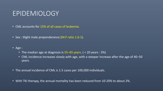
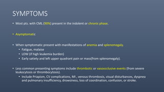
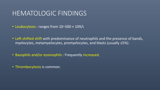
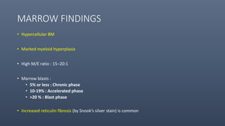
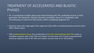
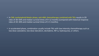
This document provides information on chronic myeloid leukemia (CML), including:
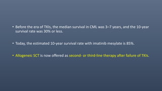
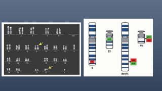
1. CML is driven by the BCR-ABL1 fusion gene resulting from the Philadelphia chromosome, and was historically treated with tyrosine kinase inhibitors (TKIs) like imatinib which improved 10-year survival to 85%.
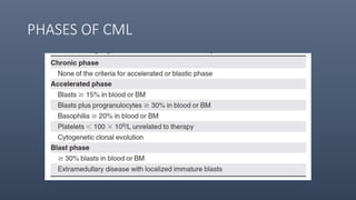
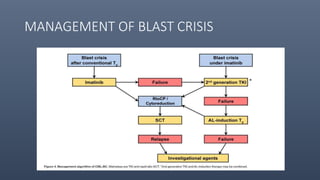
2. CML progresses through chronic, accelerated, and blast phases; TKIs can stabilize the disease in chronic phase and reduce transformation rates.
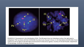
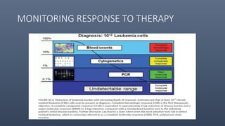
3. Diagnosis involves detecting the Philadelphia chromosome through cytogenetics of bone marrow samples and monitoring molecular response to TKIs is now standard practice.
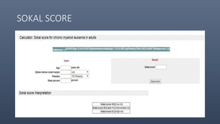
Chronic myeloid leukemia is a cancer of the white blood cells caused by a fusion of the BCR and ABL genes, which results in uncontrolled proliferation of mature and immature white blood cells. It progresses through three phases - chronic, accelerated, and blast crisis - and is characterized by excessive neutrophils, basophils and eosinophils in the blood and bone marrow. The standard diagnostic tests are detecting the Philadelphia chromosome or BCR-ABL fusion gene via cytogenetic analysis, FISH or PCR. Targeted therapy with tyrosine kinase inhibitors such as imatinib has dramatically improved survival for most CML patients.
This document discusses the approach to evaluating and diagnosing a hypotonic infant. It begins by defining hypotonia and noting that determining the cause can be challenging. A detailed history and physical exam are important to localize the cause as central or peripheral. Differential diagnosis involves considering central nervous system, genetic, infectious, metabolic, and muscular causes. Basic lab tests include screening for infection and metabolic/genetic disorders. Imaging, EMG/NCV, muscle biopsy and genetic testing can further evaluate potential peripheral and muscular etiologies. The case presented is of a newborn with hypotonia, absent reflexes, and no family history or dysmorphic features to suggest a cause.
This document provides information on various types of hematological cancers and disorders. It discusses chronic lymphocytic leukemia (CLL), the most common leukemia in adults. It affects B cells and can cause immune incompetence. Prolymphocytic leukemia (PLL) and hairy cell leukemia (HCL) are also summarized. Multiple myeloma (MM) is described as plasma cell proliferation in bone marrow causing anemia and bone pain. Myeloproliferative disorders include polycythemia vera (PV), essential thrombocythemia (ET), idiopathic myelofibrosis (IM) and chronic myelogenous leukemia (CML). Diagnosis and clinical features are summarized for each condition.
MPNs are clonal hematopoietic stem cell disorders characterized by overproduction of one or more myeloid cell lineages in the bone marrow and blood. The key subtypes include CML, PV, PMF, and ET. CML is driven by the Philadelphia chromosome and BCR-ABL1 fusion gene. PV is characterized by elevated red blood cell counts and the JAK2 V617F mutation in over 95% of cases. Without treatment, MPNs can progress to more advanced stages including myelofibrosis, leukemia, or transformation. Molecular testing plays an important role in diagnosis and classification.
This document discusses the diagnostic approach and classification of myeloproliferative neoplasms according to the 2008 WHO criteria. It describes the differential diagnosis and clinical features of polycythemia vera, essential thrombocythemia, and primary myelofibrosis. It discusses diagnostic criteria, disease course, prognosis, and treatment approaches for these chronic myeloproliferative disorders.
The document discusses hypotonia in infants and provides details on:
- The differential diagnosis of hypotonia includes both benign and serious conditions.
- Hypotonia can be caused by central nervous system issues or peripheral nervous system issues. Central causes account for 60-80% of cases.
- The evaluation of an infant with hypotonia includes a detailed history, physical exam focusing on tone and strength, and initial screening tests. Further testing may include imaging, genetic testing, and metabolic testing depending on exam findings.
The document provides an overview of chronic leukemias, focusing on chronic myeloid leukemia (CML). It defines CML and describes its epidemiology, etiology, pathogenesis, clinical presentation, diagnosis, and treatment. CML results from a genetic abnormality that fuses the BCR and ABL genes. It progresses through chronic, accelerated, and blast crisis phases if left untreated. Treatment options include stem cell transplantation, chemotherapy with tyrosine kinase inhibitors like imatinib, and management of symptoms.
- Genotyping plays an important role in the diagnosis and management of thalassemia. Laboratory diagnosis requires tests like complete blood count, hemoglobin electrophoresis, and DNA analysis to identify mutations.
- Over 1,800 mutations have been identified, with several common ones in different regions. Genotype also influences disease severity, with some mutations causing milder forms when combined with alpha-thalassemia.
- Several techniques can be used for DNA analysis including allele-specific PCR, reverse dot-blot, Sanger sequencing, and next-generation sequencing. While more advanced methods like NGS improve accuracy, considerations like cost must be made. Overall, genotyping aids in precise diagnosis, treatment decisions and genetic
Approach to pancytopenia .Dr ABHIJEET BARUA MD PGT.KOL.MED.CLG.ABHIJEET BARUA
Pancytopenia is a reduction in red blood cells, white blood cells, and platelets caused by decreased bone marrow production or destruction of blood cells. Evaluation of pancytopenia involves examining the complete blood count, peripheral smear, and bone marrow aspiration and biopsy to determine if the cause is aplasia, deficiencies, infections, infiltrative disorders, or primary bone marrow diseases like myelodysplastic syndrome. Management depends on the underlying etiology based on history, examination, and specific diagnostic tests.
Monoclonal gammopathies of undetermined significanceDrChirag Parmar
Monoclonal gammopathies of undetermined significance (MGUS) is an asymptomatic pre-malignant disorder characterized by a monoclonal protein level <3 g/dL, bone marrow plasma cells <10%, and no organ damage from the plasma cell disorder. MGUS has a risk of progression to multiple myeloma, amyloidosis, or other related disorders. Larger M-protein level, IgM or IgA type, and abnormal bone marrow plasma cells or serum free light chain ratio indicate a higher risk of progression. MGUS is typically detected incidentally during workup for suspected multiple myeloma. Management involves monitoring for progression through regular bloodwork and testing.
The document discusses Myelodysplastic/Myeloproliferative Neoplasms (MDS/MPN), a group of rare chronic myeloid neoplasms characterized by features of both myelodysplasia and myeloproliferation. It describes the key subtypes including Chronic Myelomonocytic Leukemia (CMML), Juvenile Myelomonocytic Leukemia (JMML), and Atypical Chronic Myeloid Leukemia (aCML). CMML is defined by persistent monocytosis and can be subdivided based on blast percentage. JMML mainly affects children and is characterized by leukocytosis with monocytosis. aCML involves dysplastic neutrophilic
This document discusses the classification of acute myeloid leukemia (AML). It provides an overview of classifications proposed over time including the French-American-British (FAB) classification from 1976 based on morphology and cytochemistry. The 2008 World Health Organization classification expanded genetic subtypes and incorporated cytogenetic and molecular information along with morphology. Accurate classification involves evaluating blast percentage, cell morphology, dysplasia, immunophenotyping, cytogenetics and molecular genetics to identify distinct biological entities of AML.
The document discusses myeloproliferative disorders (MPDs), which are clonal stem cell disorders characterized by increased blood cell counts and enlarged spleen and bone marrow. It focuses on chronic myeloid leukemia (CML), describing it as a MPD caused by a genetic mutation that results in uncontrolled white blood cell growth. CML progresses through chronic, accelerated, and blast phases, with symptoms ranging from fatigue to organ enlargement. Diagnosis involves blood and bone marrow tests detecting elevated white and platelet counts and the Philadelphia chromosome genetic abnormality associated with CML.
This document discusses Myelodysplastic Syndromes (MDS), including definitions, classification systems, predisposing factors, cytogenetic abnormalities, theories of pathophysiology, clinical manifestations, laboratory/pathology findings, and morphological abnormalities. Key points:
- MDS are clonal stem cell disorders characterized by ineffective hematopoiesis leading to cytopenias from defects in maturation. There have been many historical names and classification schemes, now defined by WHO.
- Predisposing factors include aging, genetic syndromes, chemotherapy/radiation, and environmental exposures. Cytogenetic abnormalities impact prognosis.
- Pathophysiology involves genetic/epigenetic changes, telomere dysfunction, altered micro
This document provides information on Chronic Myeloid Leukemia (CML), including its pathogenesis, diagnosis, staging, and management. It notes that CML is associated with the Philadelphia chromosome containing the BCR-ABL oncogene. Diagnosis involves cytogenetic testing showing the translocation and RT-PCR detecting BCR-ABL copies. CML progresses through chronic, accelerated, and blast crisis phases. First-line treatment is typically imatinib, nilotinib, or dasatinib, with switching to another tyrosine kinase inhibitor or allogeneic stem cell transplant for advanced or resistant disease.
This document discusses embryonal brain tumors in children, focusing on medulloblastoma. It provides details on the origin, epidemiology, pathology, molecular pathogenesis, clinical presentation, evaluation, treatment and prognosis of medulloblastoma. It also briefly discusses other embryonal brain tumors seen in children, including atypical teratoid/rhabdoid tumor, supratentorial primitive neuroectodermal tumor, embryonal tumors with multilayered rosettes, and pineoblastoma. The key information provided includes that medulloblastoma is the most common malignant brain tumor in children, arises from the cerebellum, and has distinct molecular subgroups associated with different clinical behaviors and outcomes.
Chronic myeloid leukemia is a type of cancer that affects white blood cells. It is caused by a genetic abnormality that results in excessive production of white blood cells. There are typically no symptoms in the early chronic phase, but later symptoms may include fatigue, fever, enlarged spleen, and weight loss. Diagnosis involves blood and bone marrow tests to detect the genetic abnormality. Treatment primarily uses targeted drug therapies such as imatinib to control the disease. Without treatment, it may progress to more advanced phases with worse symptoms.
This document describes myelodysplastic syndromes (MDS), a group of clonal hematopoietic stem cell disorders characterized by ineffective hematopoiesis leading to peripheral blood cytopenias. It discusses the history of MDS classification including the French-American-British (FAB) and World Health Organization (WHO) systems. Key features of the different MDS subtypes are outlined along with associated cytogenetic abnormalities, predisposing factors, theories of pathophysiology, clinical manifestations, and diagnostic evaluation.
1. Polycythemia vera (PV) is a chronic myeloproliferative neoplasm characterized by an overproduction of red blood cells without an identifiable stimulus. It commonly presents with erythrocytosis, splenomegaly, thrombosis, and pruritus.
2. The main cause of PV is a mutation in the JAK2 gene, but some patients have mutations in exon 12. Diagnosis requires tests to distinguish absolute from relative erythrocytosis. Treatment focuses on phlebotomy and medications to control symptoms and prevent complications.
3. Primary myelofibrosis (PMF) is a chronic myeloproliferative neoplasm involving clonal proliferation and
Chronic myeloid leukemia (CML) is a type of leukemia characterized by the Philadelphia chromosome, which is formed from the translocation of chromosomes 9 and 22. This translocation results in the BCR-ABL fusion gene which leads to increased proliferation of myeloid cells. CML progresses from a chronic phase to accelerated and blast crisis phases without treatment. Imatinib is now the standard first-line treatment for CML, achieving high rates of response. Resistance can develop through BCR-ABL mutations requiring other tyrosine kinase inhibitors. Allogeneic stem cell transplant remains the only potentially curative option but has risks.
Paroxysmal Nocturnal Hemoglobinuria (PNH) is a rare, acquired hemolytic anemia caused by a loss of proteins that regulate the complement pathway, making red blood cells sensitive to complement-mediated lysis. It is characterized by intravascular hemolysis that is worse at night due to relative acidosis during sleep. Affected cells lack proteins like CD59 and CD55 on their surface. Diagnosis involves flow cytometry to detect absent or decreased CD59 and CD55.
Multiple myeloma is a cancer of plasma cells that produce abnormal antibodies. It causes bone destruction and can damage the kidneys and suppress the bone marrow. While the cause is unknown, risk factors include age, family history, and exposure to radiation. Symptoms include bone pain, fatigue, recurrent infection, and kidney problems. Diagnosis involves blood and urine tests and a bone marrow biopsy. Staging uses tests such as MRI, blood tests, and bone surveys. Treatment may include chemotherapy, steroids, radiation, stem cell transplants, and newer drugs that target specific pathways in myeloma cells. While not yet curable, novel agents have improved survival rates and quality of life compared to conventional chemotherapy alone.
This document defines and describes pancytopenia and various bone marrow conditions that can cause pancytopenia, including constitutional (inherited) and acquired aplastic anemia. Constitutional pancytopenias are inherited disorders resulting in low blood cell counts. Fanconi anemia is described as the most common constitutional disorder, caused by mutations on FANC genes involved in DNA repair. Acquired aplastic anemia has various etiologies including radiation, drugs, chemicals, viruses, immune diseases, and idiopathic causes. Bone marrow biopsy and chromosomal breakage studies are used in diagnosis. Treatment depends on severity but may include blood transfusions, hematopoietic stem cell transplant, growth factors, steroids, androg
Multiple myeloma is a cancer of plasma cells that are found in the bone marrow. It is characterized by an overproduction of abnormal plasma cells which produce a monoclonal protein known as M protein. Symptoms include bone pain or fractures, anemia, kidney problems, and infections. Diagnosis involves blood and urine tests to detect the M protein as well as imaging tests and a bone marrow biopsy. Treatment depends on disease severity and transplant eligibility and may include chemotherapy, steroids, and stem cell transplantation.
The document discusses myelodysplastic syndromes (MDS), a group of stem cell malignancies characterized by ineffective hematopoiesis, cytopenias, and a risk of progression to acute myeloid leukemia. MDS arises from clonal mutations in hematopoietic stem cells and most commonly affects older adults. Diagnosis involves blood tests, bone marrow biopsy and aspiration showing dysplastic changes, and cytogenetic analysis to identify chromosomal abnormalities associated with prognosis. MDS ranges from indolent to aggressive disease depending on factors like karyotype and bone marrow blast percentage.
who 5th hematolymphoid B cell neoplasm.pptxDrKomalBhairo
The WHO 2022 update on lymphoid malignancies made several changes to the classification of B cell neoplasms including recognizing new entities, refining existing entities, and standardizing nomenclature. Key changes included recognizing splenic B-cell lymphoma/leukaemia with prominent nucleoli as a distinct entity, reclassifying B-cell prolymphocytic leukaemia, updating the classification of Burkitt lymphoma to recognize EBV-positive and EBV-negative subtypes, and grouping diffuse large B-cell lymphomas into 17 specific entities or the category of high-grade B-cell lymphoma, NOS based on genetic and phenotypic features.
Chronic myeloid leukemia (CML) is a clonal stem cell disorder caused by the BCR-ABL1 fusion gene from the Philadelphia chromosome. CML progresses through chronic, accelerated, and blast phases. It is characterized by excessive proliferation of myeloid cells. Diagnosis involves detecting the Philadelphia chromosome via cytogenetics or molecular testing. Treatment with tyrosine kinase inhibitors targets the BCR-ABL1 fusion protein and controls disease progression.
Chronic myeloid leukemia (CML) is a stem cell disorder caused by the Philadelphia chromosome, which results from the fusion of the BCR gene on chromosome 22 and the ABL gene on chromosome 9. This fusion produces the BCR-ABL protein which exhibits uncontrolled tyrosine kinase activity, driving excessive proliferation of CML cells. CML progresses through chronic, accelerated and blast crisis phases as additional genetic mutations accumulate. Tyrosine kinase inhibitors (TKIs) target the BCR-ABL protein and have significantly improved survival, with a 10-year survival of 85% with TKI therapy. Monitoring response through cytogenetics, FISH and molecular testing guides treatment decisions such as changing or adding other TKIs.
This document provides an overview of chronic myelogenous leukemia (CML) for primary care physicians. It discusses the epidemiology, clinical manifestations, molecular pathophysiology, natural history, diagnosis, and treatment of CML. Key points include: CML represents 15-20% of adult leukemias, with the median age of onset being 45-55 years. The Philadelphia chromosome, resulting from a translocation, produces a Bcr-abl fusion gene that drives uncontrolled proliferation. CML progresses through chronic, accelerated, and blast phases if left untreated. Tyrosine kinase inhibitors like imatinib revolutionized treatment by targeting the Bcr-abl protein. Imatinib induces high rates of remission but side effects can include
Learning Objectives:
Introduction
Definition of CML
Philadelphia Chromosome
Normal Granulopoiesis
Pathogenesis of CML
Aetiology
Incidence
Clinical Features
Phases of CML
Lab Diagnosis of CML
Course & Prognosis
Differential Diagnosis
Brief Overview of Treatment
chronic myeloid leukemia, CML, epidemiology, BCR ABL1 gene, philadelphia chromosome, t(9;22), CML incidence, etiology of CML, pathophysiology of CML, phases of CML, treatment of CML, Allogenic stem cell transplant, TKI therapy for CML, Sokal index for CML,
Chronic myeloid leukemia (CML) is a type of leukemia defined by the presence of the BCR-ABL1 fusion gene. It results from a translocation of chromosomes 9 and 22 that produces the Philadelphia chromosome. CML progresses through chronic, accelerated, and blast phases defined by increasing blast cell counts. Treatment is with tyrosine kinase inhibitors such as imatinib, which have improved survival rates to around 85% compared to older therapies. Monitoring response to therapy involves assessing cytogenetic and molecular response levels over time.
This document provides an overview of chronic myeloid leukemia (CML), including its molecular genetics, clinical manifestations, diagnosis, treatment options and outcomes. CML results from a fusion of the BCR and ABL genes, forming the Philadelphia chromosome and BCR-ABL fusion protein. It progresses through chronic, accelerated and blast crisis phases if left untreated. Tyrosine kinase inhibitors are now the standard first-line treatment and can control the disease long-term in chronic phase, while allogeneic stem cell transplant remains the only potential cure. Resistance and disease progression remain challenges.
The document summarizes recent changes to the World Health Organization's classification of myeloid neoplasms and acute leukemias based on advances since the 2008 classification. Key changes include new entities recognized based on unique biomarkers identified by gene expression analysis and sequencing. Entities were modified to better incorporate prognostic markers. Notable revisions include changes to criteria for chronic myeloid leukemia, myeloproliferative neoplasms, and myelodysplastic/myeloproliferative neoplasms. The classification of myelodysplastic syndromes was also updated to focus more on dysplasia levels than specific cytopenias. Recognition of myeloid neoplasms with germline predisposition was another major change.
This document discusses chronic myeloid leukemia (CML). It defines CML as a stem cell disease characterized by excessive blood granulocytes and the Philadelphia chromosome - a translocation between chromosomes 9 and 22. CML progresses through chronic, accelerated and blast crisis phases. Symptoms include fatigue and splenomegaly. Diagnosis involves blood tests, bone marrow biopsy and detecting the Philadelphia chromosome. Treatment includes tyrosine kinase inhibitors, interferon and chemotherapy.
Chronic myeloid leukemia (CML) is a type of leukemia caused by a genetic abnormality known as the Philadelphia chromosome. This abnormality results from the translocation of parts of chromosomes 9 and 22, producing the BCR-ABL fusion gene which encodes a tyrosine kinase that drives excessive white blood cell growth. CML progresses through three phases - chronic, accelerated, and blast crisis - and is typically diagnosed based on blood tests and the presence of the Philadelphia chromosome or BCR-ABL gene. Treatment involves tyrosine kinase inhibitors like imatinib, with the goal of achieving cytogenetic and molecular responses to control the disease.
A myeloprolifrative stem cell disorder resulting in
Proliferation of all haematopoietic lineages but
manifestation Predominantly in the granulocytic series.
The disease occurs chiefly between 30 and 80 years, with
A peak incidence at the 55 years.
*accounts for 20% of all leukaemis.
*found in all races.
*the aetiology is unknown.
This document provides an overview of the myeloid malignancies, including myeloproliferative neoplasms (MPNs), myelodysplastic syndrome (MDS), and acute myeloid leukemia (AML). It describes key characteristics of each condition, such as increased mature cells in MPNs, decreased blood cells in MDS, and presence of immature cells in AML. Diagnostic criteria and classification systems for AML, including the 2008 WHO classification, are reviewed. Risk stratification in AML and standard treatment approaches are also summarized. Two clinical cases are then presented and discussed in detail.
1. Chronic myeloproliferative disorders are a group of stem cell disorders characterized by overproduction of mature white blood cells, red blood cells, or platelets.
2. Chronic myeloid leukemia is caused by the Philadelphia chromosome which forms the BCR-ABL fusion gene. It progresses through chronic, accelerated, and blast crisis phases.
3. Other myeloproliferative neoplasms discussed include polycythemia vera, essential thrombocythemia, primary myelofibrosis, and chronic lymphocytic leukemia.
The document discusses the classification and treatment of various types of leukemia. It begins by defining leukemia as the neoplastic proliferation of white blood cells. Leukemias are classified as either lymphoid or myeloid and as either acute or chronic depending on the affected cell type and disease progression. The four major types are described as acute lymphocytic leukemia, acute myelogenous leukemia, chronic lymphocytic leukemia and chronic myelogenous leukemia. Treatment options are discussed including chemotherapy regimens, bone marrow transplantation, supportive care and goals of treatment.
This document provides an overview of chronic lymphocytic leukemia (CLL). It defines CLL as the accumulation of mature B lymphocytes in the blood, bone marrow, lymph nodes, and spleen. CLL most commonly affects older adults and has an unknown cause. The document outlines the clinical presentation, diagnostic criteria, staging systems, complications, treatment approaches, and poor prognostic factors of CLL. It also discusses new therapies that aim to induce complete remission and eliminate minimal residual disease to improve survival outcomes in CLL patients.
Leukemia is the most common pediatric malignancy, accounting for 1/3 of all childhood cancers. It is defined as the malignant clonal proliferation of lymphoid or myeloid precursor cells in the bone marrow and infiltration of other organs. The presentation includes general symptoms like fever, fatigue, and pallor as well as hematological effects from bone marrow invasion including anemia, neutropenia, and thrombocytopenia. Diagnosis involves blood tests, bone marrow examination, and other investigations. Treatment consists of induction chemotherapy followed by consolidation and maintenance therapy to achieve remission and prevent relapse.
Acute myeloid leukemia (AML) is characterized by proliferation of immature myeloid cells in the bone marrow. The patient presented with AML-M2 subtype with FLT3-ITD mutation, resulting in neutropenia, thrombocytopenia, and oral candidiasis. Bone marrow tests found 61% blasts positive for myeloid markers and the FLT3 mutation. The goals of induction chemotherapy are to reduce morbidity and mortality by inducing remission and preventing relapse while managing complications of the disease and treatment.
This document provides an overview of acute myeloid leukemia (AML). It discusses the etiology, classification, clinical features, laboratory findings, treatment including induction chemotherapy, post-remission therapy such as stem cell transplantation, and prognostic factors of AML. The key points are that AML is a cancer of the myeloid line of blood cells, its incidence increases with age, and treatment involves induction chemotherapy to achieve remission followed by post-remission therapy to prevent relapse.
The document summarizes chronic leukemias, focusing on chronic myeloid leukemia (CML). CML results from a genetic abnormality that fuses the BCR and ABL genes, producing the Philadelphia chromosome. This leads to excessive proliferation of white blood cells. CML progresses through chronic, accelerated, and blast crisis phases if left untreated. Treatment options include stem cell transplantation, chemotherapy like imatinib, and supportive care. Imatinib induces remission in most chronic phase CML patients but does not cure the disease.
Leukemia are neoplastic disorders of the hematopoietic system characterized by aberrant or arrested differentiation. There are two main types - acute and chronic leukemias. Acute leukemias are further classified as myeloid or lymphoid based on the lineage of the malignant cells. Chromosomal abnormalities are detected in the majority of acute leukemia cases and correlate with specific disease subtypes and clinical outcomes. Treatment involves induction chemotherapy followed by consolidation therapy and stem cell transplantation for eligible patients, with cure rates varying based on disease risk factors.
This document discusses megaloblastic anemia caused by vitamin B12 or folic acid deficiency. It covers the historical background, structures and roles of vitamin B12 and folic acid, causes and clinical presentations of deficiency, hematological findings, and neurological effects. Diagnosis involves assessing macrocytic anemia and hematological features showing megaloblastic changes. Treatment involves replacing the deficient vitamin.
This document summarizes IgA nephropathy (IgAN), the most common primary glomerulonephritis globally. Key points include: IgAN is characterized by deposition of IgA in the mesangium. Clinical presentations range from asymptomatic hematuria to rapidly progressive glomerulonephritis. Prognosis depends on factors like proteinuria level and hypertension. Treatment involves renin-angiotensin system blockade, glucocorticoids, and immunosuppression. The Oxford classification uses pathological features to predict prognosis.
The document provides information on the management of Hepatitis B. It discusses the virology of HBV, epidemiology, natural history, goals of treatment, criteria for treatment, treatment options and monitoring. Key points include:
- HBV is a DNA virus that causes both acute and chronic hepatitis. Approximately 240 million people globally have chronic HBV infection.
- Natural history depends on when infection is acquired. Risk of chronic infection is highest with infection at birth.
- Treatment goals are to prevent progression of disease and development of complications like cirrhosis and liver cancer.
- Treatment is recommended for those with HBeAg-positive chronic hepatitis B with HBV DNA >20,000 IU/mL and elevated
This document discusses several hematological emergencies including hyperleucocytosis and leukostasis, spinal cord compression, human antibody infusion reactions, and neutropenic enterocolitis.
Hyperleucocytosis and leukostasis refer to an extremely elevated white blood cell count that can lead to decreased tissue perfusion. Prompt treatment is needed to reduce mortality. Spinal cord compression occurs when cancer metastasizes to the spine and compresses the spinal cord, most often from lung, breast, or prostate cancers. Symptoms include back pain and neurological deficits. Human antibody infusion reactions are immune-mediated responses that can occur with monoclonal antibody therapies and range from mild to life-threatening. Neutropenic enterocolitis is inflammation
This document discusses hematological emergencies and tumor lysis syndrome. It provides classifications of hematological emergencies and describes tumor lysis syndrome, including its causes, risk factors, clinical manifestations, and treatments like hypouricemic drugs, hydration, and renal replacement therapy. It also covers superior vena cava syndrome, its causes, clinical features, grading of severity, diagnosis using imaging, and treatments including supportive care, stenting, and glucocorticoids.
This document provides information on febrile neutropenia, including:
- It is a common and serious complication of cancer chemotherapy, especially in those with hematologic malignancies.
- Initial evaluation of febrile neutropenic patients includes assessing infection risk factors and sites, as well as collecting blood and other cultures.
- High-risk patients require intravenous empirical antibiotic therapy in the hospital, while low-risk patients may be treated orally or as outpatients.
- Empirical therapy typically involves a broad-spectrum beta-lactam with coverage against pseudomonas, with vancomycin or other anti-gram positive coverage only added if clinically indicated. Therapy is continued until marrow recovery from neutropenia
This document discusses the neurological manifestations of diabetes mellitus (DM). It notes that diabetic neuropathy is the most common complication of DM, affecting approximately 50% of patients. It classifies and describes various types of diabetic neuropathies including distal symmetric polyneuropathy, autonomic neuropathy, and focal neuropathies. It also discusses diabetic complications such as hypoglycemia, diabetic ketoacidosis, and cerebrovascular disease. Finally, it mentions some genetic disorders and congenital malformations associated with DM.
Osteoporosis is a progressive bone disease characterized by low bone mass and deterioration of bone tissue, leading to fragile bones that are prone to fractures. It is diagnosed based on bone mineral density measurements. Risk factors include older age, female sex, smoking, excessive alcohol use, low body weight, vitamin D and calcium deficiency, lack of exercise, and certain medical conditions or medications. Treatment focuses on lifestyle modifications to reduce risk factors as well as pharmacological therapies to prevent bone loss and reduce fracture risk. Common medications include bisphosphonates, calcitonin, estrogen therapy, SERMs, teriparatide, and denosumab.
Constrictive pericarditis is usually caused by fibrosis and scarring of the pericardium due to prior infection, surgery, radiation, or other inflammatory processes. Tuberculosis is a common cause in developing countries. The scarring restricts cardiac filling, leading to elevated pressures in all chambers. Patients experience symptoms of right and left heart failure like edema and dyspnea. Diagnosis involves echocardiography, cardiac catheterization and MRI/CT. Treatment is surgical removal of the pericardium (pericardiectomy), which carries risks but is usually definitive. Diuretics and other medications can help symptoms but do not treat the underlying condition.
1) The document discusses pericardial diseases, beginning with the anatomy and functions of the pericardium.
2) It then covers pericarditis, including classifications, presentations, and management. Empirical anti-inflammatory therapy including NSAIDs and colchicine is recommended for acute idiopathic pericarditis.
3) Recurrent pericarditis is identified as the most common complication, occurring in 15-30% of cases, and requiring prolonged anti-inflammatory treatment.
1. Urinary tract infections are very common, affecting around 150 million people per year globally, with higher rates in women.
2. Risk factors include catheter use, which increases risk 10-fold, and hospital-acquired UTIs account for 40% of all hospital infections.
3. Symptoms depend on location, from cystitis in the bladder presenting with dysuria and urgency, to pyelonephritis in the kidneys causing fever and flank pain. Diagnosis involves urinalysis and culture.
A 74-year-old man presented with fever, increased shortness of breath, jaundice, and dark urine. Laboratory tests found hemolytic anemia with a positive direct antiglobulin test. Further evaluation indicated a diagnosis of autoimmune hemolytic anemia (AIHA). AIHA occurs when a person's immune system attacks their own red blood cells. The patient's AIHA was treated with corticosteroids, which are the first-line treatment for warm antibody AIHA.
This document discusses diabetic foot, which is an important but often neglected entity. Around 25% of people with diabetes will develop a diabetic foot ulcer in their lifetime, which can lead to amputation and increased mortality. Proper management of diabetic foot involves optimal diabetes and blood pressure control, foot care including regular inspection and debridement of wounds, offloading of pressure areas, and treatment of infections. Assessing the foot involves testing for loss of sensation and vascular status, classifying any wounds, and looking for signs of infection.
Von Willebrand disease is the most common inherited bleeding disorder caused by quantitative or qualitative deficiencies in von Willebrand factor. It is classified into types 1, 2A, 2B, 2M, 2N, and 3 based on pathophysiology. Diagnosis involves testing VWF activity, ristocetin cofactor activity, and multimer analysis. Treatment depends on severity but may include desmopressin, VWF concentrates, or antifibrinolytics. Hemophilia is caused by factor VIII or IX deficiency and classified by severity. Treatment involves factor replacement or bypassing agents for bleeding episodes and immune tolerance induction for inhibitors. Complications include hemarthroses, target joints, and
This document discusses the approach and management of thrombocytopenia and immune thrombocytopenic purpura (ITP). It defines thrombocytopenia and its causes, provides diagnostic criteria for ITP, and outlines treatment approaches including corticosteroids, IVIG, anti-D, thrombopoietin receptor agonists, splenectomy, rituximab, and experimental therapies. It also addresses management of severe bleeding, pregnancy-associated thrombocytopenia, and thrombocytopenia in the settings of HIV and hepatitis C infection.
This document discusses cardio-renal syndrome (CRS), beginning with four case studies. It then covers the classification of CRS into five types based on whether cardiac or renal dysfunction occurs first and the duration. The pathophysiology of each type is complex, involving neurohormonal activation and other factors beyond just low blood flow. Novel biomarkers provide more accurate assessment of kidney injury than serum creatinine alone. Current management includes diuretics, ACE inhibitors, and other therapies, but future directions may include ultrafiltration, vaptans, adenosine antagonists, and hypertonic saline. CRS indicates communication between the heart and kidneys, leading to worse outcomes, so accurate diagnosis and coordinated treatment are important
1) Aplastic anemia is a rare bone marrow failure syndrome characterized by pancytopenia and a hypocellular bone marrow. It can be acquired, usually from toxic exposures, or constitutional, caused by inherited bone marrow failure syndromes.
2) Severe aplastic anemia, defined by very low blood counts, requires immediate treatment to prevent life-threatening infections and bleeding. Treatment options include immunosuppressive therapy with antithymocyte globulin and cyclosporine or allogeneic bone marrow transplantation.
3) Bone marrow transplantation from an HLA-matched sibling donor is the treatment of choice for young patients with severe aplastic anemia, as it reduces the risks of relapse and development of
Our backs are like superheroes, holding us up and helping us move around. But sometimes, even superheroes can get hurt. That’s where slip discs come in.
Histololgy of Female Reproductive System.pptxAyeshaZaid1
Dive into an in-depth exploration of the histological structure of female reproductive system with this comprehensive lecture. Presented by Dr. Ayesha Irfan, Assistant Professor of Anatomy, this presentation covers the Gross anatomy and functional histology of the female reproductive organs. Ideal for students, educators, and anyone interested in medical science, this lecture provides clear explanations, detailed diagrams, and valuable insights into female reproductive system. Enhance your knowledge and understanding of this essential aspect of human biology.
Rasamanikya is a excellent preparation in the field of Rasashastra, it is used in various Kushtha Roga, Shwasa, Vicharchika, Bhagandara, Vatarakta, and Phiranga Roga. In this article Preparation& Comparative analytical profile for both Formulationon i.e Rasamanikya prepared by Kushmanda swarasa & Churnodhaka Shodita Haratala. The study aims to provide insights into the comparative efficacy and analytical aspects of these formulations for enhanced therapeutic outcomes.
Hiranandani Hospital in Powai, Mumbai, is a premier healthcare institution that has been serving the community with exceptional medical care since its establishment. As a part of the renowned Hiranandani Group, the hospital is committed to delivering world-class healthcare services across a wide range of specialties, including kidney transplantation. With its state-of-the-art facilities, advanced medical technology, and a team of highly skilled healthcare professionals, Hiranandani Hospital has earned a reputation as a trusted name in the healthcare industry. The hospital's patient-centric approach, coupled with its focus on innovation and excellence, ensures that patients receive the highest standard of care in a compassionate and supportive environment.
- Video recording of this lecture in English language: https://youtu.be/kqbnxVAZs-0
- Video recording of this lecture in Arabic language: https://youtu.be/SINlygW1Mpc
- Link to download the book free: https://nephrotube.blogspot.com/p/nephrotube-nephrology-books.html
- Link to NephroTube website: www.NephroTube.com
- Link to NephroTube social media accounts: https://nephrotube.blogspot.com/p/join-nephrotube-on-social-media.html
Local Advanced Lung Cancer: Artificial Intelligence, Synergetics, Complex Sys...Oleg Kshivets
Overall life span (LS) was 1671.7±1721.6 days and cumulative 5YS reached 62.4%, 10 years – 50.4%, 20 years – 44.6%. 94 LCP lived more than 5 years without cancer (LS=2958.6±1723.6 days), 22 – more than 10 years (LS=5571±1841.8 days). 67 LCP died because of LC (LS=471.9±344 days). AT significantly improved 5YS (68% vs. 53.7%) (P=0.028 by log-rank test). Cox modeling displayed that 5YS of LCP significantly depended on: N0-N12, T3-4, blood cell circuit, cell ratio factors (ratio between cancer cells-CC and blood cells subpopulations), LC cell dynamics, recalcification time, heparin tolerance, prothrombin index, protein, AT, procedure type (P=0.000-0.031). Neural networks, genetic algorithm selection and bootstrap simulation revealed relationships between 5YS and N0-12 (rank=1), thrombocytes/CC (rank=2), segmented neutrophils/CC (3), eosinophils/CC (4), erythrocytes/CC (5), healthy cells/CC (6), lymphocytes/CC (7), stick neutrophils/CC (8), leucocytes/CC (9), monocytes/CC (10). Correct prediction of 5YS was 100% by neural networks computing (error=0.000; area under ROC curve=1.0).
Clinic ^%[+27633867063*Abortion Pills For Sale In Tembisa Central19various
Clinic ^%[+27633867063*Abortion Pills For Sale In Tembisa Central Clinic ^%[+27633867063*Abortion Pills For Sale In Tembisa CentralClinic ^%[+27633867063*Abortion Pills For Sale In Tembisa CentralClinic ^%[+27633867063*Abortion Pills For Sale In Tembisa CentralClinic ^%[+27633867063*Abortion Pills For Sale In Tembisa Central
Does Over-Masturbation Contribute to Chronic Prostatitis.pptxwalterHu5
In some case, your chronic prostatitis may be related to over-masturbation. Generally, natural medicine Diuretic and Anti-inflammatory Pill can help mee get a cure.
8 Surprising Reasons To Meditate 40 Minutes A Day That Can Change Your Life.pptxHolistified Wellness
We’re talking about Vedic Meditation, a form of meditation that has been around for at least 5,000 years. Back then, the people who lived in the Indus Valley, now known as India and Pakistan, practised meditation as a fundamental part of daily life. This knowledge that has given us yoga and Ayurveda, was known as Veda, hence the name Vedic. And though there are some written records, the practice has been passed down verbally from generation to generation.
2. LAYOUT
• WHO classification of MPN
• CML
• Introduction
• Diagnosis
• TKIs
• Monitoring of therapy
• Treatment
• Investigational therapies
3. INTRODUCTION OF CML
• MPN characterized by
Share an origin in a multipotent hematopoietic progenitor cell;
Overproduction of one or more of the formed elements of the blood without significant
dysplasia;
Predilection to extramedullary hematopoiesis, myelofibrosis, and
Transformation at varying rates to acute leukemia.
6. INTRODUCTION
• CML is a clonal hematopoietic stem cell disorder.
• The salient biologic features of CML cells are as follows: increased proliferation, resistance to
apoptosis, perturbed interaction with bone marrow stromal cells, and genetic instability
• The disease is driven by the BCR-ABL1 chimeric gene product, resulting from a reciprocal
balanced translocation between the long arms of chromosomes 9 and 22, t(9;22) (q34;q11.2),
cytogenetically detected as the Philadelphia chromosome.
• Untreated, the course of CML may be biphasic or triphasic, with an early indolent or chronic
phase, followed often by an accelerated phase and a terminal blastic phase.
7. • Before the era of TKIs, the median survival in CML was 3–7 years, and the 10-year
survival rate was 30% or less.
• Today, the estimated 10-year survival rate with imatinib mesylate is 85%.
• Allogeneic SCT is now offered as second- or third-line therapy after failure of TKIs.
8. EPIDEMIOLOGY
• CML accounts for 15% of all cases of leukemia.
• Sex : Slight male preponderance (M:F ratio 1.6:1).
• Age :
• The median age at diagnosis is 55–65 years. ( < 20 years : 3%)
• CML incidence increases slowly with age, with a steeper increase after the age of 40–50
years.
• The annual incidence of CML is 1.5 cases per 100,000 individuals.
• With TKI therapy, the annual mortality has been reduced from 10-20% to about 2%.
13. Transformation to Accelerated and Blastic
Phase
• CCA/Ph+ is present in 70% to 80% of cases and includes a number of nonrandom abnormalities,
the most common of which are +8 (34% of cases with CCA/Ph+), +Ph (30%), i(17q) (20%), +19
(13%), -Y (8% of males), +21 (7%), +17 (5%), and monosomy 7 (5%).
• CML-BP : Pre-B-lymphoid (25%), Myeloid (70%), or Indeterminate (5%) phenotype
14. ETIOLOGY
• No familial associations.
• No associations exist with exposures to benzene or other toxins, fertilizers, insecticides, or
viruses.
• Not a frequent secondary leukemia following therapy of other cancers with alkylating agents
and/or radiation.
• Exposure to ionizing radiation
• Dose dependent risk of CML
• Peaks at 5–10 years after exposure
• The median time to development of CML among atomic bomb survivors was 6.3 years.
15. PATHOPHYSIOLOGY
• The t(9;22) (q34;q11.2) is present in more than 90% of classical CML cases.
• It is present in hematopoietic cells (myeloid, erythroid, megakaryocytes, and monocytes; less
often mature B lymphocytes; rarely mature T lymphocytes, but not stromal cells), but not in other
cells in the human body.
• BCR-ABL1 fusion gene product Codes for a novel oncoprotein of MW 210 kDa, referred to as
p210BCR-ABL1 aka Major BCR Exhibits constitutive kinase activity Excessive proliferation and
reduced apoptosis of CML cells.
• Minor BCR region (mBCR) : p190BCR-ABL1
• Micro-BCR (μ-BCR) : Larger p230BCR-ABL1 , a/w more indolent CML course.
16. Cont..
• BCR-ABL1 TKIs bind to the BCR-ABL1 kinase domain (KD), preventing the activation of
transformation pathways and inhibiting downstream signaling.
• The cause of the BCR-ABL1 molecular rearrangement is unknown.
• Atypical CML or chronic myelomonocytic leukemia.
• Do not respond to TKI therapy
• Poor prognosis with a median survival of about 2–3 years.
• The transition of CML from chronic to accelerated-blastic phase are poorly understood.
17. Cont..
• BCR-ABL1 itself induces genetic instability that leads to the acquisition of additional mutations
and eventually to blastic transformation.
• This is supported by the effect of TKIs on the ability to stabilize the CML genome, leading to a
much reduced transformation rate.
• Resistance to TKIs :
• Various mechanisms
• The most clinically relevant one is the development of different ABL1 kinase domain
mutations that prevent the binding of TKIs to the catalytic site (ATP binding site) of the
kinase.
19. CLINICAL PRESENTATION
• Depends upon the availability of health care.
• Easy access to health care : 50–60% of pts. diagnosed on routine blood tests and
have minimal symptoms at presentation, s/a fatigue.
• Limited access to health care : Present with high CML burden including
splenomegaly, anemia, and related symptoms (abdominal pain, weight loss,
fatigue).
20. SYMPTOMS
• Most pts. with CML (90%) present in the indolent or chronic phase.
• Asymptomatic
• When symptomatic present with manifestations of anemia and splenomegaly.
• Fatigue, malaise
• LOW (if high leukemia burden)
• Early satiety and left upper quadrant pain or mass(from splenomegaly).
• Less common presenting symptoms include thrombotic or vasoocclusive events (from severe
leukocytosis or thrombocytosis).
• Include Priapism, CV complications, MI , venous thrombosis, visual disturbances, dyspnea
and pulmonary insufficiency, drowsiness, loss of coordination, confusion, or stroke.
21. Cont..
• High basophil counts may be associated with histamine overproduction causing pruritus,
diarrhea, flushing, and even GI ulcers.
• Accelerated or blastic phase
• Unexplained fever
• Significant weight loss
• Severe fatigue
• Bone and joint aches
• Bleeding and thrombotic events
• Infections
22.
23. PHYSICAL FINDINGS
• Pallor (30-50%)
• Splenomegaly (20–70%)
• Hepatomegaly (10–20%)
• Lymphadenopathy (5–10%)
• Extramedullary disease (skin or subcutaneous lesions)
• Pruritus or scratch marks
24. HEMATOLOGIC FINDINGS
• Leukocytosis : ranges from 10–500 × 109/L
• Left-shifted shift with predominance of neutrophils and the presence of bands,
myelocytes, metamyelocytes, promyelocytes, and blasts (usually ≤5%).
• Basophils and/or eosinophils : Frequently increased.
• Thrombocytosis is common.
25.
26.
27. Cont..
• Thrombocytopenia is rare and, when present, suggests a worse prognosis,
disease acceleration, or an unrelated etiology.
• Anemia : 1/3rd of pts.
• Biochemical abnormalities
• Low LAP score
• High levels of vitamin B12, uric acid, LDH, and lysozyme.
28. MARROW FINDINGS
• Hypercellular BM
• Marked myeloid hyperplasia
• High M/E ratio : 15–20:1
• Marrow blasts :
• 5% or less : Chronic phase
• 10-19% : Accelerated phase
• >20 % : Blast phase
• Increased reticulin fibrosis (by Snook’s silver stain) is common
29. • Findings in CML Transformation Progression of CML is usually a/w leukocytosis resistant to
therapy, increasing anemia, fever and constitutional symptoms, and increased blasts and
basophils in the peripheral blood or marrow.
30.
31. CYTOGENETICS and MOLECULAR FINDINGS
• Classical CML (90% cases): t(9;22)(q34;q11.2) ;Philadelphia chromosome
• Variant Ph : involves ≥3 translocations that include chromosomes 9 and 22 and one or more
other chromosomes.
• Masked Ph : involves translocations between chromosome 9 and a chromosome other than 22.
• Monitoring patients on TKI therapy by cytogenetics, FISH, and molecular studies has become an
important standard practice to assess
• Response to therapy
• Compliance
• Treatment resistance
• Change TKI therapy and
• Study mutational studies.
32. • Other Philadelphia chromosome-positive malignancies
Acute precursor B cell lymphoblastic leukemia (ALL): 20-30 pc
Childhood ALL : 5-10%
Adult AML : 1%
33. Cont..
• FISH and PCR
• Advantage
Diagnosis of CML
Estimate the CML burden in patients on TKI therapy.
Can be done on peripheral blood samples Less painful and more convenient.
• Disadvantage
May not detect additional chromosomal abnormalities (clonal evolution)
Can be falsely positive at low levels or falsely negative because of technical issues
• A diagnosis of CML must always rely on a marrow analysis with routine cytogenetics.
• Presence of the Ph chromosome.
• Detects clonal evolution, i.e., chromosomal abnormalities in the Ph-positive cells (which may be
prognostic)
• Quantifies the percentage of marrow blasts and basophils.
34.
35. PROGNOSIS AND COURSE
• Before the imatinib era, the annual mortality in CML was 10% in the first 2 years and 15–20%
thereafter. The median survival time in CML was 3–7 years.
• The estimated 8- to 10-year survival rate with TKI is now 85%, or 93% if only CML-related deaths
are considered.
• Use of 2nd generation TKIs as frontline therapy have reduced the incidence of transformation in
the first 2–3 years from 6–8% with imatinib to 2–4% with nilotinib or dasatinib.
• Pts usually develop resistance in the form of cytogenetic relapse, followed by hematologic relapse
and subsequent transformation.
36. • CML Prognostic models
• Sokal
• Hasford
• European Treatment and Outcome Study [EUTOS].
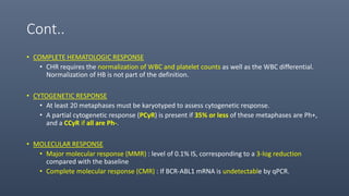
• Achievement of complete cytogenetic response has become the major therapeutic
endpoint and is the only endpoint associated with improvement in survival.
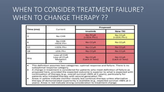
• The lack of achievement of major or complete molecular responses should not be
considered as “failure” of a particular TKI therapy and/or an indication to change the
TKI or to consider allogeneic SCT.
40. Tyrosine Kinase Inhibitors(TKIs): Drug
revolution
• Imatinib mesylate introduced in 2001
• 1st generation TKI : Imatinib
• 2nd -generation TKIs : Dasatinib, Nilotinib, and Bosutinib
• 3rd -generation TKI : Ponatinib
• Imatinib, dasatinib , bosutinib, and ponatinib are approved for the treatment of CML transformation
(accelerated and blastic phase) along with chronic phase, whereas nilotinib is only approved for chronic and
accelerated phase.
41. Cont..
• The sixth approved agent is OMACETAXINE
• A protein synthesis inhibitor with presumed more selective inhibition of the synthesis of the
BCR-ABL1 oncoprotein.
• It is approved for the treatment of chronic- and accelerated-phase CML after failure of two or
more TKIs, at 1.25 mg/m2 subcutaneously twice a day for 14 days for induction and for 7 days
for consolidation-maintenance.
• Potency of newer generation TKI as compared to imatinib : Nilotinib - 30 times ; Dasatinib : 300
times ; Bosutinib : 30–50 times
• PONATINIB is effective against wild-type and mutant BCRABL1 clones.
• It is unique in being the only currently available BCRABL1 TKI that is active against T315I, a
gatekeeper mutant resistant to other 4 TKIs
42. AGENT APPROVED INDICATIONS DOSE SCHEDULE NOTABLE TOXICITIES
IMATINIB MESYLATE All phases 400 mg OD See A/E in next slide
DASATINIB All phases First-line: 100 mg OD
Salvage: 140 mg OD
Myelosuppression;
pleural and pericardial
effusions; pulmonary
HTN
NILOTINIB All phases except blastic
phase
First-line: 300 mg BD
Salvage: 400 mg BD
Diabetes; VOD ;
pancreatitis
BOSUTINIB All phases except
frontline
500 mg OD Diarrhea
PONATINIB All phases except
frontline
45 mg daily Skin rashes, pancreatitis;
VOD (10–20%)
OMACETAXINE
MEPESUCCINATE
Failure ≥2 tyrosine
kinase
inhibitors
1.25 mg/m2 S/C BD for
14 days for induction; 7
days of maintenance
every month
Myelosuppression
44. TRIALS ON TKIs
• In two RCTs, one comparing nilotinib 300 mg twice daily or 400 mg twice daily with imatinib
(ENEST-nd) and the other comparing dasatinib 100 mg daily with imatinib (DASISION);the
second-generation TKIs were associated with better outcomes
• Higher rates of complete cytogenetic responses (85–87% vs 77–82%),
• Major molecular responses (65–76% vs 46–63%),
• Undetectable BCR-ABL1 transcripts (IS) (32–37% vs 15–30%), and
• Lower rates of transformation to accelerated-blastic phase (2–4% vs 6%).
• Neither study showed a survival benefit with second-generation TKIs
45.
46. Choice and Timing of Allogeneic SCT
• Allogeneic SCT was considered first-line CML therapy before 2000.
• Now SCT is considered 2nd or 3rd line therapy.
• Among pts. who present with or evolve to blastic phase, combinations of chemotherapy and TKIs
should be used to induce remission, f/b allogeneic SCT as soon as possible.
• Pts with T315I mutations at relapse should be offered ponatinib and considered for allogeneic SCT
(because of the short F/U with ponatinib).
47. • Pts with mutations involving Y253H, E255K/V, and F359V/C/I respond better to dasatinib or
bosutinib.
• Patients with mutations involving V299L, T315A, and F317L /F/I/C respond better to nilotinib.
• Pts with clonal evolution, unfavorable mutations, or lack of major/complete cytogenetic
response within 1 year of salvage TKI therapy have short remission durations and should
consider allogeneic SCT as more urgent in the setting of salvage.
49. Cont..
• COMPLETE HEMATOLOGIC RESPONSE
• CHR requires the normalization of WBC and platelet counts as well as the WBC differential.
Normalization of HB is not part of the definition.
• CYTOGENETIC RESPONSE
• At least 20 metaphases must be karyotyped to assess cytogenetic response.
• A partial cytogenetic response (PCyR) is present if 35% or less of these metaphases are Ph+,
and a CCyR if all are Ph-.
• MOLECULAR RESPONSE
• Major molecular response (MMR) : level of 0.1% IS, corresponding to a 3-log reduction
compared with the baseline
• Complete molecular response (CMR) : If BCR-ABL1 mRNA is undetectable by qPCR.
51. CML PHASE USE OF TKI CONSIDERATION FOR USE OF SCT
Accelerated or Blastic Interim therapy to achieve minimal
CML burden
As soon as possible (exception: de
novo accelerated phase)
Imatinib failure in chronic
phase; T315I mutation
Ponatinib to achieve minimal CML
burden
Depends on longer term follow-up
results of ponatinib efficacy
Imatinib failure in chronic
phase; no clonal evolution, no
mutations, good initial
response
Second-line kinase inhibitors long-
term
Third-line after second line TKI
failure
Imatinib failure in chronic
phase; clonal evolution or
mutations, or no cytogenetic
response to second-line KI
Interim therapy to achieve minimal
CML burden
Second-line
Older patients (≥65–70 years)
after imatinib failure in
chronic phase
Salvage TKIs as longer term
therapy
May forgo allogeneic SCT in favor of
good quality of life and survival
in chronic phase
52. TREATMENT OF ACCELERATED AND BLASTIC
PHASES
• Pts. in accelerated or blastic phase may receive therapy with TKIs, preferably second- or third-
generation TKIs (dasatinib, nilotinib, bosutinib, ponatinib), alone or in combination with
chemotherapy, to reduce the CML burden, before undergoing allogeneic SCT.
• Response rates with single-agent TKIs range from 30 to 50% in accelerated phase and from 20 to
30% in blastic phase.
• CML lymphoid blastic phase, the combination of anti-ALL chemotherapy with TKIs results in
complete response rates of 60–70% and median survival times of 2–3 years (compared with
historical response rates of 40–50% and median survival times of 12–18 months).
53. • In CML nonlymphoid blastic phase, anti-AML chemotherapy combined with TKIs results in CR
rates of 30–50% and median survival times of 9–12 months (compared with historical response
rates of 20–30% and median survival times of 3–5 months).
• In accelerated phase, combination usually include TKIs with low-intensity chemotherapy such as
low-dose cytarabine, low-dose idarubicin, decitabine, INF-α, hydroxyurea, or others.