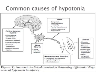
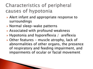
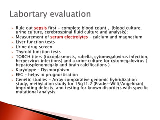
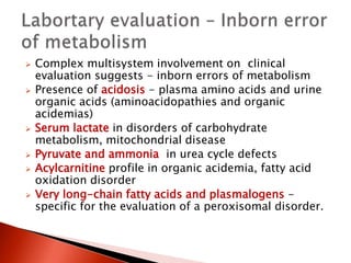
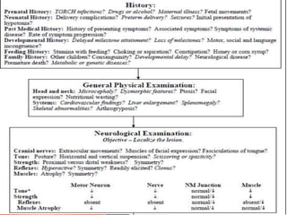
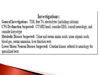
Gopakumar.H
Specialist Neonatology Trainee
Adelaide
 Sign of both benign and
serious conditions
 Exhaustive differential diagnosis
 Rare disorder
 Overwhelming advances in diagnosis and
management
 Differential diagnosis of
hypotonia in infants.
 Describe the differences between
central and peripheral causes of
hypotonia.
 Evaluation of hypotonia in infants.
Tone is the resistance of muscle to
stretch. Clinicians test two kinds of tone:
phasic and postural.
Phasic tone - The rapid contraction in
response to a high-intensity stretch , as
in tendon reflex response .
Postural tone - It is the prolonged
contraction of antigravity muscles in
response to the low-intensity stretch of
gravity. When postural tone is depressed,
the trunk and limbs cannot maintain
themselves against gravity and the infant
appears floppy.
The maintenance of normal tone requires intact central and
peripheral nervous system . Hence hypotonia is a common
symptom of neurological dysfunction and occurs in diseases
of the brain, spinal cord, nerves, and muscles.
Motor unit - One anterior
horn cell and all the muscle
fibers that it innervates
make up a motor unit . The
motor unit is the unit of
force. Therefore, weakness
is a symptom of all motor
unit disorders.
 Neuronopathy - A
primary disorder of the
anterior horn cell body
 Neuropathy - a
primary disorder of the
axon or its myelin
covering
 Myopathy - a primary
disorder of the muscle
fiber
 Two categories - Central and peripheral disorders .
 Peripheral causes include abnormalities in the motor
unit , specifically in the anterior horn cell (ie, spinal
muscular atrophy), peripheral nerve , neuromuscular
junction , and muscle
 Central causes account for 60% to 80% of hypotonia
cases and the peripheral causes occur in 15% to 30%.
 Considerable overlap of involvement and clinical
manifestations
 Cerebral insult – Hypoxic ischemic encephalopathy ,
intracranial haemorrhage
 Brain malformations
 Chromosomal disorders – Praderwilli syndrome , Down
syndrome
 Peroxisomal disorders – cerebrohepatorenal syndrome (
Zellweger’s syndrome) , Neonatal adrenoleukodystrophy
 Other genetic defects – familial dysautonomia ,
oculocerebrorenal syndrome ( Lowe syndrome )
 Neurometabolic disorders – Acid maltase deficiency ,
infantile GM1 gangliosidosis
 Drug effects ( ex Maternal Benzodiazepines )
 Benign congenital hypotonia
 Infantile spinal muscular atrophy
 Traumatic myelopathy ( esp following breech
delivery )
 Hypoxic ischemic myelopathy
 Infantile neuronal degeneration
 Congenital hypomyelinating neuropathy
 Giant axonal neuropathy
 Charcot marie tooth disease
 Dejerine sottas disease
 Myasthenia gravis ( Transient acquired
neonatal myasthenia ,congenital myasthenia )
 Infantile botulism
 Magnesium toxicity
 Aminoglycoside toxicity
 Congenital myopathy
 Nemaline myopathy
 Central core disease
 Myotubular myopathy
 Congenital fiber type disproportion myopathy
 Multicore myopathy
 Congenital muscular dystrophy with merosin deficiency
 Congenital muscular dystrophy without merosin deficiency
 Congenital muscular dystrophy with brain malformations
or intellectual disability
 Dystrophinopathies
 Walker Warburg disease
 Muscle – eye – brain disease
 Fukuyama disease
 Congenital muscular dystrophy with cerebellar atrophy /
hypoplasia
 Congenital muscular dystrophy with occipital agyria
 Early infantile facioscapulohumeral dystrophy Congenital
myotonic dystrophy
 Disorders of glycogen metabolism ( ex Acid
maltase deficiency )
 Severe neonatal phosphofructokinase deficiency
 Severe neonatal phophorylase deficiency
 Primary carnitine deficiency
 Peroxisomal disorders
 Neonatal adrenoleukodystrophy
 Cerebrohepatorenal syndrome ( zellweger )
 Disorders of creatine metabolism
 Cytochrome c oxidase deficiency
The most common central cause of hypotonia is
hypoxic encephalopathy / cerebral palsy in the
young infant. However, this dysfunction may
progress in later infancy to hypertonia.
The most common neuromuscular causes,
although still rare, are congenital myopathies,
congenital myotonic dystrophy, and spinal
muscular atrophy.
Disorders with both central and peripheral
manifestations ex acid maltase deficiency (Pompe
disease).
 Identify cause and the timing of onset
 Maternal exposures to toxins or infections
suggest a central cause
 Information on fetal movement in utero, fetal
presentation, and the amount of amniotic
fluid.
 Low Apgar scores may suggest floppiness
from birth
 Breech delivery or cervical
position – cervical spinal cord
trauma
 A term infant who is born healthy but
develops floppiness after 12 to 24 hours –
suspect inborn error of metabolism
 Infants suffering central injury usually
develop increased tone and deep tendon
reflexes.
 Central congenital hypotonia does not worsen
with time but may become more readily
apparent
 Motor delay with normal social and language
development decreases the likelihood of
brain pathology.
 Loss of milestones increases the index of
suspicion for neurodegenerative disorders.
A dietary/feeding history may point to diseases
of the neuromuscular junction, which may
present with sucking and swallowing
difficulties that ‘fatigue’ or ‘get worse’ with
repetition.
 Developmental delay (a chromosomal
abnormality)
 Delayed motor milestones (a congenital
myopathy) and
 Premature death (metabolic or muscle
disease).
 Any significant family history – affected parents or siblings,
consanguinity, stillbirths, childhood deaths
 Maternal disease – myotonic dystrophy
 Pregnancy and delivery history – drug or teratogen exposure
 Decreased fetal movements
 Abnormal presentation
 Polyhydramnios/ oligohydramnios
 Apgar scores
 Resuscitation requirements
 Cord gases
 History since delivery
◦ Respiratory effort
◦ Ability to feed
◦ Level of alertness
◦ Level of spontaneous activity
◦ Character of cry

 When lying supine, all hypotonic
infants look much the same,
regardless of the underlying cause
or location of the abnormality
within the nervous system.
 Lack spontaneous movement
 Full abduction of the legs places the
lateral surface of the thighs against
the examining table, and the arms
lie either extended at the sides of
the body or flexed at the elbow with
the hands beside the head.
Hip dislocation - The forceful
contraction of muscles pulling
the femoral head into the
acetabulum is a requirement
of normal hip joint formation.
 Pectus excavatum
indicates long standing
long-standing weakness
of the chest wall
muscles
 Infants who lie
motionless eventually
develop flattening of the
occiput and loss of hair
on the portion of the
scalp that is in constant
contact with the crib
sheet.
 Hip subluxation or
arthrogryposis suggest
hypotonia in utero .
Arthrogryposis varies in severity
from clubfoot, the most common
manifestation, to symmetrical
flexion deformities of all limb joints.
Joint contractures - a nonspecific consequence
of intrauterine immobilization.
As a rule, newborns with arthrogryposis who
require respiratory assistance do not survive
extubation unless the underlying disorder is
myasthenia.
High-pitched or unusual-sounding cry -
suggests CNS pathology
A weak cry - diaphragmatic weakness
Fatigable cry - congenital myasthenic
syndrome.
 A comprehensive neurologic evaluation
 Assessment for dysmorphic features
 Evaluation of the parents – may point towards
specific diagnosis as in myotonic dystrophy .
 Detailed neurologic assessment - tone,
strength, and reflexes
 Assessment of tone – begin by examining
posture, and movement. A floppy infant often
lies with limbs abducted and extended.
Traction response
Vertical suspension
Horizontal suspension
Further evaluation
Of
Hypotonia
Normal infant - keeps the
head erect, maintains the
back straight, and flexes
the elbow, hip, knee, and
ankle joints
Baby suspended in the
prone position with the
examiner’s palm
underneath the chest
Hyptonia - infants drape over
the examiner's hands, with
the head and legs hanging
limply
 The most sensitive measure of postural tone
 Grasp the hands and pull the infant toward a
sitting position
 A normal term infant lifts the head from the
surface immediately with the body
 When attaining the sitting position, the head
is erect in the midline for a few seconds.
 During traction, the examiner should feel the
infant pulling back against traction and
observe flexion at the elbow, knee, and ankle.
 The traction response is not present in
premature newborns of less than 33 weeks'
gestation
 The presence of more than minimal head lag
and of failure to counter traction by flexion of
the limbs in the term newborn is abnormal
and indicates hypotonia.
 By 1 month, normal infants lift the head
immediately and maintain it in line with the
trunk.
 The examiner places both hands in the infant's
axillae and, without grasping the thorax, lifts
straight up
 The muscles of the shoulders should have
sufficient strength to press down against the
examiner's hands and allow the infant to suspend
vertically without falling through
 Normal response – Head erect in the midline with
flexion at the knee, hip, and ankle joints.
 When a hypotonic infant is suspended vertically,
the head falls forward, the legs dangle, and the
infant may slip through the examiner's hands
because of weakness in the shoulder muscles
 Decreased resistance to flexion and extension
of the extremities
 Exaggerated hip abduction & ankle
dorsiflexion
 Oral-motor dysfunction
 Poor respiratory efforts
 Gastroesophageal reflux
 Note the distribution of weakness ex .face is
spared versus the trunk and extremities.
 Deep tendon reflexes (DTRs) often normal /
hyperactive in central conditions
 Clonus and primitive reflexes may persist
 DTRs - normal, decreased, or absent in
peripheral disorders
Course of hypotonia - fluctuating, static, or progressive
discriminates between a static encephalopathy (as is seen in
intellectual disability) and a degenerative neurologic condition
(eg, spinal muscular atrophy).
Distribution of hypotonia – Ex Face involvement
Distribution of hypotonia
Ex facial involvement
Usually spares extraocular muscles, while
diseases of the neuromuscular junction may be
characterized by ptosis and extraocular muscle
weakness .
 Hepatosplenomegaly – storage disorders,
congenital infections
 Renal cysts, high forehead, wide fontanelles –
Zellweger’s syndrome
 Hepatomegaly, retinitis pigmentosa – neonatal
adrenoleukodystrophy
 Congenital cataracts, glaucoma –
oculocerebrorenal (Lowe) syndrome
 Abnormal odour – metabolic disorders
 Hypopigmentation, undesceded testes – Prader
Willi
 Dysmorphic features
 Depressed level of consciousness or lethargy
 Abnormal eye movements or inability to track visually
 Early onset seizures
 Apnea
 Exaggerated irregular breathing patterns.
 Predominant axial weakness
 Normal strength with hypotonia
 scissoring on vertical suspension
 Fisting of the hands
 Hyperactive or normal reflexes
 Malformations of other organs
 Hypoxic ischemic encephalopathy,
teratogens, and metabolic disorders may
evolve into hyperreflexia and hypertonia, but
most syndromes do not.
 Infants who have experienced central injury
usually develop increased tone and deep
tendon reflexes
 Hypotonia,
 Generalized weakness
 Absent reflexes,
 Feeding difficulties
Classic infantile form of spinal muscular
atrophy
Fasciculations of the tongue as well as an
intention tremor.
Affected infants have alert, inquisitive faces but
profound distal weakness.
 Alert infant and appropriate response to
surroundings
 Normal sleep-wake patterns
 Associated with profound weakness
 Hypotonia and hyporeflexia / areflexia
 Other features - muscle atrophy, lack of
abnormalities of other organs, the presence
of respiratory and feeding impairment, and
impairments of ocular or facial movement
A systematic approach to a child
who has hypotonia, paying
attention to the history
and clinical examination, is
paramount in localizing the
problem to a specific region of
the nervous system.
 Rule out sepsis first - complete blood count , (blood culture,
urine culture, cerebrospinal fluid culture and analysis);
 Measurement of serum electrolytes – calcium and magnesium
 Liver function tests
 Urine drug screen
 Thyroid function tests
 TORCH titers (toxoplasmosis, rubella, cytomegalovirus infection,
herpesvirus infections) and a urine culture for cytomegalovirus (
hepatosplenomegaly and brain calcifications )
 Karyotype – Dysmorphism
 EEG – helps in prognostication
 Genetic studies - Array comparative genomic hybridization
study, methylation study for 15q11.2 (Prader-Willi/Angelman)
imprinting defects, and testing for known disorders with specific
mutational analysis
 Complex multisystem involvement on clinical
evaluation suggests - inborn errors of metabolism
 Presence of acidosis - plasma amino acids and urine
organic acids (aminoacidopathies and organic
acidemias)
 Serum lactate in disorders of carbohydrate
metabolism, mitochondrial disease
 Pyruvate and ammonia in urea cycle defects
 Acylcarnitine profile in organic acidemia, fatty acid
oxidation disorder
 Very long-chain fatty acids and plasmalogens -
specific for the evaluation of a peroxisomal disorder.
MRI
Delineate structural malformations
Neuronal migration defects
Abnormal signals in the basal ganglia (mitochondrial
abnormalities) or brain stem defects (Joubert syndrome)
Deep white matter changes can be seen in Lowe syndrome, a
peroxisomal defect
Abnormalities in the corpus callosum may occur in Smith- Lemli-
Opitz syndrome
Heterotopias may be seen in congenital muscular dystrophy.
Magnetic resonance spectroscopy
Magnetic resonance spectroscopy also can be
revealing for metabolic disease.
 Diagnosis mainly by history and clinical
examination
 Molecular genetics – CTG repeats, deletions
in SMN gene
 Nerve conduction studies and muscle biopsy
(Depending on clinical situation, may be
delayed until around 6 months of age as
neonatal results are difficult to interpret)
 Creatine kinase (levels need to be interpreted
with caution in the newborn, as levels tend to
be high at birth and increase in the first 24
hours, they also increase with acidosis).
 Repeat after few days , if initial value is
elevated
 Elevated in muscular dystrophy but not in
spinal muscular atrophy or in many
myopathies.
 Specific DNA testing -
for myotonic dystrophy
and for spinal
muscular atrophy (
SMN gene )
 Electrophysiological
studies - Shows
abnormalities in
nerves, myopathies,
and disorders of the
neuromuscular
junction
 Normal EMG usually
suggest central
hypotonia , with few
exceptions
 Helps to differentiate a primary myopathy from a
neurogenic disorder
 Helps to differentiate myopathies from muscular
dystrophies
 Useful in the work-up of undiagnosed weakness
 Provide the diagnosis of specific muscular conditions, such
as a muscular dystrophy, metabolic or storage
myopathies, and inflammatory myopathies.
 Helps to differentiate active from inactive and acute from
chronic conditions.
 Additional clues can be derived from ultrastructural
changes seen with the electron microscope.
 Various biochemical and genetic studies can be performed
on fresh or frozen muscle tissue to measure enzyme levels
and perform DNA studies for certain genetic diseases
 Hematoxylin and eosin (H&E)
 Trichrome , PAS (for glycogen)
 Oil red O (ORO) (for lipids)
 Acid phosphatise (for lysosomal activity)
 Congo red and cresyl violet (for amyloid)
 Myosin ATP ase Staining is useful for fiber-type
differentiation
 Oxidative markers, such as nicotinamide adenine
dinucleotide reductase (NADH), succinate dehydrogenase
(SDH), and cytochrome C oxidase(COX), are most effective
in the diagnosis of enzymatic deficiencies
 Myophosphorylase and myoadenylate deaminase (AMPAD)
for enzyme deficiencies acetylcholinesterase silver stain,
may be required in certain cases to show the motor
endplates
Muscular dystrophy - subgroup of myopathies
characterized by muscle degeneration and
regeneration. Clinically, muscular dystrophies
are typically progressive, because the muscles'
ability to regenerate is eventually lost, leading
to progressive weakness, often leading to use of
a wheel chair and eventually death, usually
related to respiratory weakness
Congenital myopathies - do not show
evidence for either a progressive
dystrophic process (i.e., muscle death)
or inflammation, but instead
characteristic microscopic changes are
seen in association with reduced
contractile ability of the muscles.
Muscle dystrophies
Versus
Congenital myopathies
 Mainly supportive – feeding ,
neurodevelopment
 Physiotherapy
 Specific treatment – Pompe disease ( enzyme
replacement therapy )
Floppy infant
Floppy infant
Floppy infant
Floppy infant

Floppy infant

  • 1.
  • 2.
     Sign ofboth benign and serious conditions  Exhaustive differential diagnosis  Rare disorder  Overwhelming advances in diagnosis and management
  • 3.
     Differential diagnosisof hypotonia in infants.  Describe the differences between central and peripheral causes of hypotonia.  Evaluation of hypotonia in infants.
  • 4.
    Tone is theresistance of muscle to stretch. Clinicians test two kinds of tone: phasic and postural. Phasic tone - The rapid contraction in response to a high-intensity stretch , as in tendon reflex response . Postural tone - It is the prolonged contraction of antigravity muscles in response to the low-intensity stretch of gravity. When postural tone is depressed, the trunk and limbs cannot maintain themselves against gravity and the infant appears floppy.
  • 5.
    The maintenance ofnormal tone requires intact central and peripheral nervous system . Hence hypotonia is a common symptom of neurological dysfunction and occurs in diseases of the brain, spinal cord, nerves, and muscles.
  • 6.
    Motor unit -One anterior horn cell and all the muscle fibers that it innervates make up a motor unit . The motor unit is the unit of force. Therefore, weakness is a symptom of all motor unit disorders.  Neuronopathy - A primary disorder of the anterior horn cell body  Neuropathy - a primary disorder of the axon or its myelin covering  Myopathy - a primary disorder of the muscle fiber
  • 8.
     Two categories- Central and peripheral disorders .  Peripheral causes include abnormalities in the motor unit , specifically in the anterior horn cell (ie, spinal muscular atrophy), peripheral nerve , neuromuscular junction , and muscle  Central causes account for 60% to 80% of hypotonia cases and the peripheral causes occur in 15% to 30%.  Considerable overlap of involvement and clinical manifestations
  • 9.
     Cerebral insult– Hypoxic ischemic encephalopathy , intracranial haemorrhage  Brain malformations  Chromosomal disorders – Praderwilli syndrome , Down syndrome  Peroxisomal disorders – cerebrohepatorenal syndrome ( Zellweger’s syndrome) , Neonatal adrenoleukodystrophy  Other genetic defects – familial dysautonomia , oculocerebrorenal syndrome ( Lowe syndrome )  Neurometabolic disorders – Acid maltase deficiency , infantile GM1 gangliosidosis  Drug effects ( ex Maternal Benzodiazepines )  Benign congenital hypotonia
  • 10.
     Infantile spinalmuscular atrophy  Traumatic myelopathy ( esp following breech delivery )  Hypoxic ischemic myelopathy  Infantile neuronal degeneration
  • 11.
     Congenital hypomyelinatingneuropathy  Giant axonal neuropathy  Charcot marie tooth disease  Dejerine sottas disease
  • 12.
     Myasthenia gravis( Transient acquired neonatal myasthenia ,congenital myasthenia )  Infantile botulism  Magnesium toxicity  Aminoglycoside toxicity
  • 13.
     Congenital myopathy Nemaline myopathy  Central core disease  Myotubular myopathy  Congenital fiber type disproportion myopathy  Multicore myopathy
  • 14.
     Congenital musculardystrophy with merosin deficiency  Congenital muscular dystrophy without merosin deficiency  Congenital muscular dystrophy with brain malformations or intellectual disability  Dystrophinopathies  Walker Warburg disease  Muscle – eye – brain disease  Fukuyama disease  Congenital muscular dystrophy with cerebellar atrophy / hypoplasia  Congenital muscular dystrophy with occipital agyria  Early infantile facioscapulohumeral dystrophy Congenital myotonic dystrophy
  • 15.
     Disorders ofglycogen metabolism ( ex Acid maltase deficiency )  Severe neonatal phosphofructokinase deficiency  Severe neonatal phophorylase deficiency  Primary carnitine deficiency  Peroxisomal disorders  Neonatal adrenoleukodystrophy  Cerebrohepatorenal syndrome ( zellweger )  Disorders of creatine metabolism  Cytochrome c oxidase deficiency
  • 16.
    The most commoncentral cause of hypotonia is hypoxic encephalopathy / cerebral palsy in the young infant. However, this dysfunction may progress in later infancy to hypertonia. The most common neuromuscular causes, although still rare, are congenital myopathies, congenital myotonic dystrophy, and spinal muscular atrophy. Disorders with both central and peripheral manifestations ex acid maltase deficiency (Pompe disease).
  • 19.
     Identify causeand the timing of onset  Maternal exposures to toxins or infections suggest a central cause  Information on fetal movement in utero, fetal presentation, and the amount of amniotic fluid.  Low Apgar scores may suggest floppiness from birth  Breech delivery or cervical position – cervical spinal cord trauma
  • 20.
     A terminfant who is born healthy but develops floppiness after 12 to 24 hours – suspect inborn error of metabolism  Infants suffering central injury usually develop increased tone and deep tendon reflexes.  Central congenital hypotonia does not worsen with time but may become more readily apparent
  • 21.
     Motor delaywith normal social and language development decreases the likelihood of brain pathology.  Loss of milestones increases the index of suspicion for neurodegenerative disorders.
  • 22.
    A dietary/feeding historymay point to diseases of the neuromuscular junction, which may present with sucking and swallowing difficulties that ‘fatigue’ or ‘get worse’ with repetition.
  • 23.
     Developmental delay(a chromosomal abnormality)  Delayed motor milestones (a congenital myopathy) and  Premature death (metabolic or muscle disease).
  • 24.
     Any significantfamily history – affected parents or siblings, consanguinity, stillbirths, childhood deaths  Maternal disease – myotonic dystrophy  Pregnancy and delivery history – drug or teratogen exposure  Decreased fetal movements  Abnormal presentation  Polyhydramnios/ oligohydramnios  Apgar scores  Resuscitation requirements  Cord gases  History since delivery ◦ Respiratory effort ◦ Ability to feed ◦ Level of alertness ◦ Level of spontaneous activity ◦ Character of cry 
  • 25.
     When lyingsupine, all hypotonic infants look much the same, regardless of the underlying cause or location of the abnormality within the nervous system.  Lack spontaneous movement  Full abduction of the legs places the lateral surface of the thighs against the examining table, and the arms lie either extended at the sides of the body or flexed at the elbow with the hands beside the head.
  • 26.
    Hip dislocation -The forceful contraction of muscles pulling the femoral head into the acetabulum is a requirement of normal hip joint formation.  Pectus excavatum indicates long standing long-standing weakness of the chest wall muscles  Infants who lie motionless eventually develop flattening of the occiput and loss of hair on the portion of the scalp that is in constant contact with the crib sheet.  Hip subluxation or arthrogryposis suggest hypotonia in utero .
  • 27.
    Arthrogryposis varies inseverity from clubfoot, the most common manifestation, to symmetrical flexion deformities of all limb joints. Joint contractures - a nonspecific consequence of intrauterine immobilization. As a rule, newborns with arthrogryposis who require respiratory assistance do not survive extubation unless the underlying disorder is myasthenia.
  • 28.
    High-pitched or unusual-soundingcry - suggests CNS pathology A weak cry - diaphragmatic weakness Fatigable cry - congenital myasthenic syndrome.
  • 29.
     A comprehensiveneurologic evaluation  Assessment for dysmorphic features  Evaluation of the parents – may point towards specific diagnosis as in myotonic dystrophy .
  • 30.
     Detailed neurologicassessment - tone, strength, and reflexes  Assessment of tone – begin by examining posture, and movement. A floppy infant often lies with limbs abducted and extended.
  • 31.
    Traction response Vertical suspension Horizontalsuspension Further evaluation Of Hypotonia
  • 32.
    Normal infant -keeps the head erect, maintains the back straight, and flexes the elbow, hip, knee, and ankle joints Baby suspended in the prone position with the examiner’s palm underneath the chest Hyptonia - infants drape over the examiner's hands, with the head and legs hanging limply
  • 33.
     The mostsensitive measure of postural tone  Grasp the hands and pull the infant toward a sitting position  A normal term infant lifts the head from the surface immediately with the body  When attaining the sitting position, the head is erect in the midline for a few seconds.  During traction, the examiner should feel the infant pulling back against traction and observe flexion at the elbow, knee, and ankle.
  • 34.
     The tractionresponse is not present in premature newborns of less than 33 weeks' gestation  The presence of more than minimal head lag and of failure to counter traction by flexion of the limbs in the term newborn is abnormal and indicates hypotonia.  By 1 month, normal infants lift the head immediately and maintain it in line with the trunk.
  • 35.
     The examinerplaces both hands in the infant's axillae and, without grasping the thorax, lifts straight up  The muscles of the shoulders should have sufficient strength to press down against the examiner's hands and allow the infant to suspend vertically without falling through  Normal response – Head erect in the midline with flexion at the knee, hip, and ankle joints.  When a hypotonic infant is suspended vertically, the head falls forward, the legs dangle, and the infant may slip through the examiner's hands because of weakness in the shoulder muscles
  • 36.
     Decreased resistanceto flexion and extension of the extremities  Exaggerated hip abduction & ankle dorsiflexion  Oral-motor dysfunction  Poor respiratory efforts  Gastroesophageal reflux  Note the distribution of weakness ex .face is spared versus the trunk and extremities.
  • 37.
     Deep tendonreflexes (DTRs) often normal / hyperactive in central conditions  Clonus and primitive reflexes may persist  DTRs - normal, decreased, or absent in peripheral disorders
  • 38.
    Course of hypotonia- fluctuating, static, or progressive discriminates between a static encephalopathy (as is seen in intellectual disability) and a degenerative neurologic condition (eg, spinal muscular atrophy). Distribution of hypotonia – Ex Face involvement Distribution of hypotonia Ex facial involvement
  • 40.
    Usually spares extraocularmuscles, while diseases of the neuromuscular junction may be characterized by ptosis and extraocular muscle weakness .
  • 41.
     Hepatosplenomegaly –storage disorders, congenital infections  Renal cysts, high forehead, wide fontanelles – Zellweger’s syndrome  Hepatomegaly, retinitis pigmentosa – neonatal adrenoleukodystrophy  Congenital cataracts, glaucoma – oculocerebrorenal (Lowe) syndrome  Abnormal odour – metabolic disorders  Hypopigmentation, undesceded testes – Prader Willi
  • 43.
     Dysmorphic features Depressed level of consciousness or lethargy  Abnormal eye movements or inability to track visually  Early onset seizures  Apnea  Exaggerated irregular breathing patterns.  Predominant axial weakness  Normal strength with hypotonia  scissoring on vertical suspension  Fisting of the hands  Hyperactive or normal reflexes  Malformations of other organs
  • 44.
     Hypoxic ischemicencephalopathy, teratogens, and metabolic disorders may evolve into hyperreflexia and hypertonia, but most syndromes do not.  Infants who have experienced central injury usually develop increased tone and deep tendon reflexes
  • 45.
     Hypotonia,  Generalizedweakness  Absent reflexes,  Feeding difficulties Classic infantile form of spinal muscular atrophy Fasciculations of the tongue as well as an intention tremor. Affected infants have alert, inquisitive faces but profound distal weakness.
  • 46.
     Alert infantand appropriate response to surroundings  Normal sleep-wake patterns  Associated with profound weakness  Hypotonia and hyporeflexia / areflexia  Other features - muscle atrophy, lack of abnormalities of other organs, the presence of respiratory and feeding impairment, and impairments of ocular or facial movement
  • 47.
    A systematic approachto a child who has hypotonia, paying attention to the history and clinical examination, is paramount in localizing the problem to a specific region of the nervous system.
  • 49.
     Rule outsepsis first - complete blood count , (blood culture, urine culture, cerebrospinal fluid culture and analysis);  Measurement of serum electrolytes – calcium and magnesium  Liver function tests  Urine drug screen  Thyroid function tests  TORCH titers (toxoplasmosis, rubella, cytomegalovirus infection, herpesvirus infections) and a urine culture for cytomegalovirus ( hepatosplenomegaly and brain calcifications )  Karyotype – Dysmorphism  EEG – helps in prognostication  Genetic studies - Array comparative genomic hybridization study, methylation study for 15q11.2 (Prader-Willi/Angelman) imprinting defects, and testing for known disorders with specific mutational analysis
  • 50.
     Complex multisysteminvolvement on clinical evaluation suggests - inborn errors of metabolism  Presence of acidosis - plasma amino acids and urine organic acids (aminoacidopathies and organic acidemias)  Serum lactate in disorders of carbohydrate metabolism, mitochondrial disease  Pyruvate and ammonia in urea cycle defects  Acylcarnitine profile in organic acidemia, fatty acid oxidation disorder  Very long-chain fatty acids and plasmalogens - specific for the evaluation of a peroxisomal disorder.
  • 51.
    MRI Delineate structural malformations Neuronalmigration defects Abnormal signals in the basal ganglia (mitochondrial abnormalities) or brain stem defects (Joubert syndrome) Deep white matter changes can be seen in Lowe syndrome, a peroxisomal defect Abnormalities in the corpus callosum may occur in Smith- Lemli- Opitz syndrome Heterotopias may be seen in congenital muscular dystrophy. Magnetic resonance spectroscopy Magnetic resonance spectroscopy also can be revealing for metabolic disease.
  • 52.
     Diagnosis mainlyby history and clinical examination  Molecular genetics – CTG repeats, deletions in SMN gene  Nerve conduction studies and muscle biopsy (Depending on clinical situation, may be delayed until around 6 months of age as neonatal results are difficult to interpret)
  • 53.
     Creatine kinase(levels need to be interpreted with caution in the newborn, as levels tend to be high at birth and increase in the first 24 hours, they also increase with acidosis).  Repeat after few days , if initial value is elevated  Elevated in muscular dystrophy but not in spinal muscular atrophy or in many myopathies.
  • 54.
     Specific DNAtesting - for myotonic dystrophy and for spinal muscular atrophy ( SMN gene )  Electrophysiological studies - Shows abnormalities in nerves, myopathies, and disorders of the neuromuscular junction  Normal EMG usually suggest central hypotonia , with few exceptions
  • 55.
     Helps todifferentiate a primary myopathy from a neurogenic disorder  Helps to differentiate myopathies from muscular dystrophies  Useful in the work-up of undiagnosed weakness  Provide the diagnosis of specific muscular conditions, such as a muscular dystrophy, metabolic or storage myopathies, and inflammatory myopathies.  Helps to differentiate active from inactive and acute from chronic conditions.  Additional clues can be derived from ultrastructural changes seen with the electron microscope.  Various biochemical and genetic studies can be performed on fresh or frozen muscle tissue to measure enzyme levels and perform DNA studies for certain genetic diseases
  • 56.
     Hematoxylin andeosin (H&E)  Trichrome , PAS (for glycogen)  Oil red O (ORO) (for lipids)  Acid phosphatise (for lysosomal activity)  Congo red and cresyl violet (for amyloid)  Myosin ATP ase Staining is useful for fiber-type differentiation  Oxidative markers, such as nicotinamide adenine dinucleotide reductase (NADH), succinate dehydrogenase (SDH), and cytochrome C oxidase(COX), are most effective in the diagnosis of enzymatic deficiencies  Myophosphorylase and myoadenylate deaminase (AMPAD) for enzyme deficiencies acetylcholinesterase silver stain, may be required in certain cases to show the motor endplates
  • 57.
    Muscular dystrophy -subgroup of myopathies characterized by muscle degeneration and regeneration. Clinically, muscular dystrophies are typically progressive, because the muscles' ability to regenerate is eventually lost, leading to progressive weakness, often leading to use of a wheel chair and eventually death, usually related to respiratory weakness Congenital myopathies - do not show evidence for either a progressive dystrophic process (i.e., muscle death) or inflammation, but instead characteristic microscopic changes are seen in association with reduced contractile ability of the muscles. Muscle dystrophies Versus Congenital myopathies
  • 58.
     Mainly supportive– feeding , neurodevelopment  Physiotherapy  Specific treatment – Pompe disease ( enzyme replacement therapy )