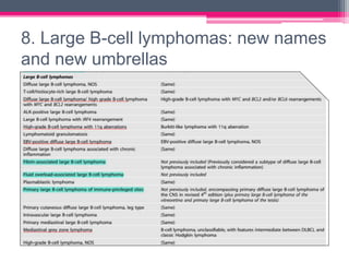
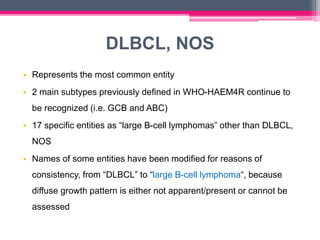
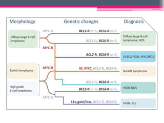
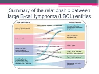
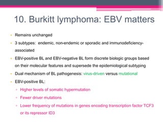
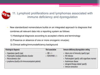
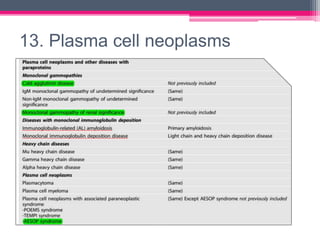
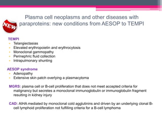
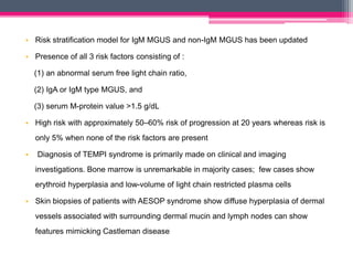
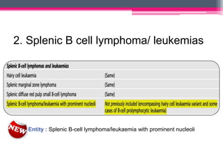
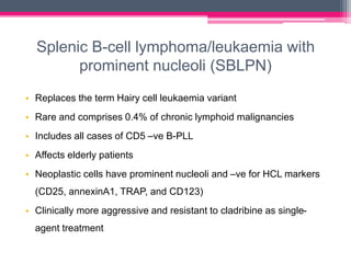
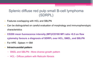
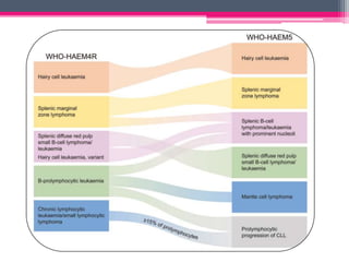
The WHO 2022 update on lymphoid malignancies made several changes to the classification of B cell neoplasms including recognizing new entities, refining existing entities, and standardizing nomenclature. Key changes included recognizing splenic B-cell lymphoma/leukaemia with prominent nucleoli as a distinct entity, reclassifying B-cell prolymphocytic leukaemia, updating the classification of Burkitt lymphoma to recognize EBV-positive and EBV-negative subtypes, and grouping diffuse large B-cell lymphomas into 17 specific entities or the category of high-grade B-cell lymphoma, NOS based on genetic and phenotypic features.
Minimal Residual Disease in Acute lymphoblastic leukemiaDr. Liza Bulsara
This document discusses minimal residual disease (MRD) in acute lymphoblastic leukemia (ALL). It provides information on several key points:
1. MRD refers to small amounts of leukemia cells that can be detected through sensitive laboratory techniques like flow cytometry and PCR, but not through standard morphology.
2. Various methods for detecting MRD are discussed, including immunophenotyping, PCR, FISH, and cytogenetics. PCR can detect a single malignant cell among 100,000 normal cells and is the most sensitive method.
3. MRD levels determined at different time points during treatment have prognostic significance and can be used for risk stratification and determining the need for treatment intensification or reduction. Monitoring
The document discusses minimal residual disease (MRD), which refers to small amounts of malignant cells that remain undetectable by conventional methods but can be detected using highly sensitive techniques like PCR. It provides an overview of techniques used for MRD detection in various hematologic malignancies, including morphology, immunophenotyping, cytogenetics, FISH, and PCR. The sensitivity and limitations of each technique is reviewed. Common genomic targets for MRD detection are discussed for several leukemias and lymphomas. The significance of accurately measuring MRD levels for prognosis, monitoring relapse risk, and guiding treatment is also summarized.
The document provides guidelines for the diagnosis, treatment indications, response assessment, and supportive management of chronic lymphocytic leukemia (CLL). It summarizes updates to the understanding of genomic alterations in CLL like TP53 mutations and their prognostic role. It also discusses clinical staging, assessment of organomegaly and lymphadenopathy, response criteria for novel drugs, and monitoring of minimal residual disease. Diagnosis of CLL requires verification through blood smear, immunophenotyping and genetics. Prognostic markers like immunoglobulin variable gene mutational status, cytogenetics, serum markers, and immunophenotype are also covered.
TAFI (thrombin-activable fibrinolysis inhibitor) plays a role in inhibiting the fibrinolytic system. It is activated by thrombin and cleaves carboxy-terminal residues on partially degraded fibrin, inhibiting plasminogen binding and activation and fibrinolysis. Variations in TAFI levels between individuals are strongly associated with single nucleotide polymorphisms in the regulatory region of the TAFI gene. TAFI is a potential target for modulating fibrinolysis and treatment of thrombotic disorders.
Chronic myeloid leukemia (CML) is a clonal stem cell disorder caused by the BCR-ABL1 fusion gene from the Philadelphia chromosome. CML progresses through chronic, accelerated, and blast phases. It is characterized by excessive proliferation of myeloid cells. Diagnosis involves detecting the Philadelphia chromosome via cytogenetics or molecular testing. Treatment with tyrosine kinase inhibitors targets the BCR-ABL1 fusion protein and controls disease progression.
Lab Diagnosis of Chronic lymphoproliferative disorders (CLPD);Flowcytometric...Dr Siddartha
Lab Diagnosis of Chronic lymphoproliferative disorders (CLPD);Flowcytometric Evaluation
Basavatarakam Indo-American Cancer Hospital and Research Institute
- A 55-year-old male presented with peripheral and central lymphadenopathy and splenomegaly but was asymptomatic. A lymph node biopsy was performed.
- Microscopic examination revealed features consistent with follicular lymphoma (FL), a neoplasm composed of follicle center B-cells which usually has at least a partially follicular pattern. FL is characterized by t(14;18) translocation and involves bone marrow in 40-70% of cases.
- FL was further classified based on the number of centroblasts per high power field according to the Mann and Berard grading system into Grade I (0-5 centroblasts/HPF), Grade II (6-15 centroblasts/
1. Polycythemia vera (PV) is a chronic myeloproliferative neoplasm characterized by an overproduction of red blood cells without an identifiable stimulus. It commonly presents with erythrocytosis, splenomegaly, thrombosis, and pruritus.
2. The main cause of PV is a mutation in the JAK2 gene, but some patients have mutations in exon 12. Diagnosis requires tests to distinguish absolute from relative erythrocytosis. Treatment focuses on phlebotomy and medications to control symptoms and prevent complications.
3. Primary myelofibrosis (PMF) is a chronic myeloproliferative neoplasm involving clonal proliferation and
Myofibroblasts are specialized cells that play an important role in wound healing, fibrosis, and tissue remodeling. They have characteristics of both fibroblasts and smooth muscle cells. Myofibroblasts secrete growth factors, cytokines, and extracellular matrix proteins that promote wound contraction, tissue repair, and the formation of new tissue. They are formed through the differentiation of various cell types and help regulate inflammation and the proliferation of epithelial, vascular and other cells through paracrine signaling. Myofibroblasts are vital for normal tissue development and wound healing but excessive accumulation can lead to fibrosis.
There are several important changes in the WHO 5th edition hemato-lymphoid with a paradigm shift towards genetic diagnosis along with morphological aspects. Precursor lesions of Clonal hematopoiesis, CHIP and CCUS are formally included, Changes include those in AML, MPN, JMML is now a part of MPN, MDS-MPN, ALAL etc.
This document discusses the classification of acute myeloid leukemia (AML). It provides an overview of classifications proposed over time including the French-American-British (FAB) classification from 1976 based on morphology and cytochemistry. The 2008 World Health Organization classification expanded genetic subtypes and incorporated cytogenetic and molecular information along with morphology. Accurate classification involves evaluating blast percentage, cell morphology, dysplasia, immunophenotyping, cytogenetics and molecular genetics to identify distinct biological entities of AML.
Myelodysplastic syndrome according to WHO 2016Madhuri Reddy
The document defines myelodysplastic syndromes as a group of clonal stem cell diseases characterized by cytopenia, dysplasia in one or more myeloid lineages, ineffective hematopoiesis, recurrent genetic abnormalities, and an increased risk of developing acute myeloid leukemia. It discusses the epidemiology, etiology, pathophysiology, cytogenetics, morphological features, clinical features, WHO classification, differential diagnosis, variants, immunophenotyping, management, and prognosis of MDS. The document provides details on the definition, evaluation, classification, genetic abnormalities, and clinical manifestations of myelodysplastic syndromes.
This document provides background information on microsatellite instability (MSI). It discusses how microsatellites are short repetitive sequences prone to mutations during DNA replication due to slipped strand mispairing or unequal crossing over. MSI occurs when mutations inactivate DNA mismatch repair genes, leading to length alterations in microsatellite regions. This causes microsatellite instability, which is seen in certain cancers like colorectal cancer and can be assessed through testing tumor DNA for instability in microsatellite markers. Immunohistochemistry for mismatch repair proteins or direct testing for MSI can help identify tumors associated with defective mismatch repair and microsatellite instability.
This document provides an overview of Hodgkin lymphoma, including its normal structure and histology, WHO classification, and subtypes. It discusses the pathogenesis and epidemiology of Hodgkin lymphoma, describing the four main subtypes of classical Hodgkin lymphoma and nodular lymphocyte predominant Hodgkin lymphoma. Key features of each subtype are summarized, along with diagnostic criteria and differential diagnoses. Prognosis and management are also briefly covered.
This document provides an overview of myelodysplastic syndromes (MDS). MDS are a heterogeneous group of stem cell disorders characterized by cytopenias, dysplastic bone marrow, and risk of leukemia development. The FAB classification from 1982 and revised WHO classification from 2001 are discussed. Key points include defining the subtypes of MDS (such as refractory anemia or RA with excess blasts), associated features (including ring sideroblasts and cytogenetic abnormalities), and diagnostic criteria based on blood and bone marrow findings. In particular, the document highlights the isolated del(5q) abnormality associated with 5q- syndrome.
MPNs are clonal hematopoietic stem cell disorders characterized by overproduction of one or more myeloid cell lineages in the bone marrow and blood. The key subtypes include CML, PV, PMF, and ET. CML is driven by the Philadelphia chromosome and BCR-ABL1 fusion gene. PV is characterized by elevated red blood cell counts and the JAK2 V617F mutation in over 95% of cases. Without treatment, MPNs can progress to more advanced stages including myelofibrosis, leukemia, or transformation. Molecular testing plays an important role in diagnosis and classification.
This document summarizes recent changes in the classification of myeloproliferative neoplasms according to the 2016 World Health Organization classification. It discusses the reclassification of chronic myeloid leukemia as chronic myeloid leukemia and updates to the diagnostic criteria for conditions such as polycythemia vera, essential thrombocythemia, primary myelofibrosis, and chronic neutrophilic leukemia. New genetic markers and provisions for incorporating treatment response are included. Representative images of peripheral blood smears and bone marrow biopsies are also presented.
Epithelial and mesenchymal transition in invasion and metastasisAshwini Gowda
This document discusses neoplasia and the process of metastasis. It defines neoplasia as new, uncontrolled growth and describes the hallmarks of cancer cells, including autonomous growth, loss of differentiation, invasion and metastasis. It explains the multi-step process of metastasis, beginning with local invasion of tumor cells into surrounding tissue facilitated by degradation of the extracellular matrix and migration of cells. The document then discusses the vascular dissemination of tumor cells and colonization at distant sites, outlining several theories for how metastatic potential arises in tumors. Key genes and pathways involved in epithelial-mesenchymal transition and the generation of cancer stem cells are also reviewed.
Plasma cell disorders are a group of lymphoid neoplasms involving the expansion of a single plasma cell clone secreting monoclonal immunoglobulins. Multiple myeloma is a malignant plasma cell disorder characterized by proliferation of plasma cells in the bone marrow, resulting in anemia, bone lesions, hypercalcemia and renal failure. Treatment involves alkylating agents, glucocorticoids, immunomodulatory drugs and proteasome inhibitors. New targeted therapies and personalized treatment approaches based on disease risk factors are improving outcomes.
Chronic lymphoproliferative disorders (CLPDs) are a heterogeneous group of malignancies characterized by the proliferation of mature B and rarely T lymphoid cells. They are classified according to the WHO into precursor lymphoid neoplasms, mature B-cell and T-cell neoplasms, and Hodgkin's lymphoma. Common CLPDs include chronic lymphocytic leukemia/small lymphocytic lymphoma, prolymphocytic leukemia, hairy cell leukemia, and various subtypes of non-Hodgkin's lymphoma such as follicular lymphoma and mantle cell lymphoma. Diagnosis involves morphology, immunophenotyping, cytogenetics, and other analyses.
Recent updates in classification of mds and myeloid neoplasmDrChirag Parmar
The document summarizes key changes in the revised 2016 WHO classification of myeloid neoplasms and acute leukemia from the previous 2008 classification. Some notable changes include: incorporating new genetic entities for several myeloid neoplasms based on recent molecular findings; refining diagnostic criteria for certain myeloproliferative neoplasms and myelodysplastic/myeloproliferative neoplasms to improve accuracy; and stratifying chronic myelomonocytic leukemia into three subgroups based on blast percentage which provides more precise prognostication. The revision aims to incorporate new knowledge obtained since 2008 while maintaining most of the original disease categories.
This document discusses techniques used to study lymphomas, including immunophenotyping, cytogenetics, and molecular analysis. Immunophenotyping helps differentiate benign from malignant processes and B and T cell neoplasms by identifying cell surface markers. Cytogenetics identifies chromosomal translocations which are characteristic of different lymphomas. Molecular analysis finds Ig and TCR gene rearrangements in B and T cell malignancies. The document also summarizes different classifications of lymphomas and characteristics of specific lymphoma types like CLL/SLL, follicular lymphoma, DLBCL, and Burkitt's lymphoma.
NEW UPDATES IN KIDNEY TUMOR PATHOLOGY: WHO 5th EDITION.pptxAnjalyNarendran
The WHO 5th Edition updates to kidney tumour pathology include changes to established renal tumors such as papillary renal cell carcinoma and chromophobe RCC, as well as the definition of new molecularly defined renal tumors including TFEB-rearranged RCC, ELOC-mutated RCC, fumarate hydratase-deficient RCC, succinate dehydrogenase-deficient RCC, and ALK-rearranged RCC. Emerging and provisional entities are also discussed such as thyroid-like follicular carcinoma, hybrid oncocytic chromophobe tumor, eosinophilic vacuolated tumor, low-grade oncocytic tumor, and biphasic hyalinizing psam
Cytogenetic Analysis in Hematological Malignanciesspa718
The document discusses the importance of cytogenetic analysis in hematological malignancies. Some key points:
- Many hematological malignancies have clonal chromosomal abnormalities that can aid in diagnosis, classification, and risk stratification.
- Certain recurrent abnormalities are specific to certain tumor subtypes and can predict treatment response and clinical outcome.
- Genes at breakpoints of recurrent abnormalities play a role in tumorigenesis and can be treatment targets.
- Cytogenetic analysis is useful for diagnosis, risk stratification, treatment selection, and monitoring treatment response in hematological cancers like CML, AML, ALL, lymphoma, MDS, MM, and CLL.
This document summarizes the major changes in the 2016 revision of the World Health Organization classification of hematological malignancies, with a focus on lymphoid neoplasms. Key changes include: refining diagnostic criteria for several entities based on new genetic and molecular data; recognizing new provisional entities; emphasizing distinct subtypes within certain lymphomas that have different clinical behaviors and molecular profiles; and clarifying the definitions of certain pre-malignant conditions. The revision aims to improve diagnosis and help guide treatment strategies based on the most up-to-date understanding of these diseases.
The document summarizes key changes in the 5th edition of the World Health Organization (WHO) classification of lymphoid neoplasms. It introduces new entities, reclassifies some existing entities, and provides improved risk stratification for some diseases. Specifically, it adds new subtypes of B- and T-cell lymphomas, reclassifies entities like B-cell prolymphocytic leukemia, and provides updated prognostic scoring systems for diseases like chronic lymphocytic leukemia. The WHO classification aims to organize lymphoid diseases hierarchically based on specification and incorporates essential and desirable diagnostic criteria.
This document discusses chronic leukemias and myeloproliferative disorders including chronic myeloid leukemia (CML) and myelofibrosis. CML is distinguished from other myeloproliferative disorders by the presence of the Philadelphia chromosome and BCR-ABL fusion gene. CML is characterized by leukocytosis, thrombocytosis, and anemia. It progresses through chronic, accelerated, and blast phases defined by increasing blast counts and symptoms. The massive spleen seen in CML is indicative of the underlying myeloproliferative process.
an update of lymphoma classification for practicing pathologists, hematologists, oncologists, residents, and fellows; important for the prognosis and treatment of lymphoma patients.
This document discusses malignancies of lymphoid cells, which range from indolent to aggressive cancers. They arise from immune cells at different stages, resulting in diverse morphologies, immunologies, and clinical presentations. Some present as leukemia primarily involving bone marrow and blood, while others present as lymphomas involving solid tumors. Their etiologies are often uncertain but include viral infections, radiation exposure, and genetic abnormalities. Prognosis depends on factors like the specific malignancy, its stage and genetic characteristics, and the patient's age and health. Evaluation and treatment are tailored to the individual malignancy's characteristics and prognosis.
This document summarizes recent updates to the 2016 WHO classification of non-Hodgkin's lymphomas compared to the 2008 classification. It discusses revisions to categories of mature B-cell and T/NK-cell neoplasms. Key changes include recognizing indolent variants of mantle cell lymphoma, reclassifying in situ follicular neoplasia, and identifying provisional entities such as large B-cell lymphoma with IRF4 rearrangement. Next-generation sequencing has provided insights into molecular markers that aid diagnosis, such as BRAF mutations in hairy cell leukemia and MYD88 mutations in lymphoplasmacytic lymphoma/Waldenstrom macroglobulinemia.
The document provides an overview of mature B-cell lymphomas and leukemias that involve the bone marrow and peripheral blood. It discusses several CD5 positive B-cell neoplasms including monoclonal B-cell lymphocytosis (MBL), chronic lymphocytic leukemia/small lymphocytic lymphoma (CLL/SLL), and B-cell prolymphocytic leukemia (B-PLL). CLL/SLL is characterized by small, monotonous lymphocytes and involves the blood, bone marrow, lymph nodes, and spleen. Prognostic factors in CLL/SLL include cytogenetic abnormalities, IGHV mutational status, and expression of cell surface markers. Rarely
Chronic lymphocytic leukemia (CLL) is characterized by the accumulation of mature-appearing B lymphocytes in the blood, bone marrow, lymph nodes, and spleen. It is considered a clonal B cell malignancy caused by a defect in apoptosis that allows long-lived, non-cycling lymphocytes to accumulate over time. CLL cells typically express CD5, CD19, and CD23 and have mutations that dysregulate pathways controlling cell survival and apoptosis. Prognosis depends on clinical features and genetic abnormalities - deletion of 13q or mutated IgVH correlate with better prognosis while deletion of 11q or 17p indicate poorer prognosis. CLL can transform into an aggressive lymphoma called Richter's syndrome over time.
This document provides an overview of chronic lymphocytic leukemia (CLL). It defines CLL as the accumulation of mature B lymphocytes in the blood, bone marrow, lymph nodes, and spleen. CLL most commonly affects older adults and has an unknown cause. The document outlines the clinical presentation, diagnostic criteria, staging systems, complications, treatment approaches, and poor prognostic factors of CLL. It also discusses new therapies that aim to induce complete remission and eliminate minimal residual disease to improve survival outcomes in CLL patients.
Mr. Salim, a 62-year-old man, presented with right neck and left groin swelling for 3 months along with 7-8 kg of weight loss. Biopsy revealed diffuse large B-cell non-Hodgkin lymphoma (NHL). He was diagnosed with stage IV NHL and treated with rituximab and CHOP chemotherapy. The presentation discusses lymphadenopathy causes, lymphoma types and differences between Hodgkin and non-Hodgkin lymphomas, risk factors, investigations and treatments. Key points include distinguishing reactive from tumoral lymph nodes, indolent versus aggressive NHL subtypes, common genetic abnormalities in lymphomas, and involvement of Epstein-Barr virus in certain malignancies.
This slide presentation summarizes the case of a 50-year-old man with fatigue and weight loss who was found to have lymphocytosis, anemia, hepatosplenomegaly, and lymphadenopathy. Peripheral blood smear, bone marrow biopsy, flow cytometry and cytogenetic testing supported a diagnosis of chronic lymphocytic leukemia (CLL) stage III. He was started on chemoimmunotherapy but did not improve, so he was considered for bone marrow transplantation. The presentation provides details on the epidemiology, clinical features, diagnostic criteria, prognostic factors, treatment approaches and histological transformation of CLL.
Learning Objectives:
Introduction
Definition of CML
Philadelphia Chromosome
Normal Granulopoiesis
Pathogenesis of CML
Aetiology
Incidence
Clinical Features
Phases of CML
Lab Diagnosis of CML
Course & Prognosis
Differential Diagnosis
Brief Overview of Treatment
This document discusses mature lymphoproliferative disorders. It covers their classification, stages of maturation, B-cell development and lymphomagenesis. Molecular features of lymphomas include genetic alterations, infection, antigen stimulation and immunosuppression. Chromosomal translocations can activate proto-oncogenes by juxtaposing regulatory sequences. Tumor suppressor genes are also inactivated through deletion and mutation. Somatic hypermutation may introduce genetic changes involved in lymphomagenesis.
- Diffuse large B-cell lymphoma (DLBCL) can be classified into molecular subtypes including germinal center B-cell-like (GCB) and activated B-cell-like (ABC) subtypes, as well as double hit lymphomas that have a poor prognosis.
- New targeted agents in combination with R-CHOP have shown promising results for DLBCL subtypes, with proteasome inhibitors and immunomodulatory agents showing more activity in ABC DLBCL and BCL6 inhibitors, HDAC inhibitors, and etoposide showing potential in GCB DLBCL.
- For double hit and MYC-positive lymphomas, intensive chemotherapy such as dose-adjusted
Hodgkin Lymphoma is a type of lymphoma that is highly curable. It most commonly affects young adults and presents with slow growing lymph node enlargement. There are two main types - classical Hodgkin lymphoma and nodular lymphocyte predominant Hodgkin lymphoma. Treatment involves chemotherapy, with or without radiation therapy, depending on the stage and risk factors. Prognosis and treatment are determined by staging tests and prognostic scoring systems. New treatments involving immunotherapy are showing promise for relapsed or refractory cases.
The 2016 revision of the WHO classification of lymphoid neoplasms made several important changes compared to the previous 2008 classification. For chronic lymphocytic leukemia/small lymphocytic lymphoma, it clarified the concept of monoclonal B-cell lymphocytosis as a precursor lesion. For follicular lymphoma, it introduced the terminology of pediatric-type follicular lymphoma and duodenal-type follicular lymphoma. For mantle cell lymphoma, it described the role of SOX11 overexpression and identified CCND2 translocations in cyclin D1-negative cases. It also provided updates for other lymphomas such as diffuse large B-cell lymphoma.
Lymphomas are primary malignancies of lymph nodes and the peripheral lymphatics.
Neoplastic proliferative process of the lymphopoietic portion of the lymphoid system that involves cells of either the lymphocytic or histiocytic series in varying degrees of differentiation & occurs in an essentially homogenous population of a single cell type.
The first lymphoma type recognised was by Dr Thomas Hodgkin in 1832. In 1865 Dr Samuel Wilks recognised additional cases, rediscovered the report by Hodgkin, and designated this neoplasm as ‘Hodgkin disease’.
Hodgkin lymphoma (HL) represents about 10% of all lymphomas.
HL is distinct from other non-Hodgkin lymphomas, clinically by the contiguous spread of tumour along the lymphoid system, and morphologically by the presence of a spectrum of neoplastic cells, including mononuclear Hodgkin (H) cells, classic multinucleated Reed–Sternberg (RS) cells, and mummified (degenerating) cells against an inflammatory background.
The background inflammatory cells actively attracted by HL tumour cells may include T cells, B cells, histiocytes, plasma cells, neutrophils, eosinophils and mast cells.
The etiology of HD is unknown. Infectious agents, especially the Epstein-Barr virus (EBV), may be involved in the pathogenesis.
In as many as 50% of HD cases, the tumor cells are EBV-positive. EBV positivity is higher with mixed cellularity Hodgkin disease (60–70%) than the nodular sclerosis Hodgkin disease (15–30%).
Epstein–Barr virus (EBV), also called human herpes virus 4 (HHV-4), is a member of the herpes family and is one of the most common viruses in humans.
In immunocompetent hosts, EBV-infected B cells are in a resting state under host T-cell immune surveillance.
In hosts with immune dysfunction, EBV-infected cells in the reservoir may be reactivated and proliferate.
In EBV-infected cells, based on the viral proteins expressed, three latency transcription programs of EBV are designated: growth program (latency III) with expression of EBV nuclear antigens 1–6 (EBNA1-6), latent membrane proteins (LMP1, 2A and 2B); default program (latency II) expressing EBNA1, LMP1 and LMP2A; and latency program (latency I), with none or only expression of LMP2A.
In EBV-positive cases, usually all HRS cells are positive, indicating that the infection was an early event in lymphoma development.
The EBV+ HRS cells typically show an EBV latency II gene expression profile, meaning expression of the viral proteins EBV nuclear antigen 1 (EBNA1) and latent membrane proteins 1 and 2a (LMP1 and LMP2a).
EBNA1 is essential for replication of the episomal viral genome in proliferating cells. LMP1 mimics an active CD40 receptor and hence stimulates NF- B and PI3K/AKT activity.
As BCR and CD40 signalling are main survival signals for GC B cells, EBV infection of GC B cells may be a way how GC B cells with destructive mutations survive and become HRS precursor cells.
The document discusses the classification and treatment of various types of leukemia. It begins by defining leukemia as the neoplastic proliferation of white blood cells. Leukemias are classified as either lymphoid or myeloid and as either acute or chronic depending on the affected cell type and disease progression. The four major types are described as acute lymphocytic leukemia, acute myelogenous leukemia, chronic lymphocytic leukemia and chronic myelogenous leukemia. Treatment options are discussed including chemotherapy regimens, bone marrow transplantation, supportive care and goals of treatment.
diffuse large B cell lymphoma recent molecular classification
molecular classification and their time frame with references
Recent advantages of DLBCL and thier implication in therapy
Similar to who 5th hematolymphoid B cell neoplasm.pptx (20)
Gender and Mental Health - Counselling and Family Therapy Applications and In...PsychoTech Services
A proprietary approach developed by bringing together the best of learning theories from Psychology, design principles from the world of visualization, and pedagogical methods from over a decade of training experience, that enables you to: Learn better, faster!
How to Download & Install Module From the Odoo App Store in Odoo 17Celine George
Custom modules offer the flexibility to extend Odoo's capabilities, address unique requirements, and optimize workflows to align seamlessly with your organization's processes. By leveraging custom modules, businesses can unlock greater efficiency, productivity, and innovation, empowering them to stay competitive in today's dynamic market landscape. In this tutorial, we'll guide you step by step on how to easily download and install modules from the Odoo App Store.
Andreas Schleicher presents PISA 2022 Volume III - Creative Thinking - 18 Jun...EduSkills OECD
Andreas Schleicher, Director of Education and Skills at the OECD presents at the launch of PISA 2022 Volume III - Creative Minds, Creative Schools on 18 June 2024.
THE SACRIFICE HOW PRO-PALESTINE PROTESTS STUDENTS ARE SACRIFICING TO CHANGE T...indexPub
The recent surge in pro-Palestine student activism has prompted significant responses from universities, ranging from negotiations and divestment commitments to increased transparency about investments in companies supporting the war on Gaza. This activism has led to the cessation of student encampments but also highlighted the substantial sacrifices made by students, including academic disruptions and personal risks. The primary drivers of these protests are poor university administration, lack of transparency, and inadequate communication between officials and students. This study examines the profound emotional, psychological, and professional impacts on students engaged in pro-Palestine protests, focusing on Generation Z's (Gen-Z) activism dynamics. This paper explores the significant sacrifices made by these students and even the professors supporting the pro-Palestine movement, with a focus on recent global movements. Through an in-depth analysis of printed and electronic media, the study examines the impacts of these sacrifices on the academic and personal lives of those involved. The paper highlights examples from various universities, demonstrating student activism's long-term and short-term effects, including disciplinary actions, social backlash, and career implications. The researchers also explore the broader implications of student sacrifices. The findings reveal that these sacrifices are driven by a profound commitment to justice and human rights, and are influenced by the increasing availability of information, peer interactions, and personal convictions. The study also discusses the broader implications of this activism, comparing it to historical precedents and assessing its potential to influence policy and public opinion. The emotional and psychological toll on student activists is significant, but their sense of purpose and community support mitigates some of these challenges. However, the researchers call for acknowledging the broader Impact of these sacrifices on the future global movement of FreePalestine.
Level 3 NCEA - NZ: A Nation In the Making 1872 - 1900 SML.pptHenry Hollis
The History of NZ 1870-1900.
Making of a Nation.
From the NZ Wars to Liberals,
Richard Seddon, George Grey,
Social Laboratory, New Zealand,
Confiscations, Kotahitanga, Kingitanga, Parliament, Suffrage, Repudiation, Economic Change, Agriculture, Gold Mining, Timber, Flax, Sheep, Dairying,
3. WHO-HAEM5
• Systemic evolution of prior classifications
• Hierarchical system:
• Provisional entities not created - lack
evidence
• Non neoplastic mimickers of lymphoid
neoplasms – added (to prevent over-
diagnosis)
• Germline tumour predisposition syndromes/
immunodeficiency syndromes
• Recognizes increasing importance of
genetic and molecular data
Category
Eg: Mature B cell
Family/ class
Eg: Large B cell lymphoma
Entity/ type
Eg: DLBCL,
NOS
Subtype
Eg: DLBCL,
NOS
4. Desirable
Criteria Essential
Criteria
Aid in confirmation
and refinement of the
diagnosis, and
usually require the
application of
advanced techniques
Minimal criteria to allow
the diagnosis of an entity
as universally as possible,
although molecular criteria
are inevitably included for
some entities
5. Tumour-like lesions with B-cell
predominance
• IgG4-related disease
• Progressive transformation of germinal centers
• Infectious mononucleosis
• Florid reactive lymphoid hyperplasia/lymphoma-like lesion of the female genital tract
• Systemic lupus erythematosus
6. B-lymphoblastic
leukaemias/lymphomas (B-ALL)
• Classified according to ploidy changes, such as hyperdiploidy and
hypodiploidy, as well as chromosomal rearrangements or the presence of
other genetic drivers
• Nomenclature focuses on molecular events rather than cytogenetic
alterations
• B-ALL, NOS is reserved for cases that cannot be classified even after
comprehensive testing
• TCF3::HLF fusion is distinct from B-ALL with TCF3::PBX1 fusion and
associated with aggressive behaviour
• B-ALL with BCR::ABL1-like features is now an entity
• B-ALL with ETV6::RUNX1-like features – new entity
7.
8. Mature B cell neoplasms
• Comprises of 12 families:
1. Pre-neoplastic and neoplastic small lymphocytic
proliferations
Monoclonal B-cell
Lymphocytosis(MBL)
Chronic Lymphocytic
Leukaemia/Small
Lymphocytic Lymphoma
(CLL/SLL)
B-PLL is no longer recognized as an entity
9. Monoclonal B cell lymphocytosis
Low count MBL
Clonal CLL/SLL -
phenotype B-cell
Count below 0.5
x 109/L
No other
features
diagnostic of B-
LPD
CLL/SLL type MBL
Monoclonal
CLL/SLL-
phenotype B-cell
leukemia
Count ≥0.5 x 10⁹/L
and total B-cell count
less than 5 x 10⁹/L
No other features
diagnostic of CLL/SLL
Non-CLL/SLL type
MBL
ANY monoclonal non-CLL/SLL
phenotype B-cell expansion with
no symptoms or features
diagnostic of another mature B-
cell neoplasm
All subtypes are
recognized by
immune
impairment and
increased risk of
infections
10. CLL
Essential markers
CD5, CD19, CD20, CD23 and
surface/cytoplasmic K/L light
chains
Additional markers
CD10, CD43, CD79b,
CD81, CD200 and ROR1
• del(11q), del(13q), del(17p) & Trisomy 12
• TP53 mutational status
• IgHV region SHM analysis/ BCR subset analysis
Prognostic evaluation
• Karyotypic complexity, BTK, PLCG2 and BCl2
mutational status
Desirable investigations
11. • Use of Richter transformation preferred over Richter syndrome
• B-prolymphocytic leukaemia (B-PLL) is no longer recognized
Cases that have been labeled as B-PLL include:
Prolymphocytic progression of CLL/SLL
CD5-positive non-mantle B-cell neoplasm
>15% prolymphocytes in the peripheral blood and/or bone marrow
A variant of mantle cell lymphoma, characterized by presence of IGH:: CCND1
Other cases, now classified as “splenic B-cell lymphoma/leukaemia with
prominent nucleoli”
12. 2. Splenic B cell lymphoma/ leukemias
New Entity : Splenic B-cell lymphoma/leukaemia with prominent nucleoli
13. Hairy Cell Leukaemia
• Mature B-cell neoplasm with distinctive clinicopathologic features
• BRAF p.V600E (NP_004324.2) somatic mutation in >95% of cases
• Other splenic small B-cell lymphomas usually lack BRAF mutations
14. Splenic B-cell lymphoma/leukaemia with
prominent nucleoli (SBLPN)
• Replaces the term Hairy cell leukaemia variant
• Rare and comprises 0.4% of chronic lymphoid malignancies
• Includes all cases of CD5 –ve B-PLL
• Affects elderly patients
• Neoplastic cells have prominent nucleoli and –ve for HCL markers
(CD25, annexinA1, TRAP, and CD123)
• Clinically more aggressive and resistant to cladribine as single-
agent treatment
15. Splenic diffuse red pulp small B-cell lymphoma
(SDRPL)
• Features overlapping with HCL and SBLPN
• Can be distinguished on careful evaluation of morphology and immunophenotypic
characteristics
• CD200 mean fuorescence intensity (MFI)/CD180 MFI ratio <0.5 on flow
cytometry favours a diagnosis of SDRPL over HCL, SMZL, and SBLPN
• For HPE : Spleen >> BM
• Intrasinusoidal pattern
▫ SMZL and SBLPN - More diverse growth pattern
▫ HCL - Diffuse pattern with Reticulin fibrosis
16.
17. 3. Lymphoplasmacytic Lymphoma
• Two subtypes
IgM-LPL/ Waldenström Macroglobulinaemia (WM) type [most common]
Non-WM type LPL represents around 5% of LPL and includes:
-IgG or IgA monoclonal proteins
-Non-secretory LPL
-IgM LPL without BM involvement
• MYD88 mutation : hallmark driver mutation in the vast majority of LPL
(>90%) [To differentiate NMZL and ENMZL with plasmacytoid
differentiation and Multiple myeloma]
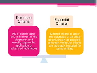
• Desirable to perform CXCR4 mutational analysis , esp. in patients planned
for Ibrutinib therapy
18. 4. Marginal Zone Lymphomas
All these entities share histological and immunophenotypic features
Trisomy 3 & 18 : common to all; 2p and 6p gain / loss of 1p and 6q - more in
NMZL
t(11;18)(q21;q21) resulting BIRC3::MALT1 fusion >> recurrent in gastric / pulm.
ENMZL
21. Classic FL (cFL)
• Follicular growth pattern (85%)
• Centrocyte/ centroblasts
• t(14;18) associated with IGH::BCL2 fusion
• Grading is optional & no longer mandatory
• Two related subtypes/ groups:
FLBL : largely equals WHO-HAEM4R FL grade 3B, and renaming
was done for reasons of consistency throughout the classification
FL with uncommon features (uFL) : 2 subsets- Blastoid / large
centrocyte & diffuse growth pattern
22. 6. Mantle cell lymphoma
Biomarkers of high-risk MCL include cytomorphology (pleomorphic or
blastoid appearance), high Ki67 proliferative index, p53 expression
and TP53 mutation
Identification of prognostic subgroups has become highly relevant due to
improved therapies
25. DLBCL, NOS
• Represents the most common entity
• 2 main subtypes previously defined in WHO-HAEM4R continue to
be recognized (i.e. GCB and ABC)
• 17 specific entities as “large B-cell lymphomas” other than DLBCL,
NOS
• Names of some entities have been modified for reasons of
consistency, from “DLBCL” to “large B-cell lymphoma“, because
diffuse growth pattern is either not apparent/present or cannot be
assessed
26. Diffuse large B-cell lymphoma/high-grade B-cell
lymphoma with MYC and BCL2 rearrangements
• Renames the entity to encompass tumours defined by dual MYC and
BCL2 rearrangements composed of large/intermediate/ blastoid cells
• Homogeneous entity with an exclusive GC GEP, close pathogenetic
relationship to FL and molecular GC-like DLBCL subsets and overlap
with BL
• Lymphoid neoplasms with dual MYC and BCL6 rearrangements
represent a more diverse spectrum- hence excluded and now classified
either as a subtype of DLBCL, NOS or HGBL, NOS according to their
cytomorphological features
27.
28. High-grade B-cell lymphoma with 11q
aberration (HGBL11q)
• Formerly known as Burkitt-like lymphoma with 11q aberration
• Aggressive MYC rearrangement-negative mature B-cell lymphoma
with a morphology similar to Burkitt lymphoma (BL)
• GEP similar to BL
• Intermediate/blastoid appearing cells- CD10+, BCL6+, BCL2-
• 11q24qter loss are more specific to this entity than the centromeric
gains
29. Large B-cell lymphomas (LBCL) of
immune-privileged sites
• Aggressive B-cell lymphomas
• Primary tumours in the central nervous system (CNS), vitreoretinal
compartment and testes of immunocompetent patient
• Arise in immune sanctuaries created by their respective anatomical
structures (e.g. blood-brain, blood-retinal and blood-testicular
barriers) and immune regulation systems within their respective
primary sites
• Share immunophenotypic and molecular features
31. Fluid overload-associated large B-cell
lymphoma
• “PEL-like lymphoma” or “HHV8-unrelated PEL-like lymphoma”
• Distinct from primary effusion lymphoma (PEL)
• Adults (elderly) without underlying immunodeficiency
• Exclusive involvement of body cavities, most commonly pleural cavity
• Underlying condition causing fluid overload such as chronic heart
failure, renal failure, protein losing enteropathy or liver failure/cirrhosis
• Mature B-cell phenotype
• EBV is positive in 13–30% of cases
• Prognosis -fairly favorable
32. Mediastinal gray zone lymphoma
(MGZL)
• Overlapping between primary mediastinal B-cell lymphoma (PMBL)
and classic Hodgkin lymphoma (CHL), especially nodular sclerosis
CHL (NSCHL)
• Replaces the term “B-cell-lymphoma, unclassifiable with features
intermediate between DLBCL and classic Hodgkin lymphoma
33. High grade B-cell lymphoma, NOS
• Aggressive mature B-cell lymphomas composed of medium-sized or
blastoid cells that do not fit into other well-defined entities
• Most frequent mutations are found in KMT2D (43%) and TP53 (30%)
• 54% of HGBL, NOS harbour “double hit” signature (DHITsig) of
LBCL/HGBL with MYC/BCL2 despite lacking rearrangements of these
genes
34. Summary of the relationship between
large B-cell lymphoma (LBCL) entities
35. 9. KSHV/HHV8-associated B-cell
lymphoid proliferations and lymphomas
• PEL/EC-PEL and KSHV/HHV8-DLBCL - HIV patients, immunodeficiency settings
• KSHV/HHV8-GLPD- elderly patients without overt immunodeficiency
• KSHV/HHV8-MCD seen in both HIV –ve and +ve patients
36. 10. Burkitt lymphoma: EBV matters
• Remains unchanged
• 3 subtypes: endemic, non-endemic or sporadic and immunodeficiency-
associated
• EBV-positive BL and EBV-negative BL form discrete biologic groups based
on their molecular features and supersede the epidemiological subtyping
• Dual mechanism of BL pathogenesis: virus-driven versus mutational
• EBV-positive BL:
▫ Higher levels of somatic hypermutation
▫ Fewer driver mutations
▫ Lower frequency of mutations in genes encoding transcription factor TCF3
or its repressor ID3
37. 11. Lymphoid proliferations and lymphomas associated with
immune deficiency and dysregulation
• New standardized nomenclature builds on an integrated approach to diagnosis that
combines all relevant data into a reporting system as follows:
1) Histological diagnosis according to accepted criteria and terminology
2) Presence or absence of one or more oncogenic virus(es)
3) Clinical setting/immunodeficiency background
38. • Poly-chemotherapy for treatment of solid tumours and haematologic
neoplasms has been largely accepted as an underlying cause for
immunodeficiency
• Primary immunodeficiencies associated with germline mutations renamed
as “inborn errors of immunity” (IEI)
39. 12. Hodgkin lymphoma
• CHL clearly defined from its
mimickers, NLPHL on the
way to, but not yet NLPBCL
41. Plasma cell neoplasms and other diseases with
paraproteins: new conditions from AESOP to TEMPI
TEMPI
• Telangiectasias
• Elevated erythropoietin and erythrocytosis
• Monoclonal gammopathy
• Perinephric fluid collection
• Intrapulmonary shunting
AESOP syndrome
• Adenopathy
• Extensive skin patch overlying a plasmacytoma
MGRS: plasma cell or B-cell proliferation that does not meet accepted criteria for
malignancy but secretes a monoclonal immunoglobulin or immunoglobulin fragment
resulting in kidney injury
CAD: AIHA mediated by monoclonal cold agglutinins and driven by an underlying clonal B-
cell lymphoid proliferation not fulfilling criteria for a B-cell lymphoma
42. • Risk stratification model for IgM MGUS and non-IgM MGUS has been updated
• Presence of all 3 risk factors consisting of :
(1) an abnormal serum free light chain ratio,
(2) IgA or IgM type MGUS, and
(3) serum M-protein value >1.5 g/dL
• High risk with approximately 50–60% risk of progression at 20 years whereas risk is
only 5% when none of the risk factors are present
• Diagnosis of TEMPI syndrome is primarily made on clinical and imaging
investigations. Bone marrow is unremarkable in majority cases; few cases show
erythroid hyperplasia and low-volume of light chain restricted plasma cells
• Skin biopsies of patients with AESOP syndrome show diffuse hyperplasia of dermal
vessels associated with surrounding dermal mucin and lymph nodes can show
features mimicking Castleman disease
43. Summary
• Tumour-like lesions with B-cell predominance
• B-ALL: B-ALL with ETV6::RUNX1-like features; B-ALL with TCF3::HLF fusion; B-ALL
with BCR::ABL1-like features
• B-PLL is no longer recognized as an entity
• “splenic B-cell lymphoma/ leukaemia with prominent nucleoli” replaces “hairy cell
leukaemia variant” and “CD5-negative B-cell prolymphocytic leukaemia”
• LPL: IgM-LPL/Waldenström Macroglobulinaemia (WM) type; Non-WM type LPL
• Marginal zone lymphomas: cytogenetic and mutational profiles differ by anatomic site,
and cutaneous MZL achieves independence
• Follicular lymphoma (FL): from classic grading to biological grouping; cFL; FLBL and
uFL.
44. • Diffuse large B-cell lymphoma/high-grade B-cell lymphoma
with MYC and BCL2 rearrangements
• High-grade B-cell lymphoma with 11q aberration (HGBL-11q)
• Large B-cell lymphomas (LBCL) of immune-privileged sites
• Fluid overload-associated large B-cell lymphoma
• Mediastinal gray zone lymphoma (MGZL)
• Burkitt lymphoma: EBV matters
• Hodgkin lymphoma: NLPHL on the way to, but not yet NLPBCL
• Plasma cell neoplasms and other diseases with paraproteins: new
conditions from AESOP to TEMPI, MGRS, CAD
Editor's Notes
In circumstances where resources are not available to reach a definitive diagnosis of an entity (or when suboptimal quality or quantity of material is limiting), a diagnostic label based on the family name of that entity can be applied.
Castleman disease is not a single disease but rather three clinicopathologically distinct entities: - unicentric Castleman disease
- idiopathic multicentric Castleman disease
- KSHV/HHV8- associated multicentric Castleman disease
(iAMP21, BCR::ABL1 fusion, KMT2A rearrangements, ETV6::RUNX1 fusion, TCF3::PBX1 fusion or IGH::IL3 fusion)
to allow for the application of differing techniques for their detection
(previously a provisional entity) shows significant benefit from targeted therapies
Use of Richter transformation preferred over Richter syndrome
Biologically distinct from HCL, despite morphological resemblance
Also absorbs all cases previously termed CD5 negative B-PLL