ntry points of glucogenicamino acids after transamination
are indicated by arrows extended from circles
the key gluconeogenicenzymes are enclosed in double
bordered boxes. oated fashion. They are interdependent; each forms a strand in the web of life. Parasitology is
the science that deals with organisms living in the human body (the host) and the medical significance of
this host-parasite relationship.
ASSOCIATION BETWEEN PARASITE AND HOST
A parasite is a living organism, which takes its nourishment and other needs from ahost; the host is an
organism which supports the parasite. The hosts vary depending on whether they harbor the various
stages in parasitic development
DIFFERENT KINDS OF PARASITES
Ectoparasite – a parasitic organism that lives on the outer surface of its host, e.g. lice, ticks, mites etc.
Endoparasites parasites that live inside the body of their host, e.g. Entamoeba histolytica.
Obligate Parasite- This parasite is completely dependent on the host during a segment or all of its life
cycle, e.g. Plasmodium spp.
Facultative parasite – an organism that exhibits both parasitic and non-parasitic modes of living and
hence does not absolutely depend on the parasitic way of life but is capable of adapting to it if paced on
a host. E.g. Naegleria fowleri
Accidental parasite – when a parasite attacks an unnatural host and survives. E.g. Hymenolepis diminuta
(rat tapeworm).
Erratic parasite - is one that wanders in to an organ in which it is not usually found. E.g. Entamoeba
histolytica in the liver or lung of humans.
Most of the parasites which live in/on the body of the host do not cause disease (non-pathogenic
parasites). In Medical parasitology we focus on most of the disease causing (pathogenic) parasites.
However, understanding parasites which do not ordinariy produce disease in heathy
(immunocompetent) individuals but do cause illness in individuals with impaired defense mechanism
(opportunistic parasites) is becoming of paramount importance because of the increasing prevaence of
HIV/AIDS in our country.
DIFFERENT KINDS OF HOSTS
Definitive host – a host that harbors a parasite in the adult stage or where the parasite undergoes a
sexual method of reproduction.
Intermediate host - harbors the arval stages of the parasite or an asexual cycle of development takes
pace. In some cases, larval development is completed in two different intermediate hosts, referred to
as first and second intermediate hosts.
Paratenic host – a host that serves as a temporary refuge and vehicle for reaching an obligatory host,
usually the definitive host, i.e. it is not necessary for the completion of the parasites life cycle.
Reservoir host – a host that makes the parasite available for the transmission to another host and is
usually not affected by the infection.
Natural host a host that is naturally infected with certain species of parasite.
Accidental host – a host that is under normal circumstances not infected with th
The study of the cell cycle focuses on mechanisms that regulate the timing and frequency of DNA duplication and cell division. As a biological concept, the cell cycle is defined as the period between successive divisions of a cell. During this period, the contents of the cell must be accurately replicated.
The cell cycle is regulated by cyclins and cyclin-dependent kinases.
How long is one cell cycle?
Depends. Eg. Skin cells every 24 hours. Some bacteria every 2 hours. Some cells every 3 months. Cancer cells very short. Nerve cells never.
Programmed cell death:
Each cell type will only do so many cell cycles then die. (Apoptosis)
This includes detailed description of the Cell Cycle and Cell Cycle regulation. Courtesy: Campbell Biology Book, And Dr, Rosemary Redfield Lectures, University of British Columbia.
Meiosis is the special type of cell division in which the number of chromosomes in daughter cells reduces to half, as compared to the parent cell. It takes place in diploid cells only, in animals at the time of gamete formation, while in plants when spores are produced.
Meiosis is the special type of cell division in which the number of chromosomes in daughter cells reduces to half, as compared to the parent cell. It takes place in diploid cells only, in animals at the time of gamete formation, while in plants when spores are produced.
This pdf is about the Schizophrenia.
For more details visit on YouTube; @SELF-EXPLANATORY;
https://www.youtube.com/channel/UCAiarMZDNhe1A3Rnpr_WkzA/videos
Thanks...!
Richard's aventures in two entangled wonderlandsRichard Gill
Since the loophole-free Bell experiments of 2020 and the Nobel prizes in physics of 2022, critics of Bell's work have retreated to the fortress of super-determinism. Now, super-determinism is a derogatory word - it just means "determinism". Palmer, Hance and Hossenfelder argue that quantum mechanics and determinism are not incompatible, using a sophisticated mathematical construction based on a subtle thinning of allowed states and measurements in quantum mechanics, such that what is left appears to make Bell's argument fail, without altering the empirical predictions of quantum mechanics. I think however that it is a smoke screen, and the slogan "lost in math" comes to my mind. I will discuss some other recent disproofs of Bell's theorem using the language of causality based on causal graphs. Causal thinking is also central to law and justice. I will mention surprising connections to my work on serial killer nurse cases, in particular the Dutch case of Lucia de Berk and the current UK case of Lucy Letby.
We present you a part of our Tampere University's team - FHAIVE!
Besides producing excellent science, they are in charge or coordinating this project as well Tampere University, Faculty of Medicine and Health Technology.
(May 29th, 2024) Advancements in Intravital Microscopy- Insights for Preclini...Scintica Instrumentation
Intravital microscopy (IVM) is a powerful tool utilized to study cellular behavior over time and space in vivo. Much of our understanding of cell biology has been accomplished using various in vitro and ex vivo methods; however, these studies do not necessarily reflect the natural dynamics of biological processes. Unlike traditional cell culture or fixed tissue imaging, IVM allows for the ultra-fast high-resolution imaging of cellular processes over time and space and were studied in its natural environment. Real-time visualization of biological processes in the context of an intact organism helps maintain physiological relevance and provide insights into the progression of disease, response to treatments or developmental processes.
In this webinar we give an overview of advanced applications of the IVM system in preclinical research. IVIM technology is a provider of all-in-one intravital microscopy systems and solutions optimized for in vivo imaging of live animal models at sub-micron resolution. The system’s unique features and user-friendly software enables researchers to probe fast dynamic biological processes such as immune cell tracking, cell-cell interaction as well as vascularization and tumor metastasis with exceptional detail. This webinar will also give an overview of IVM being utilized in drug development, offering a view into the intricate interaction between drugs/nanoparticles and tissues in vivo and allows for the evaluation of therapeutic intervention in a variety of tissues and organs. This interdisciplinary collaboration continues to drive the advancements of novel therapeutic strategies.
THE IMPORTANCE OF MARTIAN ATMOSPHERE SAMPLE RETURN.Sérgio Sacani
The return of a sample of near-surface atmosphere from Mars would facilitate answers to several first-order science questions surrounding the formation and evolution of the planet. One of the important aspects of terrestrial planet formation in general is the role that primary atmospheres played in influencing the chemistry and structure of the planets and their antecedents. Studies of the martian atmosphere can be used to investigate the role of a primary atmosphere in its history. Atmosphere samples would also inform our understanding of the near-surface chemistry of the planet, and ultimately the prospects for life. High-precision isotopic analyses of constituent gases are needed to address these questions, requiring that the analyses are made on returned samples rather than in situ.
Gliese 12 b: A Temperate Earth-sized Planet at 12 pc Ideal for Atmospheric Tr...Sérgio Sacani
Recent discoveries of Earth-sized planets transiting nearby M dwarfs have made it possible to characterize the
atmospheres of terrestrial planets via follow-up spectroscopic observations. However, the number of such planets
receiving low insolation is still small, limiting our ability to understand the diversity of the atmospheric
composition and climates of temperate terrestrial planets. We report the discovery of an Earth-sized planet
transiting the nearby (12 pc) inactive M3.0 dwarf Gliese 12 (TOI-6251) with an orbital period (Porb) of 12.76 days.
The planet, Gliese 12 b, was initially identified as a candidate with an ambiguous Porb from TESS data. We
confirmed the transit signal and Porb using ground-based photometry with MuSCAT2 and MuSCAT3, and
validated the planetary nature of the signal using high-resolution images from Gemini/NIRI and Keck/NIRC2 as
well as radial velocity (RV) measurements from the InfraRed Doppler instrument on the Subaru 8.2 m telescope
and from CARMENES on the CAHA 3.5 m telescope. X-ray observations with XMM-Newton showed the host
star is inactive, with an X-ray-to-bolometric luminosity ratio of log 5.7 L L X bol » - . Joint analysis of the light
curves and RV measurements revealed that Gliese 12 b has a radius of 0.96 ± 0.05 R⊕,a3σ mass upper limit of
3.9 M⊕, and an equilibrium temperature of 315 ± 6 K assuming zero albedo. The transmission spectroscopy metric
(TSM) value of Gliese 12 b is close to the TSM values of the TRAPPIST-1 planets, adding Gliese 12 b to the small
list of potentially terrestrial, temperate planets amenable to atmospheric characterization with JWST.
Astronomy Update- Curiosity’s exploration of Mars _ Local Briefs _ leadertele...
Meiosis : introduction and phases of meiosis.
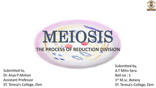
1. MEIOSIS
THE PROCESS OF REDUCTION DIVISION
Submitted by,
A.T Milin Sera
Roll no : 1
1st M.sc. Botany
ST. Teresa’s College, Ekm
Submitted to,
Dr. Arya P Mohan
Assistant Professor
ST. Teresa’s College, Ekm
3. INTRODUCTION
• The cells of a particular species have a constant number of chromosomes.
• If the gametes had the same number of chromosomes as the somatic cells, then
the zygote would have twice the diploid number of chromosomes.
• This number would go on doubling with each generation.
• However, the chromosome number remains constant from generation to
generation.
• This is because of meiotic division which reduces the chromosome number to
half, and counteracts the effect of fertilization.
• Thus fertilization and meiosis are compensating events.
Meiosis...
1
4. MEIOSIS
• Extracellular signals induce a transcriptional program that produces
meiosis-specific cell cycle.
• Such an extracellular signals inducing entry into the meiotic divisions
in mammals is retinoic acid, which is a steroid hormone.
• Meiosis consists of two cell divisions.
• The two divisions are known as the first meiotic division and the
second meiotic division.
• The stages of first meiotic division are prophase I, prometaphase I,
metaphase I, anaphase I and telophase I.
• The second meiotic division consists of prophase II, metaphase II,
anaphase II and telophase II.
Meiosis...
2
5. PHASES OF MEIOSIS
• Before entering meiosis I, a cell first go through interphase.
• The interphase preceding meiosis is important because
replication of DNA takes place during this stage.
• Replication is confined to the synthesis (S) phase of
interphase.
• It is preceded by a post-mitotic gap phase (G1) and followed
by a pre-mitotic gap phase (G2).
• During these two phases there is no replication of DNA.
PRE-MEIOTIC INTERPHASE
3
Meiosis...
6. FIRST MEIOTIC DIVISION (MEIOSIS-1)
• Cell division that reduces the chromosome number by one-half.
• It separate homologous chromosomes and produce two cells with
haploid chromosome number (n).
• For that reason it is known as Reductional Division.
• Four phases: Prophase I, Metaphase I, Anaphase I,Telophase I
PROPHASE -1
• Longest and most complex phase.
• It consists of 5 substages : Leptotene, Zygotene, Pachytene,
Diplotene, Diakinesis.
• In G2 and prophase of meiosis I, the two replicated chromatids of each
chromosome are associated with each other by cohesin complexes.
4
Meiosis...
7. LEPTOTENE
• Chromsomes become very thin
• They are not distinct, cannot be easily seen.
PROLEPTOTENE
• During this stage the chromosomes become more distinct.
• Chromosomes appear as slender threads bearing a series of
granule- like structures called chromomeres.
• Under electron microscope the leptotene chromosomes
have an axial filament.
• To this filament, chromatin fibres are attached as a series of
lateral loops.
• Cytoplasm has many polyribosomes, but few ER.
• A pre-meiotic pairing may takes place during leptotene.
5
Meiosis...
8. • Leptotene chromosomes may be irregularly arranged, or may be polarized
towards the centrioles forming a ‘bouquet’.
• In plant cells the chromosomes may sometimes form a tangle of threads, called
the synizetic knot, on one side of the nucleus.
• During leptotene the cytoplasm has many polyribosomes, but endoplasmic
vesicles are few.
• A pre-meiotic pairing may takes place during leptotene.
• Homologous chromosomes may be aligned together, but no synaptonemal
complexes are formed.
6
Meiosis...
9. ZYGOTENE
• During zygotene the chromosomes become shorter and
thicker.
• The homologous chromosome starts pairing together.
• These homologous chromosome come to lie side by side in
pairs & this pairing is called synapsis.
• The complex formed by a pair of synapsed chromosome is
called bivalent.
• Pairing may begin at the centromere and proceed towards the
ends (procentric pairing).
• They may begin at the ends and proceed towards the
centromere (proterminal pairing).
7
Meiosis...
10. • Sometimes it may occur simultaneously at all chromomeres (intermediate
condition).
• In many plant and animal species a kind of pseudosynapsis between non-
homologous regions also occurs.
• When synapsing occurs, the chromosomes do not lie side by side but rather on
top of each other with a protein called the synaptonemal complex holding them
together.
8
Meiosis...
11. SYNAPTONEMAL COMPLEX
• The synaptonemal complex is a proteinaceous
structure that is formed between paired
homologous chromosomes.
• It is a tripartite ribbon like structure.
• It consists of a dense central element, with a dense
lateral element on either side.
• The lateral elements are attached at both ends to
the nuclear membrane.
• The lateral elements are connected to the central
element by a series of lateral loops which form
transverse units or LC fibres.
9
Meiosis...
12. • Three specific components of the synaptonemal complex have been characterized:
SC protein-1 (SYCP1), SC protein-2 (SYCP2), and SC protein-3 (SYCP3).
• Lateral elements are mainly formed by SYCP3 and secondarily by SYCP2.
• Central element and transverse filaments are composed by the protein SYCP1.
• The axial filaments of leptotene chromosomes later become transformed into the
lateral elements of the SCs.
• The SCs first appear during zygotene.
• They are completed by the beginning of pachytene.
• By diplotene the SCs have mostly disappeared, but remnants may be seen even in
diakinesis.
• They are believed to be involved in chiasmata formation and crossing over.
• It stabilise the two homologous chromosomes till the crossing over is completed.
Meiosis...
10
13. PACHYTENE
• The bivalents appear as thick threads due to condensation of
chromosomes.
• Two chromatids belonging to different homologues undergo
one or more transverse breaks at the same level.
• The breaks never occur between two sister chromatids.
• The break is followed by interchange and fusion of broken
ends between two homologous chromosomes.
• This is called crossing over.
• The site where crossing over occurs forms a recombination
nodule.
• An enzyme called recombinase is involved during this phase.
11
Meiosis...
14. DIPLOTENE
• Separation of paired homologues chromosomes begin.
• The separating chromosomes are held together at one or more
points called chiasmata.
• A chiasma formed at the ends of chromosomes is called a
terminal chiasma.
• Chiasmata formed along the lengths of chromosomes are called
interstitial chiasmata.
• Terminalization of chiasmata takes place.
• The synaptonemal complexes mostly disappear during diplotene.
• Cytoplasm shows an increase in the number of endoplasmic
vesicles.
Meiosis...
12
15. DIAKINESIS
• The chromosomes become more contracted.
• The bivalents are more evenly distributed in the nucleus
and migrate towards the periphery.
• The homologues remain in contact with each other by
their terminal chiasmata.
• The nucleolus is detached from the chromosome, or
disappears.
• Remnants of the synaptonemal complexes may still be
seen.
13
Meiosis...
16. METAPHASE-1
• The chromosomes now become arranged on the
equator of the cell.
• The spindle is formed.
• Spindle fibres become attached to the centromeres of
the two homologous chromosomes.
• The two centromeres of each bivalent lie on opposite
sides of the equatorial plate.
• The nuclear membrane disappears.
• The chromosomes reach their maximum contraction.
• Spindle formation begins.
PROMETAPHASE
1
Meiosis...
14
17. ANAPHASE-1
• Separation of the two homologous chromosomes of each
bivalent begins.
• One chromosome from each bivalent begins to migrate to one
pole, the other migrates to the opposite pole.
• The sister chromatids are said to be co-oriented.
• The kinetochores of each bivalent attach to spindle microtubules
from opposite spindle poles.
• They are said to be bi-oriented.
• During Anaphase I original chromosomes separate, so reduction
in the number of chromosomes from 2n to n number.
15
Meiosis...
18. TELOPHASE-1
• The homologous chromosome complete their migration to the
two poles because of shortening of spindles.
• When the chromosomes reach the poles they persist for some
time in the condensed state.
• Later they undergo despiralization and become elongated.
• The nuclear membrane is reformed, but the nucleoli do not
reappear.
• Unlike in mitosis the spindle fibres do not disappear
completely.
16
Meiosis...
19. CYTOKINESIS
• Cytokinesis involves the formation of a cleavage furrow, resulting in the
pocketing of the cell into two cells.
• In most plant cells daughter cells are formed by the laying down of cell plate
between the two groups of chromosomes.
• In other plant cells cytokinesis does not take place until both meiotic divisions are
completed.
• At the end of Telophase I and Cytokinesis, two daughter cells are produced.
• Each daughter cell with one half of the number of chromosomes of the original
parent cell.
Meiosis...
17
20. 18
Meiosis...
MEIOSIS I STAGES MAIN EVENTS
Prophase 1 : Sub-Stages :-
Leptotene Chromosomes appear as slender threads
& Bouquet Formation
Zygotene Synapsis & synaptonemal complex
formation
Pachytene Chromosomes appear as thick threads &
Crossing over
Diplotene Chiasma formation
Diakinesis Chromosomes get thick and contracted
Metaphase 1 Chromosomes arranged in equator region
& Spindle formation
Anaphase 1 Separation of homologous chromosomes
& Beginning of chromosomes migration to
opposite poles
Telophase 1 Migration of homologous chromosomes
complete & Shortening of spindle fibres
21. INTERKINESIS OR INTRAMEIOTIC
INTERPHASE
• Interkinesis is a metabolic phase between telophase-1 & prophase-2.
• During this phase chromosomes are elongated but do not form chromatin
fibres.
• This differs from interphase I and interphase of mitosis in that there is no
duplication of DNA.
• The RNA and protein required during meiosis-2 are syntesised during this
phase.
19
Meiosis...
22. MEIOSIS -2
• During Meiosis II, two sister chromotids of each chromosome separate and
migrate to the opposite pole.
• As a result, the number of chromosomes in each of the two haploid nuclei
remains the same (i.e haploid), at the end of this division.
• Thus the second division of meiosis is often referred to as equational division.
• Sometimes, it is called as ‘Meiotic Mitosis’.
• The second meiotic division is also divided into four stages : Prophase II,
Metaphase II, Anaphase II, Telophase II.
• The 4 stages of meiosis – II is same as mitosis.
• It differs from mitosis in that DNA does not duplicate, while the centromeres do
so.
Meiosis...
20
23. 21
Meiosis...
MEIOSIS II STAGES MAIN EVENTS
Prophase 2 Chromatids with widely separated
arms & Spindle formation and
nuclear membrane disappearance
Metaphase 2 Chromosomes oriented at
equatorial plate & Spindle fibre
attachment as same as in mitosis
Anaphase 2 Centromeres divide & Two
chromatids of each chromosome
separate to opposite poles
Telophase 2 Reconstruction of nuclei. Nucleus,
centriole and chromosomes return
to interphasic condition & Nucleus
contains haploid no. of
chromosomes
Cytokinesis Dyad divides into two parts. One
parent cell produce four haploid
daughter cells, together known as
tetrad
24. REMOVAL OF COHESION
• To facilitate two consecutive chromosome segregation phases, cohesins have to be
lost from chromosome.
• During meiosis, cohesins are lost from chromosome arms by the end of meiosis I,
but a pool of cohesins around kinetochores is protected from removal.
• This pool of cohesins persists throughout meiosis I but is removed at the onset of
anaphase II.
• The mechanisms that remove cohesins during meiosis are the same as during
mitosis.
• Securin degradation releases separase, which then cleaves the cohesins.
• This allows the recombined maternal and paternal chromosomes to separate, but
each pair of chromatids remains associated at the centromere.
• During metaphase II, sister chromatids align on the metaphase II spindle and
separase is activated yet again, cleaving the residual cohesin around centromeres.
22
Meiosis...
25. Rec8
• A specialized cohesin subunit, Rec8, is necessary for the stepwise loss of cohesins
from chromosomes during meiosis.
• Rec8 is homologous to Scc1, the cohesin subunit that closes the cohesin ring in
the cohesin complex of mitotic cells.
• During early anaphase of meiosis I, Rec8 is lost from chromosome arms but is
retained at centromeres.
• During early anaphase of meiosis II, centromeric Rec8 is cleaved by separase, so
the sister chromatids can segregate.
• The mechanism that protects Rec8 from cleavage at centromeres during meiosis I
is similar to the mechanism that protects Scc1 at centromeres during mitosis.
Meiosis...
23
26. PP2A & SHUGOSHIN
• Cohesion at the centromeres is maintained because a specific isoform of protein
phosphatase 2A (PP2A).
• PP2A is localized to centromeric chromatin by members of a family of proteins
known as the Mei-S332/Shugoshin.
• PP2A keeps cohesin in a hypophosphorylated state that does not dissociate.
• During metaphase II, Mei-S332/Sgo1 dissociates from chromosomes.
• When the last kinetochore is properly associated with spindle microtubules,
Cdc20 is depressed and associates with the APC/C.
• This causes ubiquitinylation of securin.
• This releases separase activity, which cleaves Rec8, eliminating cohesion at the
centromere and allowing chromatid separation in anaphase.
24
Meiosis...
27. • Cohesin removal differs for meiosis I because when Rec8
replaces Scc1 in the cohesin complex.
• The meiotic cohesin complex can only be removed from
chromatin via the action of separase.
• Rec8 also differs from Scc1 in that it must be phosphorylated by
several protein kinases to be cleaved by separase.
• During meiosis I, the centromere-specific isoform of PP2A
targeted to centromeric chromatin by Mei-5332/Shugoshin
prevents this phosphorylation.
• The PP2A targeting factor and PP2A then dissociate from
chromosomes by metaphase II, allowing separase cleavage of
Rec8.
Meiosis...
25
28. • In mitosis and meiosis ll, sister kinetochores attach to spindle microtubules
emerging from opposite spindle poles.
• These kinetochores are said to be bi-oriented.
• This is essential for segregation of sister chromatids to different daughter cells.
• In contrast, at meiosis I metaphase, sister kinetochores attach to spindle
microtubules emerging from the same spindle pole.
• These sister kinetochores are said to be co-oriented.
• Attachment of sister kinetochores to the proper microtubules in meiosis I and II is
critical for correct meiotic segregation of chromosomes.
ORIENTATION OF KINETOCHORES
26
Meiosis...
29. • Monopolin complex is a protein complex that links with sister kinetochores
during meiosis I for their attachment to microtubules emerging from the same
spindle pole.
• In organisms where kinetochores attach to multiple microtubules, Rec8-
containing cohesins are essential for sister kinetochore co-orientation.
• These meiosis-specific cohesins impose a rigid kinetochore structure.
• This restricts the movement of sister kinetochores and thereby favoring
attachment to microtubules from the same spindle pole.
• Like during mitosis and meiosis II, correct attachment of meiosis I chromosomes
is mediated by a tension-based mechanism.
• During meiotic metaphase I, kinetochore-associated microtubules are also under
tension.
Meiosis...
27
30. • Because chiasmata generated by recombination between
homologous chromosomes and the cohesins distal to the
chiasmata prevent them from being pulled to the poles.
• Kinetochore-microtubule attachments are unstable in the
absence of tension.
• When kinetochores attach to the wrong spindle fibers
release the incorrect microtubules.
• This enables them to bind microtubules again until
attachments are made that generate tension.
• As in mitosis, once tension is generated, microtubule
attachment to the kinetochores is stabilized.
28
Meiosis...