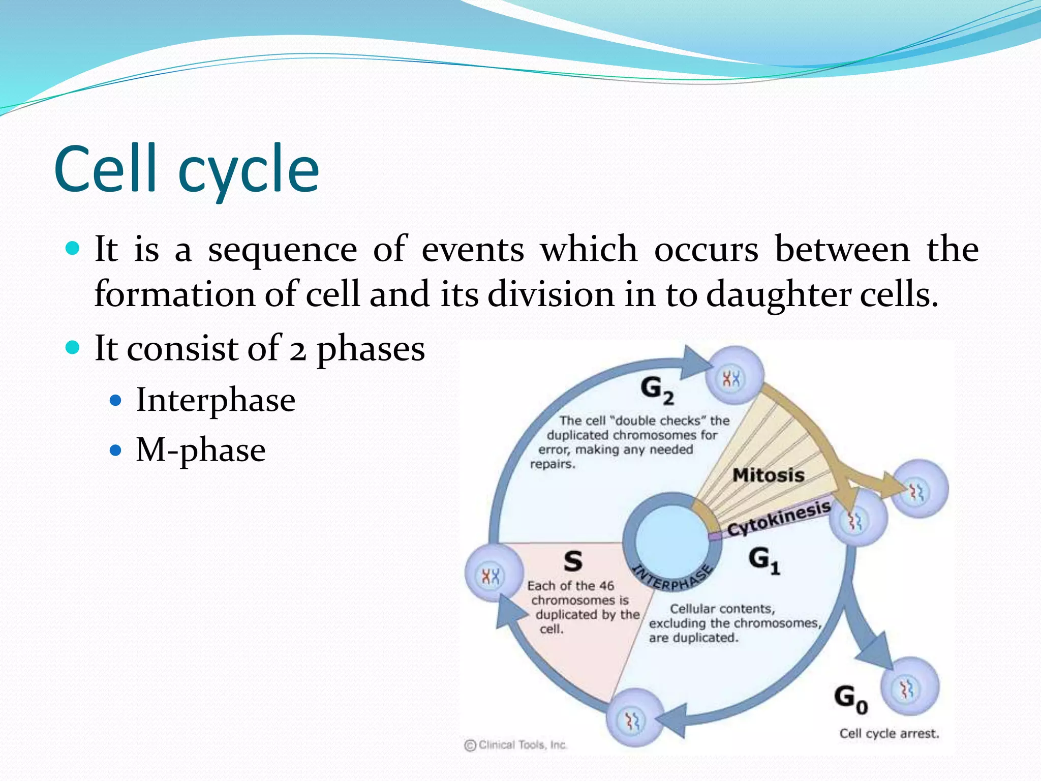
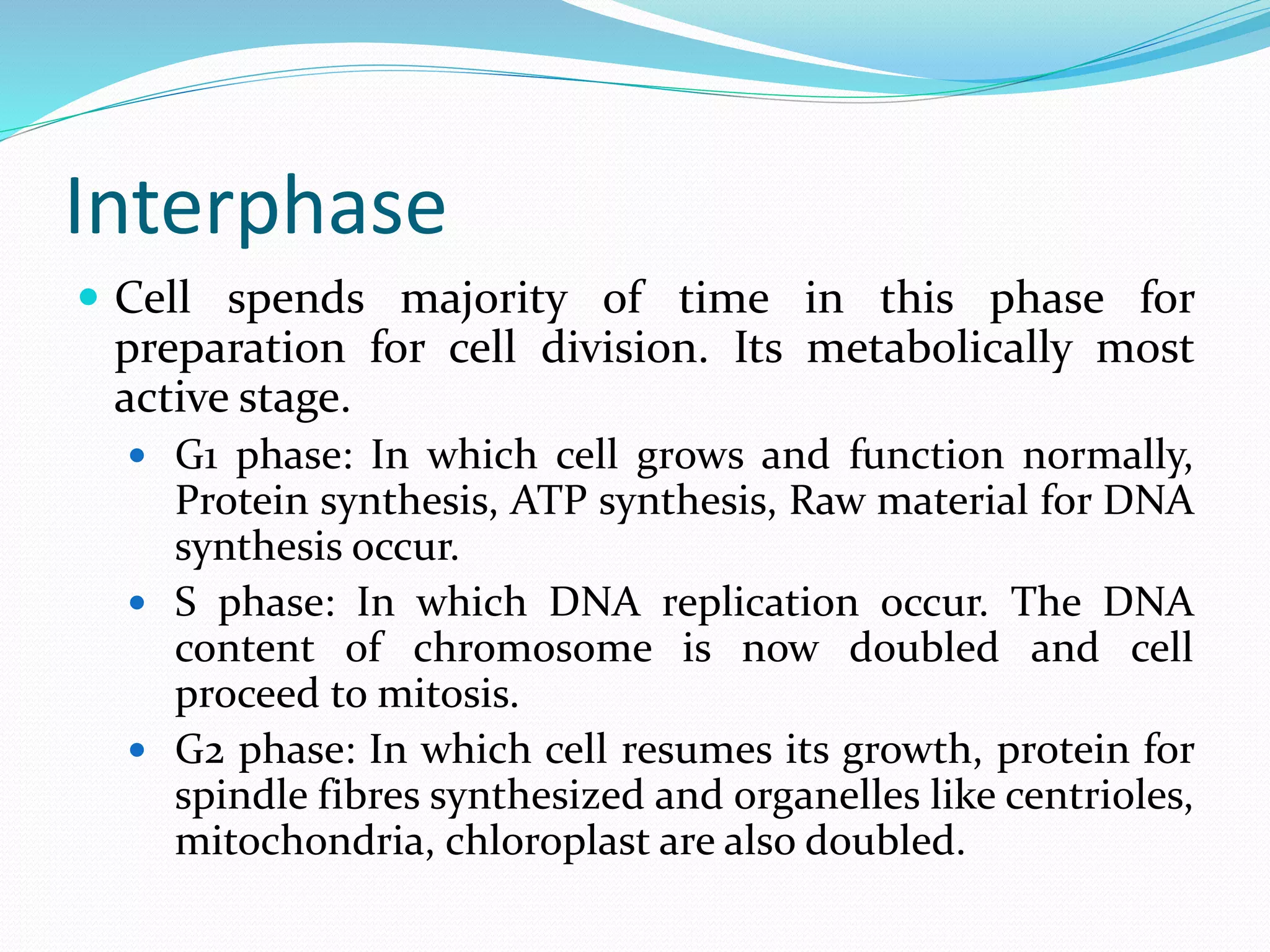
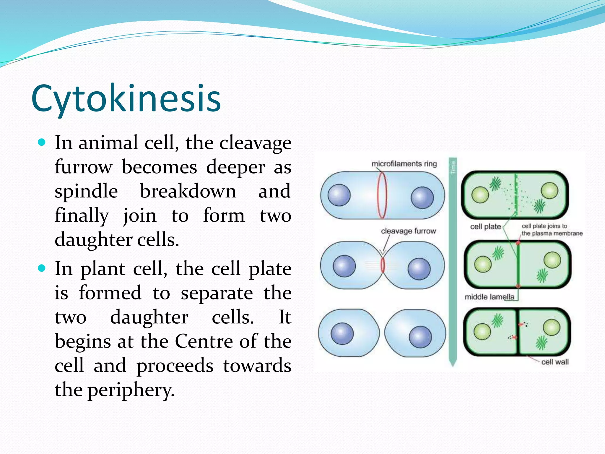
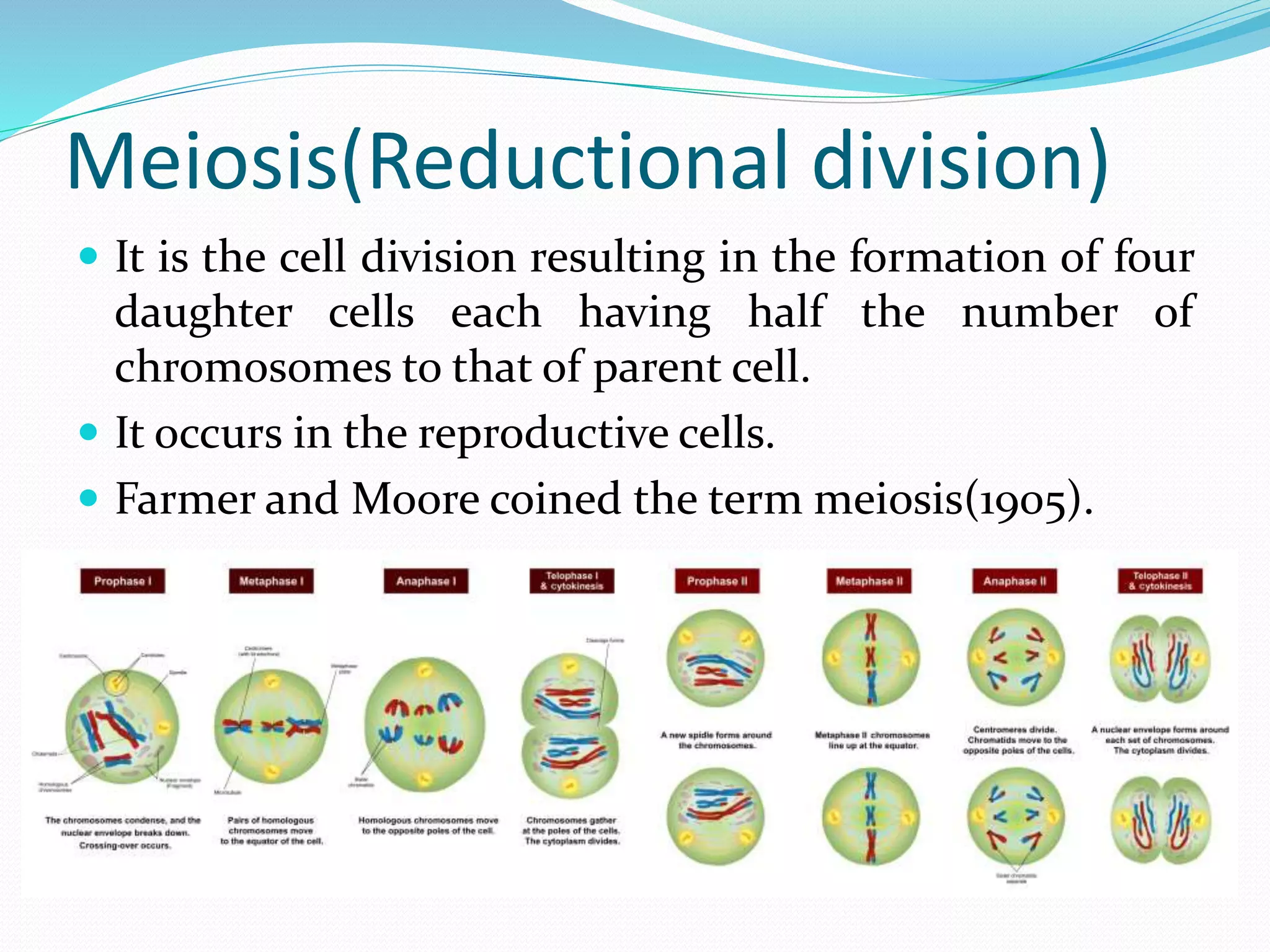
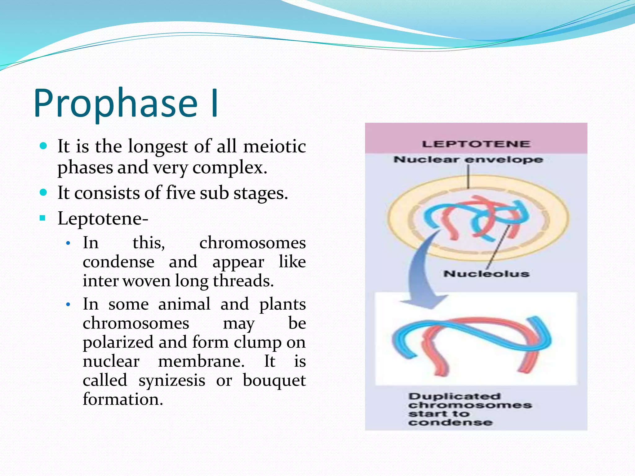
MITOSIS AND MEIOSIS
PRESENTED BY – SMRUTI RANJAN MASANTA
M.PHARM(1ST YR)
PHARMACOLOGY
CONTENT
 Cell cycle
 Interphase
 M-phase
 Mitosis
• Prophase
• Prometaphase
• Metaphase
• Anaphase
• Telophase
 Meiosis
• Meiosis-I
• Meiosis-II
Cell cycle
 It is a sequence of events which occurs between the
formation of cell and its division in to daughter cells.
 It consist of 2 phases
 Interphase
 M-phase
Interphase
 Cell spends majority of time in this phase for
preparation for cell division. Its metabolically most
active stage.
 G1 phase: In which cell grows and function normally,
Protein synthesis, ATP synthesis, Raw material for DNA
synthesis occur.
 S phase: In which DNA replication occur. The DNA
content of chromosome is now doubled and cell
proceed to mitosis.
 G2 phase: In which cell resumes its growth, protein for
spindle fibres synthesized and organelles like centrioles,
mitochondria, chloroplast are also doubled.
M-phase
 In this phase cell division takes place. Generally of two
type.
 Karyokinesis
 Mitosis
 Meiosis
 Cytokinesis
Mitosis(Equational division)
 It is a process by which a cell divides into two daughter cells
each having exactly identical number of chromosomes to
that of parent cell.
 It generally occurs in somatic (vegetative cells) cells.
 This type of division was termed as mitosis by W.
Flemming (1882).
 Stages of mitosis:
 Prophase
 Prometaphase
 Metaphase
 Anaphase
 Telophase
 cytokinesis
 (A to H) Fluorescence micrographs
of mitosis in fixed newt lung cells
stained with antibodies to reveal the
microtubules (green), and with a
dye (Hoechst 33342) to reveal the
chromosomes (blue). The spindle
forms as the separating astral MT
arrays, associated with each
centrosome (A to C), interact with
the chromosomes. Once the
chromosomes are segregated into
daughter nuclei (F and G), new MT-
based structures known as stem-
bodies form between the new nuclei
(G). These play a role in cytokinesis
(H).
Prophase
 It is the longest phase of
mitosis.
 In which chromatin
condenses into chromosome
and then thicken and shows
two distinct chromatids
which connected by
centromere.
 Centriole divide and begins
to move to opposite side of
the cell.
 Spindle fibres begins to form.
It consist of microtubules and
proteins called tubulin.
Prometaphase
 The disappearance of
nuclear membrane and
nucleolus marks the
beginning of this stage.
The distinction between
cytoplasm and
nucleoplasm is lost. It is
called extra-nuclear
mitosis or eumitosis.
 The chromosomes get
attached to spindle fibres
by their centromeres and
started to move towards
equator.
Metaphase
 At metaphase the
chromosomes have
reached the central
equatorial portion of the
spindles. They are aline to
form the equatorial
plate/metaphasic plate.
 Chromosomal spindle
fibres are attached to the
kinetochore/centromere of
chromosome.
 The continuous fibres
extends from pole to pole.
Anaphase
 Chromosomes break at
centromeres and the sister
chromatids move to opposite
side of the cell. Interzonal
fibres form due to break
down of centromere.
 The number of chromosomes
doubles.
 The shape of chromosomes is
identified, that appear like
letters
K(metacentric),L(submetace
ntric),j(acrocentric),i(telocen
tric).
Telophase
 The chromosomes reach
the opposite pole.
 Chromosomes
decondensed to form
chromatin, nuclear
membrane reappears.
 The spindle fibres
disassemble and
disappear.
 Cleavage furrow appears.
Cytokinesis
 In animal cell, the cleavage
furrow becomes deeper as
spindle breakdown and
finally join to form two
daughter cells.
 In plant cell, the cell plate
is formed to separate the
two daughter cells. It
begins at the Centre of the
cell and proceeds towards
the periphery.
Significance of mitosis
 It helps in growth.
 It helps in repair and regeneration.
 It helps in maintaining genetic stability.
 It helps in asexual reproduction in lower organisms.
Control of mitosis and mitotic poisons
 Cell area to volume ratio.
 Nucleocytoplasmic ratio(nuclear mass: cytoplasm).
 Mitogens are some substance stimulate mitotic
division
e.g.- cyclins, auxins, gibberellin, insulin, cytokinin.
 Mitotic poison are substance that inhibit mitosis.
e.g.- cyanide-stops division in prophase.
colchicine-stops division in metaphase (stops the
spindle formation).
CO-stops the krebs cycle(ATP synthesis stops).
Meiosis(Reductional division)
 It is the cell division resulting in the formation of four
daughter cells each having half the number of
chromosomes to that of parent cell.
 It occurs in the reproductive cells.
 Farmer and Moore coined the term meiosis(1905).
Stages of meiosis
 Meiosis-I
 Prophase I
 Leptotene
 Zygotene
 Pachytene
 Diplotene
 Diakinasis
 Metaphase I
 Anaphase I
 Telophase I
 Meiosis-II
 Prophase II
 Metaphase II
 Anaphase II
 Telophase II
 Fluorescent micrographs of
chromosome spreads stained for
DNA and the meiotic cohesin
subunits (A–L) SMC3 and (M–X)
SYN1 in (A–F, M–R) wild type and
(G–L, S–X) rsw4 exposed to 30°C for
2 days. In each panel, the top image
shows a merged image of the DNA
(red) and cohesin signal (green); the
bottom image only shows the
cohesin signal (green) for the same
meiotic spread. (A, G, M, S) Early
zygotene. (B, H, N, T) Pachytene.
(C, I, O, U) Diakinesis. (D, J, P, V)
Metaphase I. (E, K, Q, W) Anaphase
I. (F, L, R, X) Prometaphase
II/Metaphase II. Bar = 10 µm.
 Fluorescent micrographs of
meiocytes showing
microtubules (green) and
DNA (blue) for (A–D, I–L)
wild type and (E–H, M–P)
rsw4 exposed to 30°C for 2
days. (A, E) Metaphase I. (B,
C, F, G) anaphase I. (D, H)
Telophase I. (I, M) Metaphase
II. (J, N) Late anaphase II. (K,
O) Telophase II. (L, P) Pre-
cytokenisis. Bar = 10 µm.
Prophase I
 It is the longest of all meiotic
phases and very complex.
 It consists of five sub stages.
 Leptotene-
• In this, chromosomes
condense and appear like
inter woven long threads.
• In some animal and plants
chromosomes may be
polarized and form clump on
nuclear membrane. It is
called synizesis or bouquet
formation.
 Zygotene
• In this stage synapsis or pairing of
homologous chromosomes takes
place. One of the two
homologous chromosome from
male and other from female
parents.
• A pair of homologous
chromosome(i.e. four
chromatids) form a structure
called synaptonemal complex.
 Pachytene
• During this stage crossing over
takes place. It is an exchange of
equal segments of non-sister
chromatids belonging to two
different but homologous
chromosome.
• The chromosome pair(bivalent)
consist of four chromatids is
called tetrad. Only two of the four
chromatids takes part in crossing
over.
 Diplotene
• At the completion of crossing over
the two chromosomes are in
contact at several points along
their length. These point of
contact between homologous
chromosome are known as
chiasmata.
• The homologous chromosome
now start separating from one
another and the chiasma begin to
open. The separation begins at the
centromere and progress towards
the ends. The process is known as
terminalization.
 Diakinesis
• The chromosome continue to
contract and terminalization still
continues.
• The nuclear membrane dissociate
and disappear.
• The spindle fibres begin to appear.
Metaphase I
 The chromosomes appear
condensed. The spindle
fibres get attached to the
centromere of two
homologous
chromosomes.
 The chromosomes now
become arranged at the
equater of the cell. Due to
independent assortment
the alignment is random,
which cause diversion.
Anaphase I
 The two chromosomes
separate and move towards
the poles due to contraction
of spindle fibres. Here
centromeres do not divide
and the sister chromatids do
not separate. The entire
chromosome with two
chromatid moves to the same
pole. Hence the division is
reductional or disjunctional.
 Each pole of the cell has half
the number of chromosome
as compare to the parent cell.
Telophase I
 Nuclear membrane appears
around the group of
chromosomes at each pole of
cell. Thus the daughter nuclei
with half number of
chromosomes are formed.
 After formation of nuclei,
chromosomes pass into a
small
interphase(interkinesis)
before the second meiotic
division. In this phase DNA
replication does not occur.
Prophase II
 The chromosome
become short and thick.
 Nuclear membrane
disappear.
 The spindle apparatus is
formed.
Metaphase II
 The chromosome get
arranged on the
equatorial plate of the
cell.
 The spindle fibres
attached to the same
centromere.
Anaphase II
 The centromere divide
and the chromatids of
each chromosome
separate and move to the
poles.
Telophase II
 chromosomes form a
cluster at opposite pole
of cell. Nuclear
membrane reappear to
form two daughter
nuclei.
Cytokinesis
 In animal cell, the cleavage
furrow becomes deeper as
spindle breakdown and
finally join to form four
daughter cells.
 In plant cell, the cell plate
is formed to separate the
four daughter cells. It
begins at the Centre of the
cell and proceeds towards
the periphery.
Significance of meiosis
 It helps in haploid gamate formation and sexual
reproduction.
 It helps in maintaining the species specific
chromosome number.
 It helps in exchange of genetic material. Thus cause
variation which also leads to evolution.
References
 C.B. Power, cell biology, Himalaya publishing house,7th
edition,2017, page no-628-652.
 P. k. Gupta, cell and molecular biology, Rastogi
publications, 3rd edition,2009,page no-287-298.
 A. Bendre, P.C. Pande, concept of botany, Rastogi
publications, 8th edition, 2013,page no-2.167-2.187.
 www.slideshare.com
 www.researchgate.com
 www.studyandscore.com
 www.google.com
 www.youtube.com
Thank you

Cell division

  • 1.
    MITOSIS AND MEIOSIS PRESENTEDBY – SMRUTI RANJAN MASANTA M.PHARM(1ST YR) PHARMACOLOGY
  • 2.
    CONTENT  Cell cycle Interphase  M-phase  Mitosis • Prophase • Prometaphase • Metaphase • Anaphase • Telophase  Meiosis • Meiosis-I • Meiosis-II
  • 3.
    Cell cycle  Itis a sequence of events which occurs between the formation of cell and its division in to daughter cells.  It consist of 2 phases  Interphase  M-phase
  • 4.
    Interphase  Cell spendsmajority of time in this phase for preparation for cell division. Its metabolically most active stage.  G1 phase: In which cell grows and function normally, Protein synthesis, ATP synthesis, Raw material for DNA synthesis occur.  S phase: In which DNA replication occur. The DNA content of chromosome is now doubled and cell proceed to mitosis.  G2 phase: In which cell resumes its growth, protein for spindle fibres synthesized and organelles like centrioles, mitochondria, chloroplast are also doubled.
  • 5.
    M-phase  In thisphase cell division takes place. Generally of two type.  Karyokinesis  Mitosis  Meiosis  Cytokinesis
  • 6.
    Mitosis(Equational division)  Itis a process by which a cell divides into two daughter cells each having exactly identical number of chromosomes to that of parent cell.  It generally occurs in somatic (vegetative cells) cells.  This type of division was termed as mitosis by W. Flemming (1882).  Stages of mitosis:  Prophase  Prometaphase  Metaphase  Anaphase  Telophase  cytokinesis
  • 7.
     (A toH) Fluorescence micrographs of mitosis in fixed newt lung cells stained with antibodies to reveal the microtubules (green), and with a dye (Hoechst 33342) to reveal the chromosomes (blue). The spindle forms as the separating astral MT arrays, associated with each centrosome (A to C), interact with the chromosomes. Once the chromosomes are segregated into daughter nuclei (F and G), new MT- based structures known as stem- bodies form between the new nuclei (G). These play a role in cytokinesis (H).
  • 8.
    Prophase  It isthe longest phase of mitosis.  In which chromatin condenses into chromosome and then thicken and shows two distinct chromatids which connected by centromere.  Centriole divide and begins to move to opposite side of the cell.  Spindle fibres begins to form. It consist of microtubules and proteins called tubulin.
  • 9.
    Prometaphase  The disappearanceof nuclear membrane and nucleolus marks the beginning of this stage. The distinction between cytoplasm and nucleoplasm is lost. It is called extra-nuclear mitosis or eumitosis.  The chromosomes get attached to spindle fibres by their centromeres and started to move towards equator.
  • 10.
    Metaphase  At metaphasethe chromosomes have reached the central equatorial portion of the spindles. They are aline to form the equatorial plate/metaphasic plate.  Chromosomal spindle fibres are attached to the kinetochore/centromere of chromosome.  The continuous fibres extends from pole to pole.
  • 11.
    Anaphase  Chromosomes breakat centromeres and the sister chromatids move to opposite side of the cell. Interzonal fibres form due to break down of centromere.  The number of chromosomes doubles.  The shape of chromosomes is identified, that appear like letters K(metacentric),L(submetace ntric),j(acrocentric),i(telocen tric).
  • 12.
    Telophase  The chromosomesreach the opposite pole.  Chromosomes decondensed to form chromatin, nuclear membrane reappears.  The spindle fibres disassemble and disappear.  Cleavage furrow appears.
  • 13.
    Cytokinesis  In animalcell, the cleavage furrow becomes deeper as spindle breakdown and finally join to form two daughter cells.  In plant cell, the cell plate is formed to separate the two daughter cells. It begins at the Centre of the cell and proceeds towards the periphery.
  • 14.
    Significance of mitosis It helps in growth.  It helps in repair and regeneration.  It helps in maintaining genetic stability.  It helps in asexual reproduction in lower organisms.
  • 15.
    Control of mitosisand mitotic poisons  Cell area to volume ratio.  Nucleocytoplasmic ratio(nuclear mass: cytoplasm).  Mitogens are some substance stimulate mitotic division e.g.- cyclins, auxins, gibberellin, insulin, cytokinin.  Mitotic poison are substance that inhibit mitosis. e.g.- cyanide-stops division in prophase. colchicine-stops division in metaphase (stops the spindle formation). CO-stops the krebs cycle(ATP synthesis stops).
  • 16.
    Meiosis(Reductional division)  Itis the cell division resulting in the formation of four daughter cells each having half the number of chromosomes to that of parent cell.  It occurs in the reproductive cells.  Farmer and Moore coined the term meiosis(1905).
  • 17.
    Stages of meiosis Meiosis-I  Prophase I  Leptotene  Zygotene  Pachytene  Diplotene  Diakinasis  Metaphase I  Anaphase I  Telophase I  Meiosis-II  Prophase II  Metaphase II  Anaphase II  Telophase II
  • 18.
     Fluorescent micrographsof chromosome spreads stained for DNA and the meiotic cohesin subunits (A–L) SMC3 and (M–X) SYN1 in (A–F, M–R) wild type and (G–L, S–X) rsw4 exposed to 30°C for 2 days. In each panel, the top image shows a merged image of the DNA (red) and cohesin signal (green); the bottom image only shows the cohesin signal (green) for the same meiotic spread. (A, G, M, S) Early zygotene. (B, H, N, T) Pachytene. (C, I, O, U) Diakinesis. (D, J, P, V) Metaphase I. (E, K, Q, W) Anaphase I. (F, L, R, X) Prometaphase II/Metaphase II. Bar = 10 µm.
  • 19.
     Fluorescent micrographsof meiocytes showing microtubules (green) and DNA (blue) for (A–D, I–L) wild type and (E–H, M–P) rsw4 exposed to 30°C for 2 days. (A, E) Metaphase I. (B, C, F, G) anaphase I. (D, H) Telophase I. (I, M) Metaphase II. (J, N) Late anaphase II. (K, O) Telophase II. (L, P) Pre- cytokenisis. Bar = 10 µm.
  • 20.
    Prophase I  Itis the longest of all meiotic phases and very complex.  It consists of five sub stages.  Leptotene- • In this, chromosomes condense and appear like inter woven long threads. • In some animal and plants chromosomes may be polarized and form clump on nuclear membrane. It is called synizesis or bouquet formation.
  • 21.
     Zygotene • Inthis stage synapsis or pairing of homologous chromosomes takes place. One of the two homologous chromosome from male and other from female parents. • A pair of homologous chromosome(i.e. four chromatids) form a structure called synaptonemal complex.  Pachytene • During this stage crossing over takes place. It is an exchange of equal segments of non-sister chromatids belonging to two different but homologous chromosome. • The chromosome pair(bivalent) consist of four chromatids is called tetrad. Only two of the four chromatids takes part in crossing over.
  • 22.
     Diplotene • Atthe completion of crossing over the two chromosomes are in contact at several points along their length. These point of contact between homologous chromosome are known as chiasmata. • The homologous chromosome now start separating from one another and the chiasma begin to open. The separation begins at the centromere and progress towards the ends. The process is known as terminalization.  Diakinesis • The chromosome continue to contract and terminalization still continues. • The nuclear membrane dissociate and disappear. • The spindle fibres begin to appear.
  • 24.
    Metaphase I  Thechromosomes appear condensed. The spindle fibres get attached to the centromere of two homologous chromosomes.  The chromosomes now become arranged at the equater of the cell. Due to independent assortment the alignment is random, which cause diversion.
  • 25.
    Anaphase I  Thetwo chromosomes separate and move towards the poles due to contraction of spindle fibres. Here centromeres do not divide and the sister chromatids do not separate. The entire chromosome with two chromatid moves to the same pole. Hence the division is reductional or disjunctional.  Each pole of the cell has half the number of chromosome as compare to the parent cell.
  • 26.
    Telophase I  Nuclearmembrane appears around the group of chromosomes at each pole of cell. Thus the daughter nuclei with half number of chromosomes are formed.  After formation of nuclei, chromosomes pass into a small interphase(interkinesis) before the second meiotic division. In this phase DNA replication does not occur.
  • 27.
    Prophase II  Thechromosome become short and thick.  Nuclear membrane disappear.  The spindle apparatus is formed.
  • 28.
    Metaphase II  Thechromosome get arranged on the equatorial plate of the cell.  The spindle fibres attached to the same centromere.
  • 29.
    Anaphase II  Thecentromere divide and the chromatids of each chromosome separate and move to the poles.
  • 30.
    Telophase II  chromosomesform a cluster at opposite pole of cell. Nuclear membrane reappear to form two daughter nuclei.
  • 31.
    Cytokinesis  In animalcell, the cleavage furrow becomes deeper as spindle breakdown and finally join to form four daughter cells.  In plant cell, the cell plate is formed to separate the four daughter cells. It begins at the Centre of the cell and proceeds towards the periphery.
  • 32.
    Significance of meiosis It helps in haploid gamate formation and sexual reproduction.  It helps in maintaining the species specific chromosome number.  It helps in exchange of genetic material. Thus cause variation which also leads to evolution.
  • 33.
    References  C.B. Power,cell biology, Himalaya publishing house,7th edition,2017, page no-628-652.  P. k. Gupta, cell and molecular biology, Rastogi publications, 3rd edition,2009,page no-287-298.  A. Bendre, P.C. Pande, concept of botany, Rastogi publications, 8th edition, 2013,page no-2.167-2.187.  www.slideshare.com  www.researchgate.com  www.studyandscore.com  www.google.com  www.youtube.com
  • 34.