Download to read offline

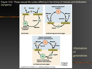
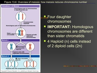
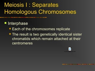
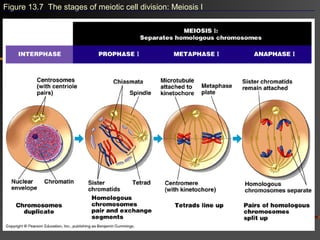
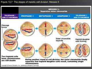
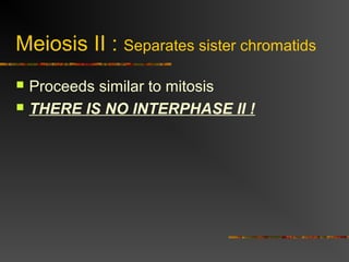
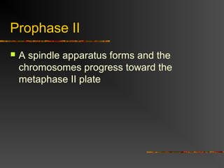
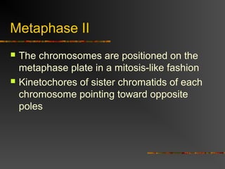
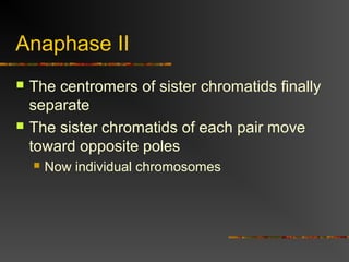

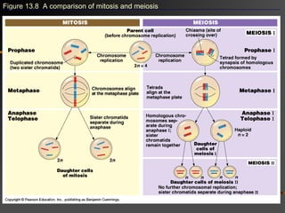
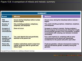
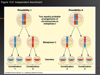
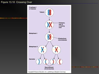
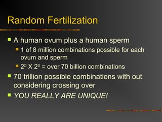


Meiosis involves two cell divisions that result in four haploid cells, each with half the number of chromosomes as the original cell. In meiosis I, homologous chromosomes pair up and separate, resulting in two cells each with one chromosome from each homologous pair. Sister chromatids remain attached. In meiosis II, sister chromatids separate, resulting in four haploid cells, each with an independent assortment of chromosomes. This process of meiosis and random fertilization leads to genetic variation between offspring.