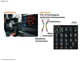
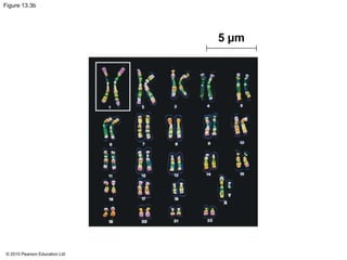
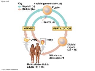
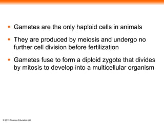
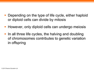
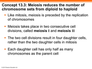
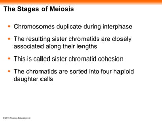
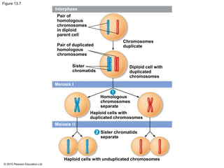
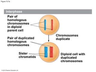
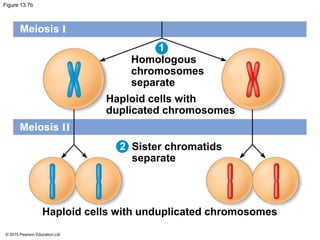
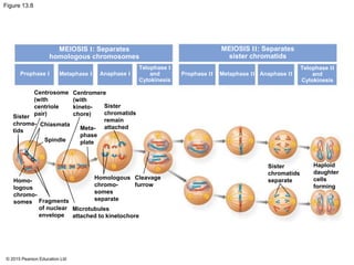
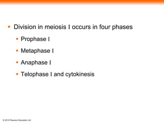
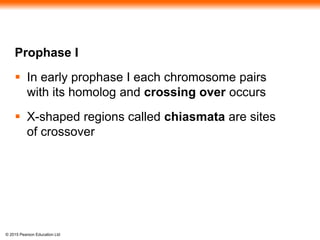
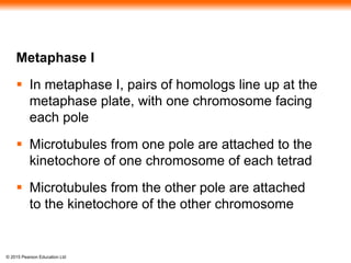
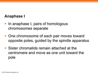
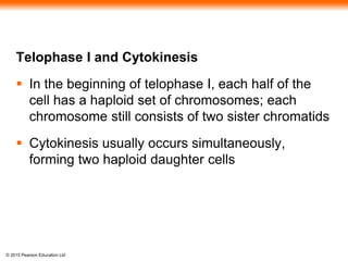
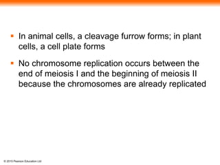
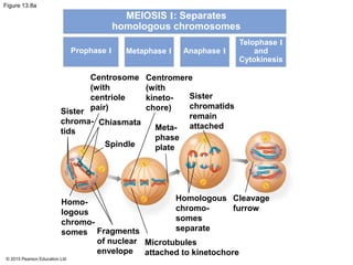
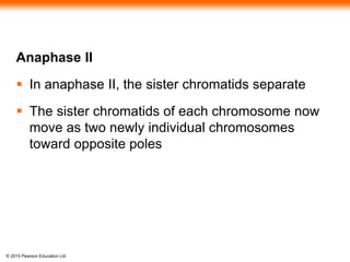
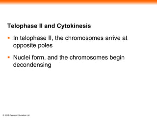
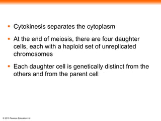
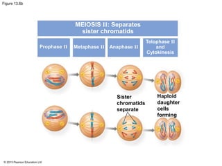
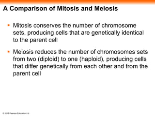
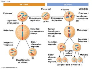
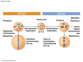
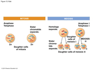
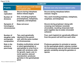
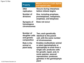
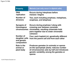
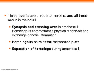
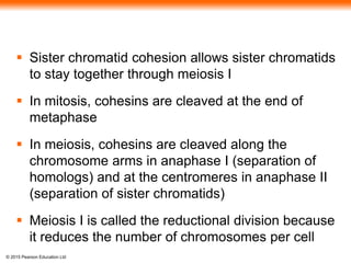
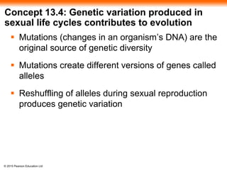
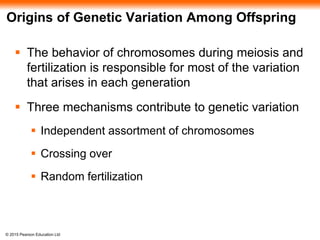
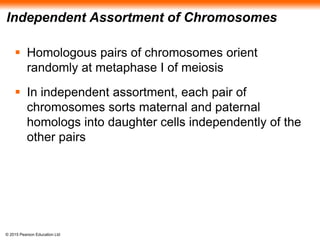
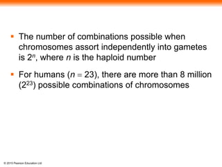
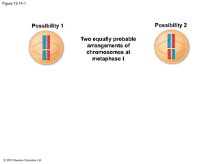
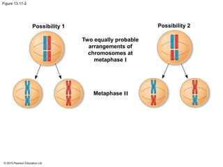
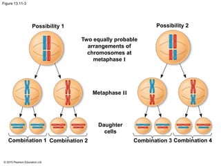
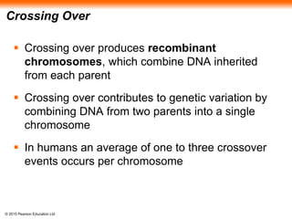
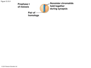
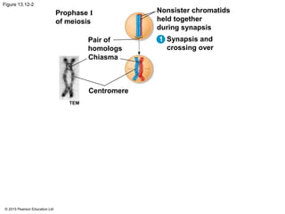
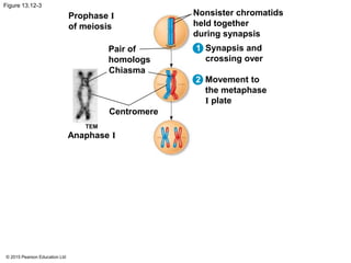
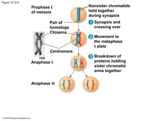
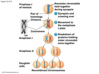
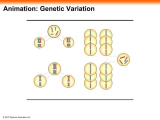
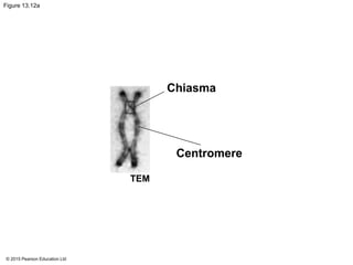
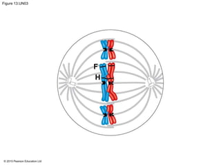
BIOLOGY
Campbell • Reece • Urry • Cain • Wasserman • Minorsky • Jackson
© 2015 Pearson Education Ltd
TENTH EDITION
Global Edition
Lecture Presentation by
Nicole Tunbridge and
Kathleen Fitzpatrick
13
Sexual Life Cycles
and Meiosis
© 2015 Pearson Education Ltd
Variations on a Theme
 Living organisms are distinguished by their ability
to reproduce their own kind
 Heredity is the transmission of traits from one
generation to the next
 Variation is demonstrated by the differences in
appearance that offspring show from parents and
siblings
 Genetics is the scientific study of heredity and
variation
© 2015 Pearson Education Ltd
Figure 13.1a
© 2015 Pearson Education Ltd
Concept 13.1: Offspring acquire genes from
parents by inheriting chromosomes
 In a literal sense, children do not inherit particular
physical traits from their parents
 It is genes that are actually inherited
© 2015 Pearson Education Ltd
Inheritance of Genes
 Genes are the units of heredity and are made up of
segments of DNA
 Genes are passed to the next generation via
reproductive cells called gametes (sperm and eggs)
 Most DNA is packaged into chromosomes
 Humans have 46 chromosomes in their somatic cells,
all cells of the body except gametes and their
precursors
 A gene’s specific position along a chromosome is
called the locus
© 2015 Pearson Education Ltd
Comparison of Asexual and Sexual
Reproduction
 In asexual reproduction, a single individual
passes all of its genes to its offspring without the
fusion of gametes
 A clone is a group of genetically identical
individuals from the same parent
 In sexual reproduction, two parents give rise to
offspring that have unique combinations of genes
inherited from the two parents
© 2015 Pearson Education Ltd
Figure 13.2
0.5 mm
Bud
Parent
(a) (b)Hydra Redwoods
© 2015 Pearson Education Ltd
Figure 13.2a
0.5 mm
Bud
Parent
(a) Hydra
© 2015 Pearson Education Ltd
Video: Hydra Budding
© 2015 Pearson Education Ltd
Figure 13.2b
(b) Redwoods
© 2015 Pearson Education Ltd
Concept 13.2: Fertilization and meiosis alternate
in sexual life cycles
 A life cycle is the generation-to-generation
sequence of stages in the reproductive history of
an organism
© 2015 Pearson Education Ltd
Sets of Chromosomes in Human Cells
 Human somatic cells have 23 pairs of
chromosomes
 A karyotype is an ordered display of the pairs of
chromosomes from a cell
 The two chromosomes in each pair are called
homologous chromosomes, or homologs
 Chromosomes in a homologous pair are the same
length and shape and carry genes controlling the
same inherited characters
© 2015 Pearson Education Ltd
Figure 13.3
TechniqueApplication
Pair of homologous
duplicated chromosomes
Centromere
Metaphase
chromosome
Sister
chromatids
5 µm
© 2015 Pearson Education Ltd
Figure 13.3a
Application
© 2015 Pearson Education Ltd
Figure 13.3b
5 µm
© 2015 Pearson Education Ltd
 The sex chromosomes, which determine the sex
of the individual, are called X and Y
 Human females have a homologous pair of X
chromosomes (XX)
 Human males have one X and one Y chromosome
 The remaining 22 pairs of chromosomes are
called autosomes
© 2015 Pearson Education Ltd
 Each pair of homologous chromosomes includes
one chromosome from each parent
 The 46 chromosomes in a human somatic cell are
two sets of 23: one from the mother and one from
the father
 A diploid cell (2n) has two sets of chromosomes
 For humans, the diploid number is 46 (2n = 46)
© 2015 Pearson Education Ltd
 In a cell in which DNA synthesis has occurred,
each chromosome is replicated
 Each replicated chromosome consists of two
identical sister chromatids
© 2015 Pearson Education Ltd
Figure 13.4
Key
Maternal set of
chromosomes (n = 3)
Paternal set of
chromosomes (n = 3)
2n = 6
Sister chromatids
of one duplicated
chromosome
Two nonsister
chromatids in
a homologous pair
Centromere
Pair of homologous
chromosomes
(one from each set)
© 2015 Pearson Education Ltd
 A gamete (sperm or egg) contains a single set of
chromosomes and is haploid (n)
 For humans, the haploid number is 23 (n = 23)
 Each set of 23 consists of 22 autosomes and a
single sex chromosome
 In an unfertilized egg (ovum), the sex
chromosome is X
 In a sperm cell, the sex chromosome may be
either X or Y
© 2015 Pearson Education Ltd
Behavior of Chromosome Sets in the Human
Life Cycle
 Fertilization is the union of gametes (the sperm
and the egg)
 The fertilized egg is called a zygote and has one
set of chromosomes from each parent
 The zygote produces somatic cells by mitosis and
develops into an adult
© 2015 Pearson Education Ltd
 At sexual maturity, the ovaries and testes produce
haploid gametes
 Gametes are the only types of human cells
produced by meiosis, rather than mitosis
 Meiosis results in one set of chromosomes in each
gamete
 Fertilization and meiosis alternate in sexual life
cycles to maintain chromosome number
© 2015 Pearson Education Ltd
Figure 13.5
Key
Haploid (n)
Diploid (2n)
Haploid gametes (n = 23)
Egg (n)
Sperm (n)
MEIOSIS FERTILIZATION
Testis
Diploid
zygote
(2n = 46)
Ovary
Mitosis and
development
Multicellular diploid
adults (2n = 46)
© 2015 Pearson Education Ltd
The Variety of Sexual Life Cycles
 The alternation of meiosis and fertilization is
common to all organisms that reproduce sexually
 The three main types of sexual life cycles differ in
the timing of meiosis and fertilization
© 2015 Pearson Education Ltd
 Gametes are the only haploid cells in animals
 They are produced by meiosis and undergo no
further cell division before fertilization
 Gametes fuse to form a diploid zygote that divides
by mitosis to develop into a multicellular organism
© 2015 Pearson Education Ltd
 Depending on the type of life cycle, either haploid
or diploid cells can divide by mitosis
 However, only diploid cells can undergo meiosis
 In all three life cycles, the halving and doubling
of chromosomes contributes to genetic variation
in offspring
© 2015 Pearson Education Ltd
Concept 13.3: Meiosis reduces the number of
chromosome sets from diploid to haploid
 Like mitosis, meiosis is preceded by the replication
of chromosomes
 Meiosis takes place in two consecutive cell
divisions, called meiosis I and meiosis II
 The two cell divisions result in four daughter cells,
rather than the two daughter cells in mitosis
 Each daughter cell has only half as many
chromosomes as the parent cell
© 2015 Pearson Education Ltd
The Stages of Meiosis
 Chromosomes duplicate during interphase
 The resulting sister chromatids are closely
associated along their lengths
 This is called sister chromatid cohesion
 The chromatids are sorted into four haploid
daughter cells
© 2015 Pearson Education Ltd
Figure 13.7
Interphase
Meiosis I
Meiosis II
Pair of
homologous
chromosomes
in diploid
parent cell
Pair of duplicated
homologous
chromosomes
Chromosomes
duplicate
Diploid cell with
duplicated
chromosomes
Sister
chromatids
Homologous
chromosomes
separate
Haploid cells with
duplicated chromosomes
Sister chromatids
separate
Haploid cells with unduplicated chromosomes
1
2
© 2015 Pearson Education Ltd
Figure 13.7a
Interphase
Pair of
homologous
chromosomes
in diploid
parent cell
Pair of duplicated
homologous
chromosomes
Chromosomes
duplicate
Diploid cell with
duplicated
chromosomes
Sister
chromatids
© 2015 Pearson Education Ltd
Figure 13.7b
Meiosis I
Meiosis II
Homologous
chromosomes
separate
Haploid cells with
duplicated chromosomes
Sister chromatids
separate
Haploid cells with unduplicated chromosomes
1
2
© 2015 Pearson Education Ltd
Figure 13.8
MEIOSIS I: Separates
homologous chromosomes
Prophase I Metaphase I Anaphase I
Telophase I
and
Cytokinesis
Prophase II Metaphase II Anaphase II
Telophase II
and
Cytokinesis
Centrosome
(with
centriole
pair)
Centromere
(with
kineto-
chore)
Sister
chromatids
remain
attachedMeta-
phase
plate
Sister
chroma-
tids
Chiasmata
Spindle
Homo-
logous
chromo-
somes Fragments
of nuclear
envelope
Microtubules
attached to kinetochore
Homologous
chromo-
somes
separate
Cleavage
furrow
Sister
chromatids
separate
Haploid
daughter
cells
forming
MEIOSIS II: Separates
sister chromatids
© 2015 Pearson Education Ltd
 Division in meiosis I occurs in four phases
 Prophase I
 Metaphase I
 Anaphase I
 Telophase I and cytokinesis
© 2015 Pearson Education Ltd
Prophase I
 In early prophase I each chromosome pairs
with its homolog and crossing over occurs
 X-shaped regions called chiasmata are sites
of crossover
© 2015 Pearson Education Ltd
Metaphase I
 In metaphase I, pairs of homologs line up at the
metaphase plate, with one chromosome facing
each pole
 Microtubules from one pole are attached to the
kinetochore of one chromosome of each tetrad
 Microtubules from the other pole are attached
to the kinetochore of the other chromosome
© 2015 Pearson Education Ltd
Anaphase I
 In anaphase I, pairs of homologous
chromosomes separate
 One chromosome of each pair moves toward
opposite poles, guided by the spindle apparatus
 Sister chromatids remain attached at the
centromere and move as one unit toward the
pole
© 2015 Pearson Education Ltd
Telophase I and Cytokinesis
 In the beginning of telophase I, each half of the
cell has a haploid set of chromosomes; each
chromosome still consists of two sister chromatids
 Cytokinesis usually occurs simultaneously,
forming two haploid daughter cells
© 2015 Pearson Education Ltd
 In animal cells, a cleavage furrow forms; in plant
cells, a cell plate forms
 No chromosome replication occurs between the
end of meiosis I and the beginning of meiosis II
because the chromosomes are already replicated
© 2015 Pearson Education Ltd
Figure 13.8a
MEIOSIS I: Separates
homologous chromosomes
Prophase I Metaphase I Anaphase I
Telophase I
and
Cytokinesis
Centrosome
(with
centriole
pair)Sister
chroma-
tids
Chiasmata
Spindle
Centromere
(with
kineto-
chore)
Sister
chromatids
remain
attachedMeta-
phase
plate
Homo-
logous
chromo-
somes Fragments
of nuclear
envelope
Microtubules
attached to kinetochore
Homologous
chromo-
somes
separate
Cleavage
furrow
© 2015 Pearson Education Ltd
BioFlix: Meiosis
© 2015 Pearson Education Ltd
Video: Meiosis I in Sperm Formation
© 2015 Pearson Education Ltd
 Division in meiosis II also occurs in four phases
 Prophase II
 Metaphase II
 Anaphase II
 Telophase II and cytokinesis
 Meiosis II is very similar to mitosis
© 2015 Pearson Education Ltd
Prophase II
 In prophase II, a spindle apparatus forms
 In late prophase II, chromosomes (each still
composed of two chromatids) move toward the
metaphase plate
© 2015 Pearson Education Ltd
Metaphase II
 In metaphase II, the sister chromatids are
arranged at the metaphase plate
 Because of crossing over in meiosis I, the two
sister chromatids of each chromosome are no
longer genetically identical
 The kinetochores of sister chromatids attach to
microtubules extending from opposite poles
© 2015 Pearson Education Ltd
Anaphase II
 In anaphase II, the sister chromatids separate
 The sister chromatids of each chromosome now
move as two newly individual chromosomes
toward opposite poles
© 2015 Pearson Education Ltd
Telophase II and Cytokinesis
 In telophase II, the chromosomes arrive at
opposite poles
 Nuclei form, and the chromosomes begin
decondensing
© 2015 Pearson Education Ltd
 Cytokinesis separates the cytoplasm
 At the end of meiosis, there are four daughter
cells, each with a haploid set of unreplicated
chromosomes
 Each daughter cell is genetically distinct from the
others and from the parent cell
© 2015 Pearson Education Ltd
Figure 13.8b
MEIOSIS II: Separates
sister chromatids
Prophase II Metaphase II Anaphase II
Telophase II
and
Cytokinesis
Sister
chromatids
separate
Haploid
daughter
cells
forming
© 2015 Pearson Education Ltd
A Comparison of Mitosis and Meiosis
 Mitosis conserves the number of chromosome
sets, producing cells that are genetically identical
to the parent cell
 Meiosis reduces the number of chromosomes sets
from two (diploid) to one (haploid), producing cells
that differ genetically from each other and from the
parent cell
© 2015 Pearson Education Ltd
Figure 13.10a
MITOSIS MEIOSIS
Prophase
Duplicated
chromosome
Metaphase
Anaphase
Telophase
2n 2n
Daughter cells
of mitosis
Sister
chromatids
separate.
Individual
chromosomes
line up.
Chromosome
duplication
Chromosome
duplication
2n = 6
Parent cell Chiasma MEIOSIS I
Prophase I
Homologous
chromosome
pair
Metaphase I
Anaphase I
Telophase I
MEIOSIS
II
Pairs of
homologous
chromosomes
line up.
Homologs
separate.
Sister
chroma-
tids
separate.
Daughter cells of meiosis II
Daughter
cells of
meiosis I
n n n n
© 2015 Pearson Education Ltd
Figure 13.10aa
MITOSIS MEIOSIS
MEIOSIS I
Prophase I
Metaphase I
Homo-
logous
chromosome
pair
ChiasmaParent cell
Prophase
Duplicated
chromosome
Metaphase
Chromosome
duplication
Chromosome
duplication
Individual
chromosomes
line up.
Pairs of
homologous
chromosomes
line up.
2n = 6
© 2015 Pearson Education Ltd
Figure 13.10ab
MITOSIS MEIOSIS
Anaphase IAnaphase
Telophase
2n 2n
n n n n
Daughter cells
of mitosis
Sister
chromatids
separate.
Homologs
separate.
Sister
chroma-
tids
separate.
Daughter cells of meiosis II
Daughter
cells of
meiosis I
MEIOSIS
II
Telophase I
© 2015 Pearson Education Ltd
Figure 13.10b
SUMMARY
Property
Mitosis (occurs in both diploid
and haploid cells)
Meiosis (can only occur in diploid cells)
DNA
replication
Number of
divisions
Synapsis of
homologous
chromosomes
Number of
daughter cells
and genetic
composition
Role in the
animal or
plant body
Occurs during interphase
before mitosis begins
One, including prophase,
prometaphase, metaphase,
anaphase, and telophase
Does not occur
Two, each genetically
identical to the parent
cell, with the same number
of chromosomes
Enables multicellular animal
or plant (gametophyte or
sporophyte) to arise from a
single cell; produces cells
for growth, repair, and, in
some species, asexual
reproduction; produces
gametes in the gametophyte
plant
Occurs during interphase before
meiosis I begins
Two, each including prophase, metaphase,
anaphase, and telophase
Occurs during prophase I along with
crossing over between nonsister
chromatids; resulting chiasmata hold
pairs together due to sister chromatid
cohesion
Four, each haploid (n); genetically different
from the parent cell and from each other
Produces gametes (in animals) or spores
(in the sporophyte plant); reduces number
of chromosomes sets by half and introduces
genetic variability among the gametes or
spores
© 2015 Pearson Education Ltd
Figure 13.10ba
Property
Mitosis (occurs in both diploid
and haploid cells)
DNA
replication
Number of
divisions
Synapsis of
homologous
chromosomes
Number of
daughter cells
and genetic
composition
Role in the
animal or
plant body
Occurs during interphase
before mitosis begins
One, including prophase,
prometaphase, metaphase,
anaphase, and telophase
Does not occur
Two, each genetically
identical to the parent
cell, with the same number
of chromosomes
Enables multicellular animal
or plant (gametophyte or
sporophyte) to arise from a
single cell; produces cells
for growth, repair, and, in
some species, asexual
reproduction; produces
gametes in the gametophyte
plant
© 2015 Pearson Education Ltd
Figure 13.10bb
Property
DNA
replication
Number of
divisions
Synapsis of
homologous
chromosomes
Number of
daughter cells
and genetic
composition
Role in the
animal or
plant body
Meiosis (can only occur in diploid cells)
Occurs during interphase before
meiosis I begins
Two, each including prophase, metaphase,
anaphase, and telophase
Occurs during prophase I along with
crossing over between nonsister
chromatids; resulting chiasmata hold
pairs together due to sister chromatid
cohesion
Four, each haploid (n); genetically different
from the parent cell and from each other
Produces gametes (in animals) or spores
(in the sporophyte plant); reduces number
of chromosomes sets by half and introduces
genetic variability among the gametes or
spores
© 2015 Pearson Education Ltd
 Three events are unique to meiosis, and all three
occur in meiosis l
 Synapsis and crossing over in prophase I:
Homologous chromosomes physically connect and
exchange genetic information
 Homologous pairs at the metaphase plate
 Separation of homologs during anaphase I
© 2015 Pearson Education Ltd
 Sister chromatid cohesion allows sister chromatids
to stay together through meiosis I
 In mitosis, cohesins are cleaved at the end of
metaphase
 In meiosis, cohesins are cleaved along the
chromosome arms in anaphase I (separation of
homologs) and at the centromeres in anaphase II
(separation of sister chromatids)
 Meiosis I is called the reductional division because
it reduces the number of chromosomes per cell
© 2015 Pearson Education Ltd
Concept 13.4: Genetic variation produced in
sexual life cycles contributes to evolution
 Mutations (changes in an organism’s DNA) are the
original source of genetic diversity
 Mutations create different versions of genes called
alleles
 Reshuffling of alleles during sexual reproduction
produces genetic variation
© 2015 Pearson Education Ltd
Origins of Genetic Variation Among Offspring
 The behavior of chromosomes during meiosis and
fertilization is responsible for most of the variation
that arises in each generation
 Three mechanisms contribute to genetic variation
 Independent assortment of chromosomes
 Crossing over
 Random fertilization
© 2015 Pearson Education Ltd
Independent Assortment of Chromosomes
 Homologous pairs of chromosomes orient
randomly at metaphase I of meiosis
 In independent assortment, each pair of
chromosomes sorts maternal and paternal
homologs into daughter cells independently of the
other pairs
© 2015 Pearson Education Ltd
 The number of combinations possible when
chromosomes assort independently into gametes
is 2n, where n is the haploid number
 For humans (n = 23), there are more than 8 million
(223) possible combinations of chromosomes
© 2015 Pearson Education Ltd
Figure 13.11-1
Possibility 1 Possibility 2
Two equally probable
arrangements of
chromosomes at
metaphase I
© 2015 Pearson Education Ltd
Figure 13.11-2
Possibility 1 Possibility 2
Two equally probable
arrangements of
chromosomes at
metaphase I
Metaphase II
© 2015 Pearson Education Ltd
Figure 13.11-3
Possibility 1 Possibility 2
Two equally probable
arrangements of
chromosomes at
metaphase I
Metaphase II
Daughter
cells
Combination 1 Combination 2 Combination 3 Combination 4
© 2015 Pearson Education Ltd
Crossing Over
 Crossing over produces recombinant
chromosomes, which combine DNA inherited
from each parent
 Crossing over contributes to genetic variation by
combining DNA from two parents into a single
chromosome
 In humans an average of one to three crossover
events occurs per chromosome
© 2015 Pearson Education Ltd
Figure 13.12-1
Prophase I
of meiosis
Pair of
homologs
Nonsister chromatids
held together
during synapsis
© 2015 Pearson Education Ltd
Figure 13.12-2
Prophase I
of meiosis
Pair of
homologs
Chiasma
Centromere
TEM
Nonsister chromatids
held together
during synapsis
Synapsis and
crossing over
1
© 2015 Pearson Education Ltd
Figure 13.12-3
Prophase I
of meiosis
Pair of
homologs
Chiasma
Centromere
TEM
Anaphase I
Nonsister chromatids
held together
during synapsis
Synapsis and
crossing over
Movement to
the metaphase
I plate
1
2
© 2015 Pearson Education Ltd
Figure 13.12-4
Prophase I
of meiosis
Pair of
homologs
Chiasma
Centromere
TEM
Anaphase I
Anaphase II
Nonsister chromatids
held together
during synapsis
Synapsis and
crossing over
Movement to
the metaphase
I plate
Breakdown of
proteins holding
sister chromatid
arms together
1
2
3
© 2015 Pearson Education Ltd
Figure 13.12-5
Prophase I
of meiosis
Pair of
homologs
Chiasma
Centromere
TEM
Anaphase I
Anaphase II
Daughter
cells
Nonsister chromatids
held together
during synapsis
Synapsis and
crossing over
Movement to
the metaphase
I plate
Breakdown of
proteins holding
sister chromatid
arms together
Recombinant chromosomes
1
2
3
© 2015 Pearson Education Ltd
Animation: Genetic Variation
© 2015 Pearson Education Ltd
Figure 13.12a
Chiasma
Centromere
TEM
© 2015 Pearson Education Ltd
Random Fertilization
 Random fertilization adds to genetic variation
because any sperm can fuse with any ovum
(unfertilized egg)
 The fusion of two gametes (each with 8.4 million
possible chromosome combinations from
independent assortment) produces a zygote with
any of about 70 trillion diploid combinations
© 2015 Pearson Education Ltd
 Crossing over adds even more variation
 Each zygote has a unique genetic identity
© 2015 Pearson Education Ltd
The Evolutionary Significance of Genetic
Variation Within Populations
 Natural selection results in the accumulation of
genetic variations favored by the environment
 Sexual reproduction contributes to the genetic
variation in a population, which originates from
mutations
 Animals that always reproduce asexually are quite
rare
 Organisms like the bdelloid rotifer increase their
genetic diversity through horizontal gene transfer
© 2015 Pearson Education Ltd
Figure 13.UN03
F
H

13 ge lecture presentation

  • 1.
    BIOLOGY Campbell • Reece• Urry • Cain • Wasserman • Minorsky • Jackson © 2015 Pearson Education Ltd TENTH EDITION Global Edition Lecture Presentation by Nicole Tunbridge and Kathleen Fitzpatrick 13 Sexual Life Cycles and Meiosis
  • 2.
    © 2015 PearsonEducation Ltd Variations on a Theme  Living organisms are distinguished by their ability to reproduce their own kind  Heredity is the transmission of traits from one generation to the next  Variation is demonstrated by the differences in appearance that offspring show from parents and siblings  Genetics is the scientific study of heredity and variation
  • 3.
    © 2015 PearsonEducation Ltd Figure 13.1a
  • 4.
    © 2015 PearsonEducation Ltd Concept 13.1: Offspring acquire genes from parents by inheriting chromosomes  In a literal sense, children do not inherit particular physical traits from their parents  It is genes that are actually inherited
  • 5.
    © 2015 PearsonEducation Ltd Inheritance of Genes  Genes are the units of heredity and are made up of segments of DNA  Genes are passed to the next generation via reproductive cells called gametes (sperm and eggs)  Most DNA is packaged into chromosomes  Humans have 46 chromosomes in their somatic cells, all cells of the body except gametes and their precursors  A gene’s specific position along a chromosome is called the locus
  • 6.
    © 2015 PearsonEducation Ltd Comparison of Asexual and Sexual Reproduction  In asexual reproduction, a single individual passes all of its genes to its offspring without the fusion of gametes  A clone is a group of genetically identical individuals from the same parent  In sexual reproduction, two parents give rise to offspring that have unique combinations of genes inherited from the two parents
  • 7.
    © 2015 PearsonEducation Ltd Figure 13.2 0.5 mm Bud Parent (a) (b)Hydra Redwoods
  • 8.
    © 2015 PearsonEducation Ltd Figure 13.2a 0.5 mm Bud Parent (a) Hydra
  • 9.
    © 2015 PearsonEducation Ltd Video: Hydra Budding
  • 10.
    © 2015 PearsonEducation Ltd Figure 13.2b (b) Redwoods
  • 11.
    © 2015 PearsonEducation Ltd Concept 13.2: Fertilization and meiosis alternate in sexual life cycles  A life cycle is the generation-to-generation sequence of stages in the reproductive history of an organism
  • 12.
    © 2015 PearsonEducation Ltd Sets of Chromosomes in Human Cells  Human somatic cells have 23 pairs of chromosomes  A karyotype is an ordered display of the pairs of chromosomes from a cell  The two chromosomes in each pair are called homologous chromosomes, or homologs  Chromosomes in a homologous pair are the same length and shape and carry genes controlling the same inherited characters
  • 13.
    © 2015 PearsonEducation Ltd Figure 13.3 TechniqueApplication Pair of homologous duplicated chromosomes Centromere Metaphase chromosome Sister chromatids 5 µm
  • 14.
    © 2015 PearsonEducation Ltd Figure 13.3a Application
  • 15.
    © 2015 PearsonEducation Ltd Figure 13.3b 5 µm
  • 16.
    © 2015 PearsonEducation Ltd  The sex chromosomes, which determine the sex of the individual, are called X and Y  Human females have a homologous pair of X chromosomes (XX)  Human males have one X and one Y chromosome  The remaining 22 pairs of chromosomes are called autosomes
  • 17.
    © 2015 PearsonEducation Ltd  Each pair of homologous chromosomes includes one chromosome from each parent  The 46 chromosomes in a human somatic cell are two sets of 23: one from the mother and one from the father  A diploid cell (2n) has two sets of chromosomes  For humans, the diploid number is 46 (2n = 46)
  • 18.
    © 2015 PearsonEducation Ltd  In a cell in which DNA synthesis has occurred, each chromosome is replicated  Each replicated chromosome consists of two identical sister chromatids
  • 19.
    © 2015 PearsonEducation Ltd Figure 13.4 Key Maternal set of chromosomes (n = 3) Paternal set of chromosomes (n = 3) 2n = 6 Sister chromatids of one duplicated chromosome Two nonsister chromatids in a homologous pair Centromere Pair of homologous chromosomes (one from each set)
  • 20.
    © 2015 PearsonEducation Ltd  A gamete (sperm or egg) contains a single set of chromosomes and is haploid (n)  For humans, the haploid number is 23 (n = 23)  Each set of 23 consists of 22 autosomes and a single sex chromosome  In an unfertilized egg (ovum), the sex chromosome is X  In a sperm cell, the sex chromosome may be either X or Y
  • 21.
    © 2015 PearsonEducation Ltd Behavior of Chromosome Sets in the Human Life Cycle  Fertilization is the union of gametes (the sperm and the egg)  The fertilized egg is called a zygote and has one set of chromosomes from each parent  The zygote produces somatic cells by mitosis and develops into an adult
  • 22.
    © 2015 PearsonEducation Ltd  At sexual maturity, the ovaries and testes produce haploid gametes  Gametes are the only types of human cells produced by meiosis, rather than mitosis  Meiosis results in one set of chromosomes in each gamete  Fertilization and meiosis alternate in sexual life cycles to maintain chromosome number
  • 23.
    © 2015 PearsonEducation Ltd Figure 13.5 Key Haploid (n) Diploid (2n) Haploid gametes (n = 23) Egg (n) Sperm (n) MEIOSIS FERTILIZATION Testis Diploid zygote (2n = 46) Ovary Mitosis and development Multicellular diploid adults (2n = 46)
  • 24.
    © 2015 PearsonEducation Ltd The Variety of Sexual Life Cycles  The alternation of meiosis and fertilization is common to all organisms that reproduce sexually  The three main types of sexual life cycles differ in the timing of meiosis and fertilization
  • 25.
    © 2015 PearsonEducation Ltd  Gametes are the only haploid cells in animals  They are produced by meiosis and undergo no further cell division before fertilization  Gametes fuse to form a diploid zygote that divides by mitosis to develop into a multicellular organism
  • 26.
    © 2015 PearsonEducation Ltd  Depending on the type of life cycle, either haploid or diploid cells can divide by mitosis  However, only diploid cells can undergo meiosis  In all three life cycles, the halving and doubling of chromosomes contributes to genetic variation in offspring
  • 27.
    © 2015 PearsonEducation Ltd Concept 13.3: Meiosis reduces the number of chromosome sets from diploid to haploid  Like mitosis, meiosis is preceded by the replication of chromosomes  Meiosis takes place in two consecutive cell divisions, called meiosis I and meiosis II  The two cell divisions result in four daughter cells, rather than the two daughter cells in mitosis  Each daughter cell has only half as many chromosomes as the parent cell
  • 28.
    © 2015 PearsonEducation Ltd The Stages of Meiosis  Chromosomes duplicate during interphase  The resulting sister chromatids are closely associated along their lengths  This is called sister chromatid cohesion  The chromatids are sorted into four haploid daughter cells
  • 29.
    © 2015 PearsonEducation Ltd Figure 13.7 Interphase Meiosis I Meiosis II Pair of homologous chromosomes in diploid parent cell Pair of duplicated homologous chromosomes Chromosomes duplicate Diploid cell with duplicated chromosomes Sister chromatids Homologous chromosomes separate Haploid cells with duplicated chromosomes Sister chromatids separate Haploid cells with unduplicated chromosomes 1 2
  • 30.
    © 2015 PearsonEducation Ltd Figure 13.7a Interphase Pair of homologous chromosomes in diploid parent cell Pair of duplicated homologous chromosomes Chromosomes duplicate Diploid cell with duplicated chromosomes Sister chromatids
  • 31.
    © 2015 PearsonEducation Ltd Figure 13.7b Meiosis I Meiosis II Homologous chromosomes separate Haploid cells with duplicated chromosomes Sister chromatids separate Haploid cells with unduplicated chromosomes 1 2
  • 32.
    © 2015 PearsonEducation Ltd Figure 13.8 MEIOSIS I: Separates homologous chromosomes Prophase I Metaphase I Anaphase I Telophase I and Cytokinesis Prophase II Metaphase II Anaphase II Telophase II and Cytokinesis Centrosome (with centriole pair) Centromere (with kineto- chore) Sister chromatids remain attachedMeta- phase plate Sister chroma- tids Chiasmata Spindle Homo- logous chromo- somes Fragments of nuclear envelope Microtubules attached to kinetochore Homologous chromo- somes separate Cleavage furrow Sister chromatids separate Haploid daughter cells forming MEIOSIS II: Separates sister chromatids
  • 33.
    © 2015 PearsonEducation Ltd  Division in meiosis I occurs in four phases  Prophase I  Metaphase I  Anaphase I  Telophase I and cytokinesis
  • 34.
    © 2015 PearsonEducation Ltd Prophase I  In early prophase I each chromosome pairs with its homolog and crossing over occurs  X-shaped regions called chiasmata are sites of crossover
  • 35.
    © 2015 PearsonEducation Ltd Metaphase I  In metaphase I, pairs of homologs line up at the metaphase plate, with one chromosome facing each pole  Microtubules from one pole are attached to the kinetochore of one chromosome of each tetrad  Microtubules from the other pole are attached to the kinetochore of the other chromosome
  • 36.
    © 2015 PearsonEducation Ltd Anaphase I  In anaphase I, pairs of homologous chromosomes separate  One chromosome of each pair moves toward opposite poles, guided by the spindle apparatus  Sister chromatids remain attached at the centromere and move as one unit toward the pole
  • 37.
    © 2015 PearsonEducation Ltd Telophase I and Cytokinesis  In the beginning of telophase I, each half of the cell has a haploid set of chromosomes; each chromosome still consists of two sister chromatids  Cytokinesis usually occurs simultaneously, forming two haploid daughter cells
  • 38.
    © 2015 PearsonEducation Ltd  In animal cells, a cleavage furrow forms; in plant cells, a cell plate forms  No chromosome replication occurs between the end of meiosis I and the beginning of meiosis II because the chromosomes are already replicated
  • 39.
    © 2015 PearsonEducation Ltd Figure 13.8a MEIOSIS I: Separates homologous chromosomes Prophase I Metaphase I Anaphase I Telophase I and Cytokinesis Centrosome (with centriole pair)Sister chroma- tids Chiasmata Spindle Centromere (with kineto- chore) Sister chromatids remain attachedMeta- phase plate Homo- logous chromo- somes Fragments of nuclear envelope Microtubules attached to kinetochore Homologous chromo- somes separate Cleavage furrow
  • 40.
    © 2015 PearsonEducation Ltd BioFlix: Meiosis
  • 41.
    © 2015 PearsonEducation Ltd Video: Meiosis I in Sperm Formation
  • 42.
    © 2015 PearsonEducation Ltd  Division in meiosis II also occurs in four phases  Prophase II  Metaphase II  Anaphase II  Telophase II and cytokinesis  Meiosis II is very similar to mitosis
  • 43.
    © 2015 PearsonEducation Ltd Prophase II  In prophase II, a spindle apparatus forms  In late prophase II, chromosomes (each still composed of two chromatids) move toward the metaphase plate
  • 44.
    © 2015 PearsonEducation Ltd Metaphase II  In metaphase II, the sister chromatids are arranged at the metaphase plate  Because of crossing over in meiosis I, the two sister chromatids of each chromosome are no longer genetically identical  The kinetochores of sister chromatids attach to microtubules extending from opposite poles
  • 45.
    © 2015 PearsonEducation Ltd Anaphase II  In anaphase II, the sister chromatids separate  The sister chromatids of each chromosome now move as two newly individual chromosomes toward opposite poles
  • 46.
    © 2015 PearsonEducation Ltd Telophase II and Cytokinesis  In telophase II, the chromosomes arrive at opposite poles  Nuclei form, and the chromosomes begin decondensing
  • 47.
    © 2015 PearsonEducation Ltd  Cytokinesis separates the cytoplasm  At the end of meiosis, there are four daughter cells, each with a haploid set of unreplicated chromosomes  Each daughter cell is genetically distinct from the others and from the parent cell
  • 48.
    © 2015 PearsonEducation Ltd Figure 13.8b MEIOSIS II: Separates sister chromatids Prophase II Metaphase II Anaphase II Telophase II and Cytokinesis Sister chromatids separate Haploid daughter cells forming
  • 49.
    © 2015 PearsonEducation Ltd A Comparison of Mitosis and Meiosis  Mitosis conserves the number of chromosome sets, producing cells that are genetically identical to the parent cell  Meiosis reduces the number of chromosomes sets from two (diploid) to one (haploid), producing cells that differ genetically from each other and from the parent cell
  • 50.
    © 2015 PearsonEducation Ltd Figure 13.10a MITOSIS MEIOSIS Prophase Duplicated chromosome Metaphase Anaphase Telophase 2n 2n Daughter cells of mitosis Sister chromatids separate. Individual chromosomes line up. Chromosome duplication Chromosome duplication 2n = 6 Parent cell Chiasma MEIOSIS I Prophase I Homologous chromosome pair Metaphase I Anaphase I Telophase I MEIOSIS II Pairs of homologous chromosomes line up. Homologs separate. Sister chroma- tids separate. Daughter cells of meiosis II Daughter cells of meiosis I n n n n
  • 51.
    © 2015 PearsonEducation Ltd Figure 13.10aa MITOSIS MEIOSIS MEIOSIS I Prophase I Metaphase I Homo- logous chromosome pair ChiasmaParent cell Prophase Duplicated chromosome Metaphase Chromosome duplication Chromosome duplication Individual chromosomes line up. Pairs of homologous chromosomes line up. 2n = 6
  • 52.
    © 2015 PearsonEducation Ltd Figure 13.10ab MITOSIS MEIOSIS Anaphase IAnaphase Telophase 2n 2n n n n n Daughter cells of mitosis Sister chromatids separate. Homologs separate. Sister chroma- tids separate. Daughter cells of meiosis II Daughter cells of meiosis I MEIOSIS II Telophase I
  • 53.
    © 2015 PearsonEducation Ltd Figure 13.10b SUMMARY Property Mitosis (occurs in both diploid and haploid cells) Meiosis (can only occur in diploid cells) DNA replication Number of divisions Synapsis of homologous chromosomes Number of daughter cells and genetic composition Role in the animal or plant body Occurs during interphase before mitosis begins One, including prophase, prometaphase, metaphase, anaphase, and telophase Does not occur Two, each genetically identical to the parent cell, with the same number of chromosomes Enables multicellular animal or plant (gametophyte or sporophyte) to arise from a single cell; produces cells for growth, repair, and, in some species, asexual reproduction; produces gametes in the gametophyte plant Occurs during interphase before meiosis I begins Two, each including prophase, metaphase, anaphase, and telophase Occurs during prophase I along with crossing over between nonsister chromatids; resulting chiasmata hold pairs together due to sister chromatid cohesion Four, each haploid (n); genetically different from the parent cell and from each other Produces gametes (in animals) or spores (in the sporophyte plant); reduces number of chromosomes sets by half and introduces genetic variability among the gametes or spores
  • 54.
    © 2015 PearsonEducation Ltd Figure 13.10ba Property Mitosis (occurs in both diploid and haploid cells) DNA replication Number of divisions Synapsis of homologous chromosomes Number of daughter cells and genetic composition Role in the animal or plant body Occurs during interphase before mitosis begins One, including prophase, prometaphase, metaphase, anaphase, and telophase Does not occur Two, each genetically identical to the parent cell, with the same number of chromosomes Enables multicellular animal or plant (gametophyte or sporophyte) to arise from a single cell; produces cells for growth, repair, and, in some species, asexual reproduction; produces gametes in the gametophyte plant
  • 55.
    © 2015 PearsonEducation Ltd Figure 13.10bb Property DNA replication Number of divisions Synapsis of homologous chromosomes Number of daughter cells and genetic composition Role in the animal or plant body Meiosis (can only occur in diploid cells) Occurs during interphase before meiosis I begins Two, each including prophase, metaphase, anaphase, and telophase Occurs during prophase I along with crossing over between nonsister chromatids; resulting chiasmata hold pairs together due to sister chromatid cohesion Four, each haploid (n); genetically different from the parent cell and from each other Produces gametes (in animals) or spores (in the sporophyte plant); reduces number of chromosomes sets by half and introduces genetic variability among the gametes or spores
  • 56.
    © 2015 PearsonEducation Ltd  Three events are unique to meiosis, and all three occur in meiosis l  Synapsis and crossing over in prophase I: Homologous chromosomes physically connect and exchange genetic information  Homologous pairs at the metaphase plate  Separation of homologs during anaphase I
  • 57.
    © 2015 PearsonEducation Ltd  Sister chromatid cohesion allows sister chromatids to stay together through meiosis I  In mitosis, cohesins are cleaved at the end of metaphase  In meiosis, cohesins are cleaved along the chromosome arms in anaphase I (separation of homologs) and at the centromeres in anaphase II (separation of sister chromatids)  Meiosis I is called the reductional division because it reduces the number of chromosomes per cell
  • 58.
    © 2015 PearsonEducation Ltd Concept 13.4: Genetic variation produced in sexual life cycles contributes to evolution  Mutations (changes in an organism’s DNA) are the original source of genetic diversity  Mutations create different versions of genes called alleles  Reshuffling of alleles during sexual reproduction produces genetic variation
  • 59.
    © 2015 PearsonEducation Ltd Origins of Genetic Variation Among Offspring  The behavior of chromosomes during meiosis and fertilization is responsible for most of the variation that arises in each generation  Three mechanisms contribute to genetic variation  Independent assortment of chromosomes  Crossing over  Random fertilization
  • 60.
    © 2015 PearsonEducation Ltd Independent Assortment of Chromosomes  Homologous pairs of chromosomes orient randomly at metaphase I of meiosis  In independent assortment, each pair of chromosomes sorts maternal and paternal homologs into daughter cells independently of the other pairs
  • 61.
    © 2015 PearsonEducation Ltd  The number of combinations possible when chromosomes assort independently into gametes is 2n, where n is the haploid number  For humans (n = 23), there are more than 8 million (223) possible combinations of chromosomes
  • 62.
    © 2015 PearsonEducation Ltd Figure 13.11-1 Possibility 1 Possibility 2 Two equally probable arrangements of chromosomes at metaphase I
  • 63.
    © 2015 PearsonEducation Ltd Figure 13.11-2 Possibility 1 Possibility 2 Two equally probable arrangements of chromosomes at metaphase I Metaphase II
  • 64.
    © 2015 PearsonEducation Ltd Figure 13.11-3 Possibility 1 Possibility 2 Two equally probable arrangements of chromosomes at metaphase I Metaphase II Daughter cells Combination 1 Combination 2 Combination 3 Combination 4
  • 65.
    © 2015 PearsonEducation Ltd Crossing Over  Crossing over produces recombinant chromosomes, which combine DNA inherited from each parent  Crossing over contributes to genetic variation by combining DNA from two parents into a single chromosome  In humans an average of one to three crossover events occurs per chromosome
  • 66.
    © 2015 PearsonEducation Ltd Figure 13.12-1 Prophase I of meiosis Pair of homologs Nonsister chromatids held together during synapsis
  • 67.
    © 2015 PearsonEducation Ltd Figure 13.12-2 Prophase I of meiosis Pair of homologs Chiasma Centromere TEM Nonsister chromatids held together during synapsis Synapsis and crossing over 1
  • 68.
    © 2015 PearsonEducation Ltd Figure 13.12-3 Prophase I of meiosis Pair of homologs Chiasma Centromere TEM Anaphase I Nonsister chromatids held together during synapsis Synapsis and crossing over Movement to the metaphase I plate 1 2
  • 69.
    © 2015 PearsonEducation Ltd Figure 13.12-4 Prophase I of meiosis Pair of homologs Chiasma Centromere TEM Anaphase I Anaphase II Nonsister chromatids held together during synapsis Synapsis and crossing over Movement to the metaphase I plate Breakdown of proteins holding sister chromatid arms together 1 2 3
  • 70.
    © 2015 PearsonEducation Ltd Figure 13.12-5 Prophase I of meiosis Pair of homologs Chiasma Centromere TEM Anaphase I Anaphase II Daughter cells Nonsister chromatids held together during synapsis Synapsis and crossing over Movement to the metaphase I plate Breakdown of proteins holding sister chromatid arms together Recombinant chromosomes 1 2 3
  • 71.
    © 2015 PearsonEducation Ltd Animation: Genetic Variation
  • 72.
    © 2015 PearsonEducation Ltd Figure 13.12a Chiasma Centromere TEM
  • 73.
    © 2015 PearsonEducation Ltd Random Fertilization  Random fertilization adds to genetic variation because any sperm can fuse with any ovum (unfertilized egg)  The fusion of two gametes (each with 8.4 million possible chromosome combinations from independent assortment) produces a zygote with any of about 70 trillion diploid combinations
  • 74.
    © 2015 PearsonEducation Ltd  Crossing over adds even more variation  Each zygote has a unique genetic identity
  • 75.
    © 2015 PearsonEducation Ltd The Evolutionary Significance of Genetic Variation Within Populations  Natural selection results in the accumulation of genetic variations favored by the environment  Sexual reproduction contributes to the genetic variation in a population, which originates from mutations  Animals that always reproduce asexually are quite rare  Organisms like the bdelloid rotifer increase their genetic diversity through horizontal gene transfer
  • 76.
    © 2015 PearsonEducation Ltd Figure 13.UN03 F H