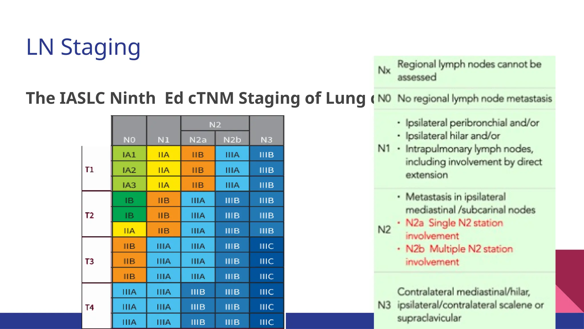
Current Evidences on Mediastinal Lymph
Node Dissection for Lung Cancer
Ruhama Yoseph (CTF)
Moderator- Dr. Seyoum
May, 2025
introduction
TNM Staging and LN Mapping
Approaches of Invasive Staging
Options of mediastinal lymph node dissection
The evidences
Introduction
● The technique of mediastinal lymph node dissection (MLND) was first
described in detail in 1951, differentiating simple pneumonectomy from
radical pneumonectomy
● MLND differed in practice from mere inspection of unopened
mediastinum to radical lymphadenectomy
● MLND as a “standard” was followed by only 57.8%
● The effect of MLND was debated
● Later technique of mediastinal staging preceding surgical resection
introduced- Mediastinoscopy became a requirement for selected group
since 1993
Introduction
Naruke
LN map
introduced
1976
Martini
Modern MLND
technique
introduced
1995
1933
Graham
First successful
pneumonectomy
Allison
Radical
Pneumonectomy
1949
Cahan
Intrathoracic LN
dissection
1951
introduction
TNM Staging and LN
Mapping
Approaches of Invasive Staging
Options of mediastinal lymph node dissection
The evidences
LN Staging
The IASLC Ninth Ed cTNM Staging of Lung ca
LN Staging
History of the LN map…
The Naruke lymph node map for the staging of lung
cancers as recommended by the Japan Lung Cancer
Society, 1978
Interpretation of lymph node levels varied because of
the lack of precise anatomic definitions.
Confusion of stations 4 Vs 10, stations 1 through 4,
station 5 Vs 6, which were later modified
The Naruke LN map
History of the LN map…
Later modifications of station
10
The Mountain-Dresler modification of LN map (1997),
originally proposed by the American Thoracic Society, was
based on structures readily identified by CT scans
History of the LN map…
The IASLC ( International Association for the study of Lung
Cancer) LN map (2007) shows clear anatomic landmarks and
addresses controversial areas that had inter-observer variation for
labeling.
Controversial areas were:
● Right paratracheal and hilar lymph nodes
● Left inferior paratracheal and aortopulmonary lymph nodes
● The subcarinal and right hilar lymph nodes.
IASLC Nodal Chart
IASLC Nodal Chart
IASLC Nodal Chart
IASLC Nodal Chart
Intraoperative view of right
superior mediastinal and hilar
nodes
Intraoperative view left superior
mediastinal, aortopulmonary and
hilar nodes
IASLC Nodal Chart
IASLC Nodal Chart
Intraoperative view of right
subcarinal area
Lower limit is lower border of
bronchus intermedius
IASLC Nodal Chart
IASLC Nodal Chart
IASLC Nodal Chart
IASLC Nodal Chart
IASLC Nodal Chart
introduction
TNM Staging and LN Mapping
Approaches of Invasive
Staging
Options of mediastinal lymph node dissection
The evidences
Approach for C-Staging of Non-metastatic Lung Cancer
Positive lymph
nodes defined us
greater than 1 cm
in short axis
Approach for C-Staging of Non-metastatic Lung
Cancer
Who needs invasive LN staging?
● Patients who fulfill criteria based on CT-PET finding, OR
● Patients who are predicted to have N2/N3 disease based on prediction
models
Invasive staging preferentially beings with EBUS, EUS, EBUS/EUS
Invasive mediastinal staging
Lymph node sampling establishes nodal involvement for patients without evidence
of extrathoracic disease.
Based on CT-PET findings, it is recommended in the following scenarios:
● Patients without evidence of distant metastasis with evidence of mediastinal
and/or hilar node enlargement ( 1 cm),
≥ regardless of FDG uptake on PET
● Patients without distant metastases with normal-sized mediastinal and/or hilar
lymph nodes (<1 cm) on CT with FDG avidity on PET
● Patients with central tumor
● Patients with tumor size greater than 3 cm
Adenopathy in Lung cancer (HAL) prediction model:
A multivariable logistic regression model to predict the likelihood of N2/N3
disease based on age, the location and histology of the primary cancer, and the
N stage by CT and PET imaging
ESTS recommends that surgical candidates with a 10% or less predicted
chance of N2 disease may proceed directly to surgery without invasive
mediastinal staging
Patients with >10% risk of N2 disease should undergo invasive mediastinal
staging before surgery
Prediction models
EBUS and/or EUS
EBUS-TBNA
Needle deployed along the working channel of the flexible scope allows sampling
of paratracheal (2R/L, 4R/L), subcarinal (7), hilar (10R/L), interlobar (11R/L), and
interlobar (12R/L).
EUS-FNA
It allows for the sampling of some mediastinal lymph nodes accessible by EBUS
(2 L, 4 L, 7, 3p) as well as some which are not accessible via EBUS (stations 8, 9)
Mediastinoscopy
To be considered when EBUS is inconclusive and/or clinical suspicion remains
high despite negative EBUS/EUS
Allows sampling of stations 2R/L, 4R/L, 10 R/L and 7 via direct visualization or
leveraging of VAM.
Left Anterior Mediastinoscopy (Chamberlain procedure) is an option to
access aortopulmonary window & para-aortic LNs (5&6).
Options of mediastinal
lymph node dissection
introduction
TNM Staging and LN Mapping
Approaches of Invasive Staging
The evidences
Sampling- Process of harvesting grossly abnormal suspicious LN (keller 2000)
Later definition was extended to include normal looking LN.
Systematic Mediastinal LN Sampling (MLNS)- Routine biopsy of one or more
representative LN at predetermined stations/levels specified by author (Keller 2000)
According to Z0300 trial sampling of the following stations fulfilled criteria of MLNS
● stations 2R, 4R,7 and 10R on the right side
● stations 5, 6, 7 and 10L on the left side
Complete Mediastinal LN Dissection (CMLND)- routine removal of all LN containing
tissue within defined anatomic landmarks/levels indicated by investigators (Keller 2000)
Terminology of MLND
Sampling
A staging procedure for patients with proven NSCLC that were candidates for
invasive staging where they undergo rigorous sampling of normal looking
mediastinal and hilar LN (e.g The Z0300 trail)
In previous studies, LN that were grossly abnormal were taken ( e.g RCT by Wu et al.)
The methods:
● EBUS-TBNA/EUS-FNA
● Mediastinoscopy
● VATS/Open thoracotomy (frozen section examination at the time of surgery)
Systematic sampling (MLNS)
Systematic invasive sampling of mediastinal LN including all specified node
stations required based on location of the tumour (using EBUS/EUS,
Mediastinoscopy, VATS or thoracotomy)
Knowledge of distribution of nodal involvement in patients with N2 disease
can be helpful
E.g. Stations 5&6 for LUL tumours, 8&9 for lower lobe tumours
Complete MLND
By principle, all the mediastinal fatty tissue containing lymphatics should be
dissected and removed systematically.
For pathological staging (pTNM), a complete resection requires a minimum of
six stations including subcarinal station, two mediastinal nodes, and three
hilar or intrapulmonary nodes.
After removal, the different nodal stations will be put in different vials with
separate labeling.
Complete MLND…
For tumors on the right, all fatty lymphatic tissue harbouring:
● Paratracheal LNs (stations 2R & 4R) removed
● Prevascular (3a) and retrotracheal (3p) LN removed
● Subcarinal (station 7) removed
● Paraesophageal and inferior pulmonary ligament LN (station 8R & 9R) removed
● N1 zone LN (stations 10R to 14R) removed along or separate from specimen
For tumors on the left, all fatty lymphatic tissue harbouring:
● Aortopulmonary window (station 5) & Para Aortic (station 6) LN removed
● Subcarinal (station 7) removed
● Paraesophageal and inferior pulmonary ligament LN (station 8L & 9L) removed
● N1 zone LN (stations 10L to 14L) removed along or separate from specimen
Complete MLND…
Complete MLND- Right Thoracotomy
Paratracheal LN (2R and 4R)
Accessed by retracting lung down &
opening pleura above the azygos vein,
developing anterior and posterior pleural
flaps
The upper boundary of dissection is the
right subclavian artery takeoff where the
recurrent laryngeal nerve is also located.
Vessels secured.
Subcarinal LN (station 7)
Approached by retracing the lung anteriorly,
opening pleura between azygos and hilum.
Nodal packet lying between carina/right
bronchus superiorly and inferior pulmonary
vein inferiorly swept off proximally along
the right main stem bronchus.
Vessels secured.
Complete MLND- Right Thoracotomy…
Inferior mediastinal LN (8R & 9R)
Tension on the RLL allows the opening of
the inferior pulmonary ligament with its
anterior and posterior leaves exposing
station 9 nodes.
The station 8 nodes are located in the
paraesophageal region which will become
evident upon exposure of the esophagus.
Hilar LN (10R)
Accessed by opening the (?visceral) pleura
anteriorly along the superior hilum and they
are located along the distal part of bronchus
posterior to the RPA and inferior to azygos.
Interlobar LN (11R) or “sump” nodes
located within bifurcation of upper
bronchus and bronchus intermedius.
Lobar LN (12R) nodes exposed when
bronchus is transected and the rest
Peripheral LN (13R & 14R) in general
included within the lobectomy specimen
Complete MLND- Right Thoracotomy…
Complete MLND- Left Thoracotomy
Aortic LN (5 & 6)
To approach LUL retracted inferiorly and
mediastinal pleura opened over the LPA
between phrenic and vagus nerves and
flaps developed towards aortic arch.
Ofnote, station 6 is situated in the
prevascular space.
Station 5 nodes are found more
posteriorly, lateral to ligamentum
arteriosum( medial to it is left paratracheal
or 4L).
Complete MLND- Left Thoracotomy…
Subcarinal LN (station 7)
Lung retracted anteriorly and pleura
opened over the groove anterior to
descending thoracic aorta retracting it
posteriorly. The left main bronchus
exposed and LN packet inferior to the
carina grasped.
Complete MLND- Left Thoracotomy…
Inferior mediastinal LN (8L & 9L)
Approach same as the right side
Hilar LN (10L)
It is found inferior to the LPA and posterior
to the superior pulmonary vein at the distal
left mainstem bronchus
introduction
TNM Staging and LN Mapping
Approaches of Invasive Staging
Options of mediastinal lymph node dissection
The evidences
Izbicki et al.
(169 patients-Stage I, II, IIIA NSCLC)
✖
● EFfectiveness of radical systematic mediastinal
lymphadenectomy in patients with resectable non-small cell
lung cancer: Results of a prospective randomized controlled
trial
Studies addressing the survival benefit of complete MLND have been
inconclusive with only one out of three previous randomized prospective trials
reporting a survival advantage ( in pre-mediastinoscope era).
Proponents argue that MLND by removing occult N2 disease, would lower
recurrence and increase survival. However, two-thirds of patients with N2
disease develop distant metastases as their first site of recurrence.
Wu et al.
(532 patients- stage I, II, IIIA NSCLC)
✅ ● A randomized trial of systematic nodal dissection in
resectable non-small cell lung cancer
Sugi et al.
(115 patients-stage I, small
peripheral NSCLC)
✖
● Systematic lymph node dissection for clinically
diagnosed peripheral non-small cell lung cancer less
than 2 cm in diameter.
Manser 2005
A Meta-analysis
A meta-analysis of relevant RCTs on this topic ( Sugi 1998,
Izbicki 1998, Wu 2002)
Conclusion- overall survival was superior (pooled analysis) in
patients with resectable stage I to IIIA NSCLC who underwent
resection and CMLND compared with those undergoing
resection and MLNS (hazard ratio 0.63, 95% CI 0.51 to 0.78, P
0.0001) and there was no statistically significant
≤
heterogeneity.
There was a significant reduction in any cancer recurrence
(local or distant) in the CMLND group (RR 0.79, 95% CI 0.66 to
0.95, P = 0.01) and there was no significant statistical
heterogeneity (P = 0.64).
Limitation- small number of participants and methodological
weakness of primary studies
Keller 2000
(Part of Intergroup Trial 0115)
Non-randomized prospective trial of 373 patients, patients
with completely resected stages II and IIIa NSCLC,median
survival was 57.5 months for those patients who had
undergone complete MLND (186) and 29.2 months for
those patients who had SS (p 0.004). However, the DFS
advantage was limited to patients with right lung tumors
(66.4 months vs 24.5 months, p < 0.001).
Conclusion- SS was as efficacious as complete MLND in
staging patients with NSCLC. However, complete MLND
identified significantly more levels of N2 disease. CMLND
was associated with improved survival with right NSCLC when
compared with SS.
Limitation- non-randomization, retrospective and relatively
small number of study units, higher stage of disease
A series of 100 patients with T1-3 N0-1 (radiologically)
NSCLC disease grouped into CMLND or MLNS groups (50
each) based on preference of surgeon
Significantly longer disease-free survival after MLND
compared with MLNS(60 months vs. 45 months, p < 0.03) in
patients with stage I and a significantly higher local
recurrence rate after MLNS (compared with CMLND 13% vs.
45%, p = 0.02) in patients with stage I disease.
Conclusion- There was no difference observed in overall
survival & DFS except in those with Stage I disease.
Increased recurrence after MLNS observed.
Limitation- non-randomized and retrospective, small sample
size
Lardinois 2005
The ACOSOG Z0300 Trial
A multi-institutional prospective RCT conducted 1111
patients (1023 eligible) with N0 or N1 early NSCLC that
were randomized into CMLND and MLNS arm revealed
that there was no statistically significant difference in
median survival (P=0.25), 5-year DFS (P=0.92) and
recurrence (local P=0.51, regional P=0.10, distant
P=0.76)
Conclusion- If systematic through pre-resection
sampling of mediastinal & hilar LN is negative, CMLND
doesn’t improve survival and recurrence rates in
patients with early stage NSCLC.
CMLND didn’t increase morbidity and mortality.
*Not applicable for patients: staged radiologically
only and/or tumours of higher stage.
A retrospective study of 1,358 patients clinically staged as N0 who
underwent curative resections and CMLND (not MLNS) was compared
to N2 disease reported by the Society of Thoracic Surgeons (STS)
database( considered to represent MLNS).
In patients who underwent segmentectomy, N2 disease was detected
more frequently(13% vs 5.3%, P<0.001) in patients who had
preoperative clinical N0 disease (EBUS,EUS, Mediastinoscopy) as
compared to the controls.
Conclusion- When CMLND is performed in patients during
pulmonary resection who are clinically node negative, more patients
are pathologically staged with N2 disease.
Limitation- Retrospective, non-randomized, assumption may be
wrong, survival not studied
Cerfolio 2012
Conclusion
Meta-analysis of studies done in pre-mediastinoscope era revealed survival advantage of
complete MLND over MLNS which may be relevant for application in the Ethiopian setup.
Nevertheless, the most recent Z0030 trial ,which has superior methodology in terms of
randomization, standardization and sample size, didn’t prove survival advantage of
complete MLND over MLNS even though the context of the study differs largely from the
practice in an average setup.
MLND is safe and superior in detecting clinically “occult” N2 disease.
Recommendation- further studies (trials) on survival advantages of CMLND Vs MLNS in
resource limited setup
THANK YOU!
1.Izbicki JR, Passlick B, Pantel K, Pichlmeier U, Hosch SB, Karg O, et al. EFfectiveness of radical systematic mediastinal lymphadenectomy in
patients with resectable non-small cell lung cancer: Results of a prospective randomized controlled trial. Annals of Surgery 1998;227(1):138-44.
2.Sugi K, Nawata K, Fujita N, Ueda K, Tanaka T, Matsuoka T, et al. Systematic lymph node dissection for clinically diagnosed peripheral non-small
cell lung cancer less than 2 cm in diameter. World Journal of Surgery 1998;22:290-5.
3.Wu YL, Huang ZF, Wang SY, Yang XN, Ou W. A randomized trial of systematic nodal dissection in resectable non-small cell lung cancer. Lung
Cancer 2002;36:1-6.
4.Manser R, Wright G, Hart D, Byrnes G, Campbell DA. Surgery for early stage non-small cell lung cancer. Cochrane Database Syst Rev. 2005 Jan
25;2005(1):CD004699. doi: 10.1002/14651858.CD004699.pub2. PMID: 15674959; PMCID: PMC8407335.
5.Keller ,Adak, Wagner and Johnson. Mediastinal Lymph Node Dissection Improves Survival in Patients With Stages II and IIIa Non Small Cell
Lung Cancer. The Society of Thoracic Surgeons. Ann Thorac Surg 2000;70:358–66
6.Lardinois D, Suter H, Hakki H, Rousson V, Betticher D, Ris HB. Morbidity, survival, and site of recurrence after mediastinal lymph-node dissection
versus systematic sampling after complete resection for non-small cell lung cancer. Ann Thorac Surg. 2005 Jul;80(1):268-74; discussion 274-5. doi:
10.1016/j.athoracsur.2005.02.005. PMID: 15975380.
7.Darling GE, Allen MS, Decker PA, Ballman K, Malthaner RA, Inculet RI, Jones DR, McKenna RJ, Landreneau RJ, Rusch VW, Putnam JB Jr.
Randomized trial of mediastinal lymph node sampling versus complete lymphadenectomy during pulmonary resection in the patient with N0 or
N1 (less than hilar) non-small cell carcinoma: results of the American College of Surgery Oncology Group Z0030 Trial. J Thorac Cardiovasc Surg.
2011 Mar;141(3):662-70. doi: 10.1016/j.jtcvs.2010.11.008. PMID: 21335122; PMCID: PMC5082844.
8.Cerfolio, Bryant, and Minnich.Complete Thoracic Mediastinal Lymphadenectomy Leads to a Higher Rate of Pathologically Proven N2 Disease in
Patients With Non-Small Cell Lung Cancer. The Society of Thoracic Surgeons. Ann Thorac Surg 2012;94:902– 6
References
References
The Radiology Assistant

Current Evidences on Mediastinal Lymph Node Dissection for Lung Cancer

  • 1.
    Current Evidences onMediastinal Lymph Node Dissection for Lung Cancer Ruhama Yoseph (CTF) Moderator- Dr. Seyoum May, 2025
  • 2.
    introduction TNM Staging andLN Mapping Approaches of Invasive Staging Options of mediastinal lymph node dissection The evidences
  • 3.
    Introduction ● The techniqueof mediastinal lymph node dissection (MLND) was first described in detail in 1951, differentiating simple pneumonectomy from radical pneumonectomy ● MLND differed in practice from mere inspection of unopened mediastinum to radical lymphadenectomy ● MLND as a “standard” was followed by only 57.8% ● The effect of MLND was debated ● Later technique of mediastinal staging preceding surgical resection introduced- Mediastinoscopy became a requirement for selected group since 1993
  • 4.
    Introduction Naruke LN map introduced 1976 Martini Modern MLND technique introduced 1995 1933 Graham Firstsuccessful pneumonectomy Allison Radical Pneumonectomy 1949 Cahan Intrathoracic LN dissection 1951
  • 5.
    introduction TNM Staging andLN Mapping Approaches of Invasive Staging Options of mediastinal lymph node dissection The evidences
  • 6.
    LN Staging The IASLCNinth Ed cTNM Staging of Lung ca
  • 7.
  • 8.
    History of theLN map… The Naruke lymph node map for the staging of lung cancers as recommended by the Japan Lung Cancer Society, 1978 Interpretation of lymph node levels varied because of the lack of precise anatomic definitions. Confusion of stations 4 Vs 10, stations 1 through 4, station 5 Vs 6, which were later modified
  • 9.
    The Naruke LNmap History of the LN map… Later modifications of station 10
  • 10.
    The Mountain-Dresler modificationof LN map (1997), originally proposed by the American Thoracic Society, was based on structures readily identified by CT scans History of the LN map…
  • 11.
    History of theLN map… The IASLC ( International Association for the study of Lung Cancer) LN map (2007) shows clear anatomic landmarks and addresses controversial areas that had inter-observer variation for labeling. Controversial areas were: ● Right paratracheal and hilar lymph nodes ● Left inferior paratracheal and aortopulmonary lymph nodes ● The subcarinal and right hilar lymph nodes.
  • 12.
  • 13.
  • 14.
  • 15.
    IASLC Nodal Chart Intraoperativeview of right superior mediastinal and hilar nodes
  • 16.
    Intraoperative view leftsuperior mediastinal, aortopulmonary and hilar nodes IASLC Nodal Chart
  • 17.
    IASLC Nodal Chart Intraoperativeview of right subcarinal area Lower limit is lower border of bronchus intermedius
  • 18.
  • 19.
  • 20.
  • 21.
  • 22.
  • 23.
    introduction TNM Staging andLN Mapping Approaches of Invasive Staging Options of mediastinal lymph node dissection The evidences
  • 24.
    Approach for C-Stagingof Non-metastatic Lung Cancer Positive lymph nodes defined us greater than 1 cm in short axis
  • 25.
    Approach for C-Stagingof Non-metastatic Lung Cancer Who needs invasive LN staging? ● Patients who fulfill criteria based on CT-PET finding, OR ● Patients who are predicted to have N2/N3 disease based on prediction models Invasive staging preferentially beings with EBUS, EUS, EBUS/EUS
  • 26.
    Invasive mediastinal staging Lymphnode sampling establishes nodal involvement for patients without evidence of extrathoracic disease. Based on CT-PET findings, it is recommended in the following scenarios: ● Patients without evidence of distant metastasis with evidence of mediastinal and/or hilar node enlargement ( 1 cm), ≥ regardless of FDG uptake on PET ● Patients without distant metastases with normal-sized mediastinal and/or hilar lymph nodes (<1 cm) on CT with FDG avidity on PET ● Patients with central tumor ● Patients with tumor size greater than 3 cm
  • 27.
    Adenopathy in Lungcancer (HAL) prediction model: A multivariable logistic regression model to predict the likelihood of N2/N3 disease based on age, the location and histology of the primary cancer, and the N stage by CT and PET imaging ESTS recommends that surgical candidates with a 10% or less predicted chance of N2 disease may proceed directly to surgery without invasive mediastinal staging Patients with >10% risk of N2 disease should undergo invasive mediastinal staging before surgery Prediction models
  • 28.
    EBUS and/or EUS EBUS-TBNA Needledeployed along the working channel of the flexible scope allows sampling of paratracheal (2R/L, 4R/L), subcarinal (7), hilar (10R/L), interlobar (11R/L), and interlobar (12R/L). EUS-FNA It allows for the sampling of some mediastinal lymph nodes accessible by EBUS (2 L, 4 L, 7, 3p) as well as some which are not accessible via EBUS (stations 8, 9)
  • 29.
    Mediastinoscopy To be consideredwhen EBUS is inconclusive and/or clinical suspicion remains high despite negative EBUS/EUS Allows sampling of stations 2R/L, 4R/L, 10 R/L and 7 via direct visualization or leveraging of VAM. Left Anterior Mediastinoscopy (Chamberlain procedure) is an option to access aortopulmonary window & para-aortic LNs (5&6).
  • 30.
    Options of mediastinal lymphnode dissection introduction TNM Staging and LN Mapping Approaches of Invasive Staging The evidences
  • 31.
    Sampling- Process ofharvesting grossly abnormal suspicious LN (keller 2000) Later definition was extended to include normal looking LN. Systematic Mediastinal LN Sampling (MLNS)- Routine biopsy of one or more representative LN at predetermined stations/levels specified by author (Keller 2000) According to Z0300 trial sampling of the following stations fulfilled criteria of MLNS ● stations 2R, 4R,7 and 10R on the right side ● stations 5, 6, 7 and 10L on the left side Complete Mediastinal LN Dissection (CMLND)- routine removal of all LN containing tissue within defined anatomic landmarks/levels indicated by investigators (Keller 2000) Terminology of MLND
  • 32.
    Sampling A staging procedurefor patients with proven NSCLC that were candidates for invasive staging where they undergo rigorous sampling of normal looking mediastinal and hilar LN (e.g The Z0300 trail) In previous studies, LN that were grossly abnormal were taken ( e.g RCT by Wu et al.) The methods: ● EBUS-TBNA/EUS-FNA ● Mediastinoscopy ● VATS/Open thoracotomy (frozen section examination at the time of surgery)
  • 33.
    Systematic sampling (MLNS) Systematicinvasive sampling of mediastinal LN including all specified node stations required based on location of the tumour (using EBUS/EUS, Mediastinoscopy, VATS or thoracotomy) Knowledge of distribution of nodal involvement in patients with N2 disease can be helpful E.g. Stations 5&6 for LUL tumours, 8&9 for lower lobe tumours
  • 34.
    Complete MLND By principle,all the mediastinal fatty tissue containing lymphatics should be dissected and removed systematically. For pathological staging (pTNM), a complete resection requires a minimum of six stations including subcarinal station, two mediastinal nodes, and three hilar or intrapulmonary nodes. After removal, the different nodal stations will be put in different vials with separate labeling.
  • 35.
    Complete MLND… For tumorson the right, all fatty lymphatic tissue harbouring: ● Paratracheal LNs (stations 2R & 4R) removed ● Prevascular (3a) and retrotracheal (3p) LN removed ● Subcarinal (station 7) removed ● Paraesophageal and inferior pulmonary ligament LN (station 8R & 9R) removed ● N1 zone LN (stations 10R to 14R) removed along or separate from specimen
  • 36.
    For tumors onthe left, all fatty lymphatic tissue harbouring: ● Aortopulmonary window (station 5) & Para Aortic (station 6) LN removed ● Subcarinal (station 7) removed ● Paraesophageal and inferior pulmonary ligament LN (station 8L & 9L) removed ● N1 zone LN (stations 10L to 14L) removed along or separate from specimen Complete MLND…
  • 37.
    Complete MLND- RightThoracotomy Paratracheal LN (2R and 4R) Accessed by retracting lung down & opening pleura above the azygos vein, developing anterior and posterior pleural flaps The upper boundary of dissection is the right subclavian artery takeoff where the recurrent laryngeal nerve is also located. Vessels secured.
  • 38.
    Subcarinal LN (station7) Approached by retracing the lung anteriorly, opening pleura between azygos and hilum. Nodal packet lying between carina/right bronchus superiorly and inferior pulmonary vein inferiorly swept off proximally along the right main stem bronchus. Vessels secured. Complete MLND- Right Thoracotomy…
  • 39.
    Complete MLND- RightThoracotomy… Inferior mediastinal LN (8R & 9R) Tension on the RLL allows the opening of the inferior pulmonary ligament with its anterior and posterior leaves exposing station 9 nodes. The station 8 nodes are located in the paraesophageal region which will become evident upon exposure of the esophagus.
  • 40.
    Hilar LN (10R) Accessedby opening the (?visceral) pleura anteriorly along the superior hilum and they are located along the distal part of bronchus posterior to the RPA and inferior to azygos. Interlobar LN (11R) or “sump” nodes located within bifurcation of upper bronchus and bronchus intermedius. Lobar LN (12R) nodes exposed when bronchus is transected and the rest Peripheral LN (13R & 14R) in general included within the lobectomy specimen Complete MLND- Right Thoracotomy…
  • 41.
    Complete MLND- LeftThoracotomy Aortic LN (5 & 6) To approach LUL retracted inferiorly and mediastinal pleura opened over the LPA between phrenic and vagus nerves and flaps developed towards aortic arch. Ofnote, station 6 is situated in the prevascular space. Station 5 nodes are found more posteriorly, lateral to ligamentum arteriosum( medial to it is left paratracheal or 4L).
  • 42.
    Complete MLND- LeftThoracotomy… Subcarinal LN (station 7) Lung retracted anteriorly and pleura opened over the groove anterior to descending thoracic aorta retracting it posteriorly. The left main bronchus exposed and LN packet inferior to the carina grasped.
  • 43.
    Complete MLND- LeftThoracotomy… Inferior mediastinal LN (8L & 9L) Approach same as the right side Hilar LN (10L) It is found inferior to the LPA and posterior to the superior pulmonary vein at the distal left mainstem bronchus
  • 44.
    introduction TNM Staging andLN Mapping Approaches of Invasive Staging Options of mediastinal lymph node dissection The evidences
  • 45.
    Izbicki et al. (169patients-Stage I, II, IIIA NSCLC) ✖ ● EFfectiveness of radical systematic mediastinal lymphadenectomy in patients with resectable non-small cell lung cancer: Results of a prospective randomized controlled trial Studies addressing the survival benefit of complete MLND have been inconclusive with only one out of three previous randomized prospective trials reporting a survival advantage ( in pre-mediastinoscope era). Proponents argue that MLND by removing occult N2 disease, would lower recurrence and increase survival. However, two-thirds of patients with N2 disease develop distant metastases as their first site of recurrence. Wu et al. (532 patients- stage I, II, IIIA NSCLC) ✅ ● A randomized trial of systematic nodal dissection in resectable non-small cell lung cancer Sugi et al. (115 patients-stage I, small peripheral NSCLC) ✖ ● Systematic lymph node dissection for clinically diagnosed peripheral non-small cell lung cancer less than 2 cm in diameter.
  • 46.
    Manser 2005 A Meta-analysis Ameta-analysis of relevant RCTs on this topic ( Sugi 1998, Izbicki 1998, Wu 2002) Conclusion- overall survival was superior (pooled analysis) in patients with resectable stage I to IIIA NSCLC who underwent resection and CMLND compared with those undergoing resection and MLNS (hazard ratio 0.63, 95% CI 0.51 to 0.78, P 0.0001) and there was no statistically significant ≤ heterogeneity. There was a significant reduction in any cancer recurrence (local or distant) in the CMLND group (RR 0.79, 95% CI 0.66 to 0.95, P = 0.01) and there was no significant statistical heterogeneity (P = 0.64). Limitation- small number of participants and methodological weakness of primary studies
  • 47.
    Keller 2000 (Part ofIntergroup Trial 0115) Non-randomized prospective trial of 373 patients, patients with completely resected stages II and IIIa NSCLC,median survival was 57.5 months for those patients who had undergone complete MLND (186) and 29.2 months for those patients who had SS (p 0.004). However, the DFS advantage was limited to patients with right lung tumors (66.4 months vs 24.5 months, p < 0.001). Conclusion- SS was as efficacious as complete MLND in staging patients with NSCLC. However, complete MLND identified significantly more levels of N2 disease. CMLND was associated with improved survival with right NSCLC when compared with SS. Limitation- non-randomization, retrospective and relatively small number of study units, higher stage of disease
  • 48.
    A series of100 patients with T1-3 N0-1 (radiologically) NSCLC disease grouped into CMLND or MLNS groups (50 each) based on preference of surgeon Significantly longer disease-free survival after MLND compared with MLNS(60 months vs. 45 months, p < 0.03) in patients with stage I and a significantly higher local recurrence rate after MLNS (compared with CMLND 13% vs. 45%, p = 0.02) in patients with stage I disease. Conclusion- There was no difference observed in overall survival & DFS except in those with Stage I disease. Increased recurrence after MLNS observed. Limitation- non-randomized and retrospective, small sample size Lardinois 2005
  • 49.
    The ACOSOG Z0300Trial A multi-institutional prospective RCT conducted 1111 patients (1023 eligible) with N0 or N1 early NSCLC that were randomized into CMLND and MLNS arm revealed that there was no statistically significant difference in median survival (P=0.25), 5-year DFS (P=0.92) and recurrence (local P=0.51, regional P=0.10, distant P=0.76) Conclusion- If systematic through pre-resection sampling of mediastinal & hilar LN is negative, CMLND doesn’t improve survival and recurrence rates in patients with early stage NSCLC. CMLND didn’t increase morbidity and mortality. *Not applicable for patients: staged radiologically only and/or tumours of higher stage.
  • 50.
    A retrospective studyof 1,358 patients clinically staged as N0 who underwent curative resections and CMLND (not MLNS) was compared to N2 disease reported by the Society of Thoracic Surgeons (STS) database( considered to represent MLNS). In patients who underwent segmentectomy, N2 disease was detected more frequently(13% vs 5.3%, P<0.001) in patients who had preoperative clinical N0 disease (EBUS,EUS, Mediastinoscopy) as compared to the controls. Conclusion- When CMLND is performed in patients during pulmonary resection who are clinically node negative, more patients are pathologically staged with N2 disease. Limitation- Retrospective, non-randomized, assumption may be wrong, survival not studied Cerfolio 2012
  • 51.
    Conclusion Meta-analysis of studiesdone in pre-mediastinoscope era revealed survival advantage of complete MLND over MLNS which may be relevant for application in the Ethiopian setup. Nevertheless, the most recent Z0030 trial ,which has superior methodology in terms of randomization, standardization and sample size, didn’t prove survival advantage of complete MLND over MLNS even though the context of the study differs largely from the practice in an average setup. MLND is safe and superior in detecting clinically “occult” N2 disease. Recommendation- further studies (trials) on survival advantages of CMLND Vs MLNS in resource limited setup
  • 52.
  • 53.
    1.Izbicki JR, PasslickB, Pantel K, Pichlmeier U, Hosch SB, Karg O, et al. EFfectiveness of radical systematic mediastinal lymphadenectomy in patients with resectable non-small cell lung cancer: Results of a prospective randomized controlled trial. Annals of Surgery 1998;227(1):138-44. 2.Sugi K, Nawata K, Fujita N, Ueda K, Tanaka T, Matsuoka T, et al. Systematic lymph node dissection for clinically diagnosed peripheral non-small cell lung cancer less than 2 cm in diameter. World Journal of Surgery 1998;22:290-5. 3.Wu YL, Huang ZF, Wang SY, Yang XN, Ou W. A randomized trial of systematic nodal dissection in resectable non-small cell lung cancer. Lung Cancer 2002;36:1-6. 4.Manser R, Wright G, Hart D, Byrnes G, Campbell DA. Surgery for early stage non-small cell lung cancer. Cochrane Database Syst Rev. 2005 Jan 25;2005(1):CD004699. doi: 10.1002/14651858.CD004699.pub2. PMID: 15674959; PMCID: PMC8407335. 5.Keller ,Adak, Wagner and Johnson. Mediastinal Lymph Node Dissection Improves Survival in Patients With Stages II and IIIa Non Small Cell Lung Cancer. The Society of Thoracic Surgeons. Ann Thorac Surg 2000;70:358–66 6.Lardinois D, Suter H, Hakki H, Rousson V, Betticher D, Ris HB. Morbidity, survival, and site of recurrence after mediastinal lymph-node dissection versus systematic sampling after complete resection for non-small cell lung cancer. Ann Thorac Surg. 2005 Jul;80(1):268-74; discussion 274-5. doi: 10.1016/j.athoracsur.2005.02.005. PMID: 15975380. 7.Darling GE, Allen MS, Decker PA, Ballman K, Malthaner RA, Inculet RI, Jones DR, McKenna RJ, Landreneau RJ, Rusch VW, Putnam JB Jr. Randomized trial of mediastinal lymph node sampling versus complete lymphadenectomy during pulmonary resection in the patient with N0 or N1 (less than hilar) non-small cell carcinoma: results of the American College of Surgery Oncology Group Z0030 Trial. J Thorac Cardiovasc Surg. 2011 Mar;141(3):662-70. doi: 10.1016/j.jtcvs.2010.11.008. PMID: 21335122; PMCID: PMC5082844. 8.Cerfolio, Bryant, and Minnich.Complete Thoracic Mediastinal Lymphadenectomy Leads to a Higher Rate of Pathologically Proven N2 Disease in Patients With Non-Small Cell Lung Cancer. The Society of Thoracic Surgeons. Ann Thorac Surg 2012;94:902– 6 References
  • 54.

Editor's Notes

  • #4 Martini- MLND as opposed to sampling gives accurate staging and long-term survival in patients with resectable lung ca with positive regional LN.
  • #6 The first TNM staging for lung cancer was developed by Mountain et al. in 1974.
  • #7 Clinical N classification may be based on radiographic findings with or without pathologic confirmation via invasive staging procedures
  • #8 Numbers were first assigned to the lymph node regions by Weinberg (station 1-15).
  • #11 It also includes grouping of lymph node stations into “zones” for the purpose of prognostic analysis
  • #12 Regional LN involvement in lung ca includes intrathoracic, scalene and supraclavicular nodes. Other less common involvements like axillary and parasternal LAP are considered metastatic.
  • #17 The upper border of the lower lobe bronchus on the left; the lower border of the bronchus intermedius on the right
  • #24 On chest CT, pathologic lymphadenopathy is defined as any lymph node measuring greater than 1 cm in the short axis on a transverse cut. CT alone is not reliable in diagnosing N-status, CT-PET has higher reliability even though some false positives can be there in TB, sarcoidosis…But the NPP (negative predictive value) is very high. Complete mediastinal lymph node dissection or systematic sampling is performed in all patients undergoing curative surgery
  • #28 “EBUS allows for needle aspiration of mediastinal, hilar, and central lung lesions under direct ultrasound visualization. While all lymph node stations are assessed, only those greater than 5 mm in the short axis require sampling.” Right paratracheal lymph nodes (stations 2R, 4R) have limited visualization with EUS. ESTS recommends combination EUS/EBUS- can access all LN except sation 5 &6
  • #29 Involves creating of a pre-tracheal tunnel (after a small transverse cervical incision) and insertion of a mediastinoscope under GA. It doesn’t allow sampling of stations 5,6 and inferior mediastinal (8,9) LNs. VAMLA and TEMLA relatively new approaches.
  • #31 According to Keller 2000- “levels 4, 7, and 10 during a right thoracotomy and at levels 5 and/or 6 and 7 during a left thoracotomy” used for SS
  • #32 According to the Z0300 trial, samples were taken from “normal” hilar and mediastinal nodes prior to randomization of patients According to Ran. trial by Wu, mediastinal nodes were sampled if they were suspicious based on their size (> 1cm) or hardness
  • #37 area bounded by the takeoff of the right upper lobe bronchus, the innominate artery, the superior vena cava and the trachea If station 4 nodes grossly involved, the azygos vein can be included in the sepcimen
  • #45 Izbicki 1998- randomized trial of 169 patients with stage I, II, IIIA NSCLC revealed no significant survival after follow up of 47.5 mo. Overall Survival & DFS improved only in subgroup of pts with N1 or single N2 ds. Sugi 1998- used similar protocol for 115 patients with stage I and for small (<2cm) T NSCLC and no difference in survival detected. Wu 2002- done in 532 patients with stage I, II or IIIA NSCLC showed median survival of 43mo in MLND and 32 mo in MLNS (p=0.0001). But in this study stage IIIA was present in 48% of MLND (compared to 28% of MLNS) that showed inconsistency between the two arms. ANd in MLNS arm LN smapled if only “hard” or >1cm.
  • #46 However the strength of this evidence is limited by the small number of participants studied to date. Furthermore interpretation is hampered by the methodological weaknesses of some of the primary studies
  • #47  ECOG 3590 (a randomized prospective trial of adjuvant therapy in patients with completely resected stages II and IIIa NSCLC). SS as defined as removal of at least one lymph node at levels 4, 7, and 10 during a right thoracotomy and at levels 5 and/or 6 and 7 during a left thoracotomy, while the latter required complete removal of all lymph nodes at those levels. Multiple N2 disease identified in 30% of MLND vs 12% of SS (P=0.001) Limitations- non-randomization (only stratification), small sample size,
  • #48 DFS defined as time from surgery to first recurrence, either local or distant or both. The overall survival was calculated from the date of surgery Limitations- non-randomization, small sample size, retrospective method of reviewing data, non-invasive staging (MLNS guided by preop or intraop findings)
  • #49 Strengths- multi-institutional, blind randomization, standardization of procedures Limitation- not applicable in the usual scenario when N2 disease status is clinically determined radiologically. Not-applicable for higher stage tumours (T3, T4, hilar LN ns N2 disease)
  • #50 This is not RCT and Survival advantages not studied here.