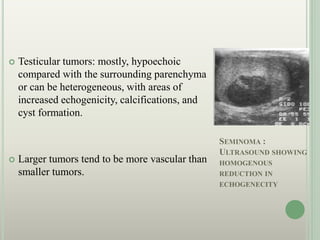
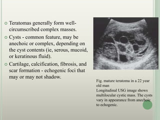
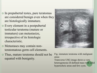
This document provides an overview of imaging in testicular tumors. It begins with the embryology and anatomy of the testes. Ultrasound is described as the primary imaging method for evaluating testicular lesions. MRI may also be used and can help characterize indeterminate lesions. The document reviews the classification, risk factors, clinical manifestations, patterns of spread, staging, and imaging appearances of various testicular tumors including seminoma, mixed germ cell tumors, and others. Imaging plays an important role in detecting tumors, staging disease, and monitoring for recurrence.


![imaging of scrotum [Repaired] [Repaired].pptx](https://cdn.slidesharecdn.com/ss_thumbnails/imagingofscrotumrepairedrepaired-230522050410-51cee1e6-thumbnail.jpg?width=640&height=640&fit=bounds)


