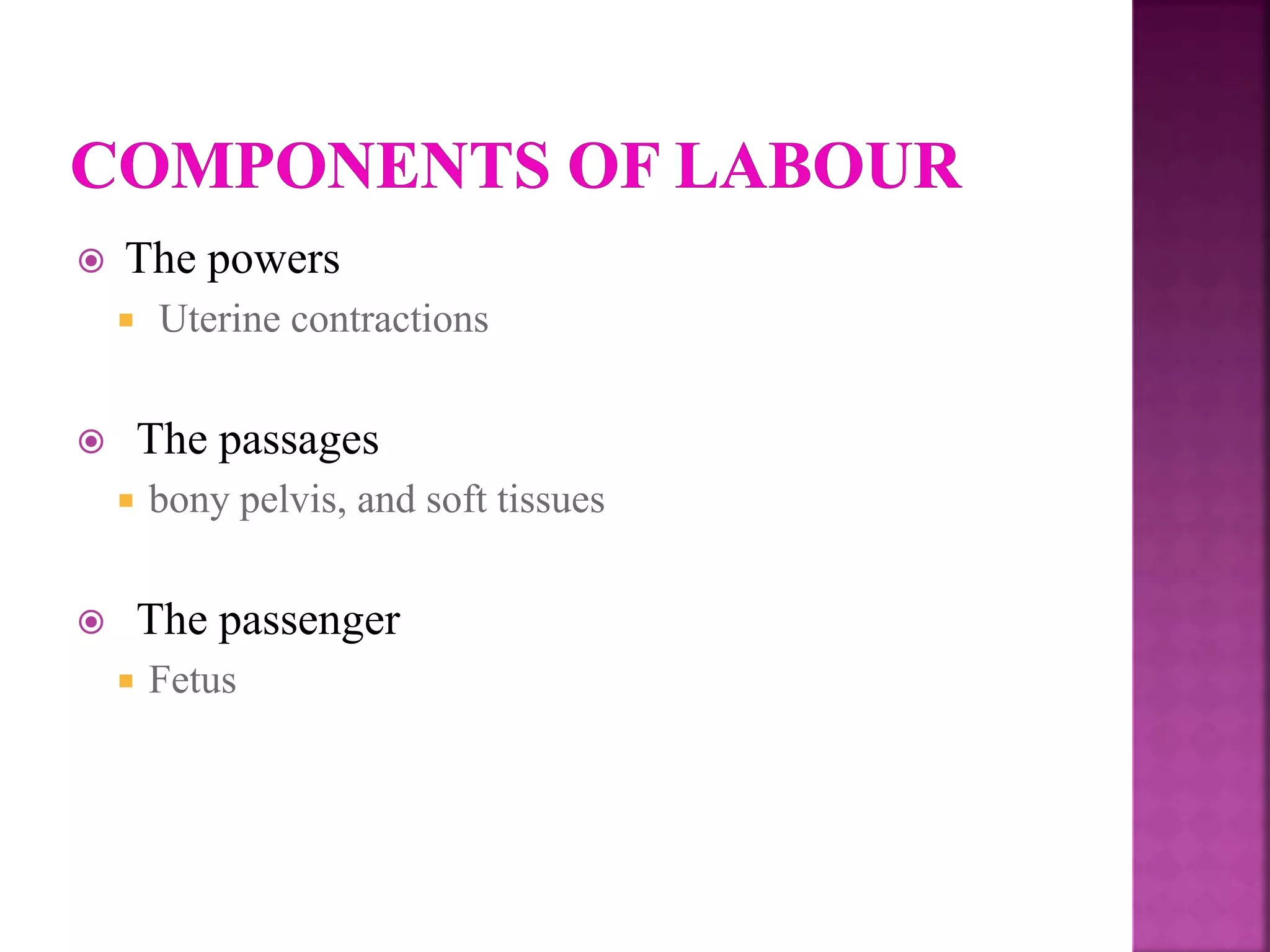
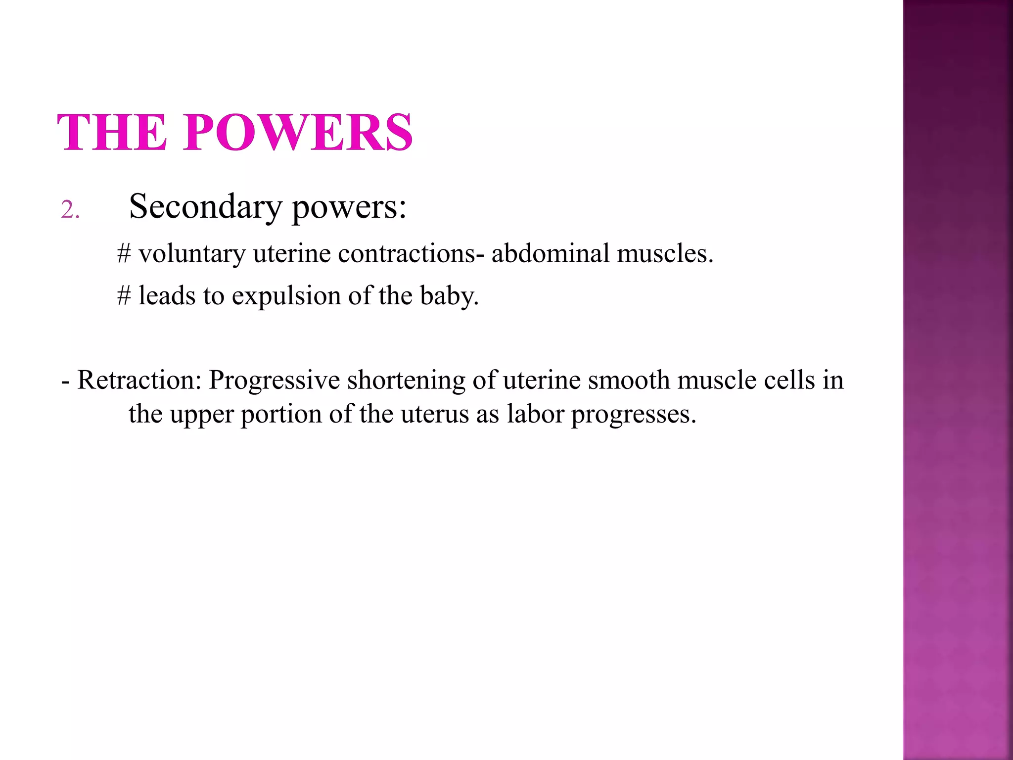
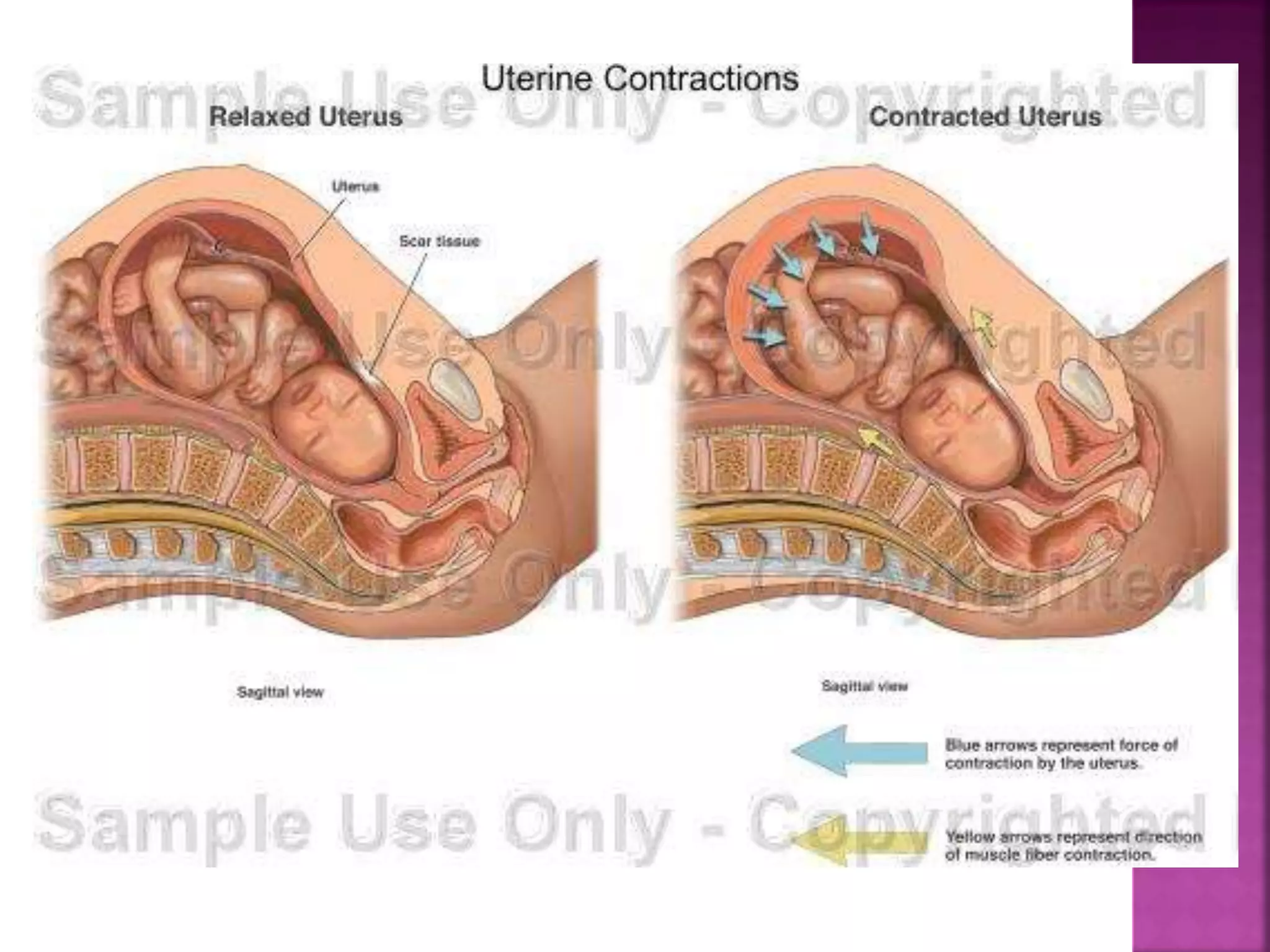
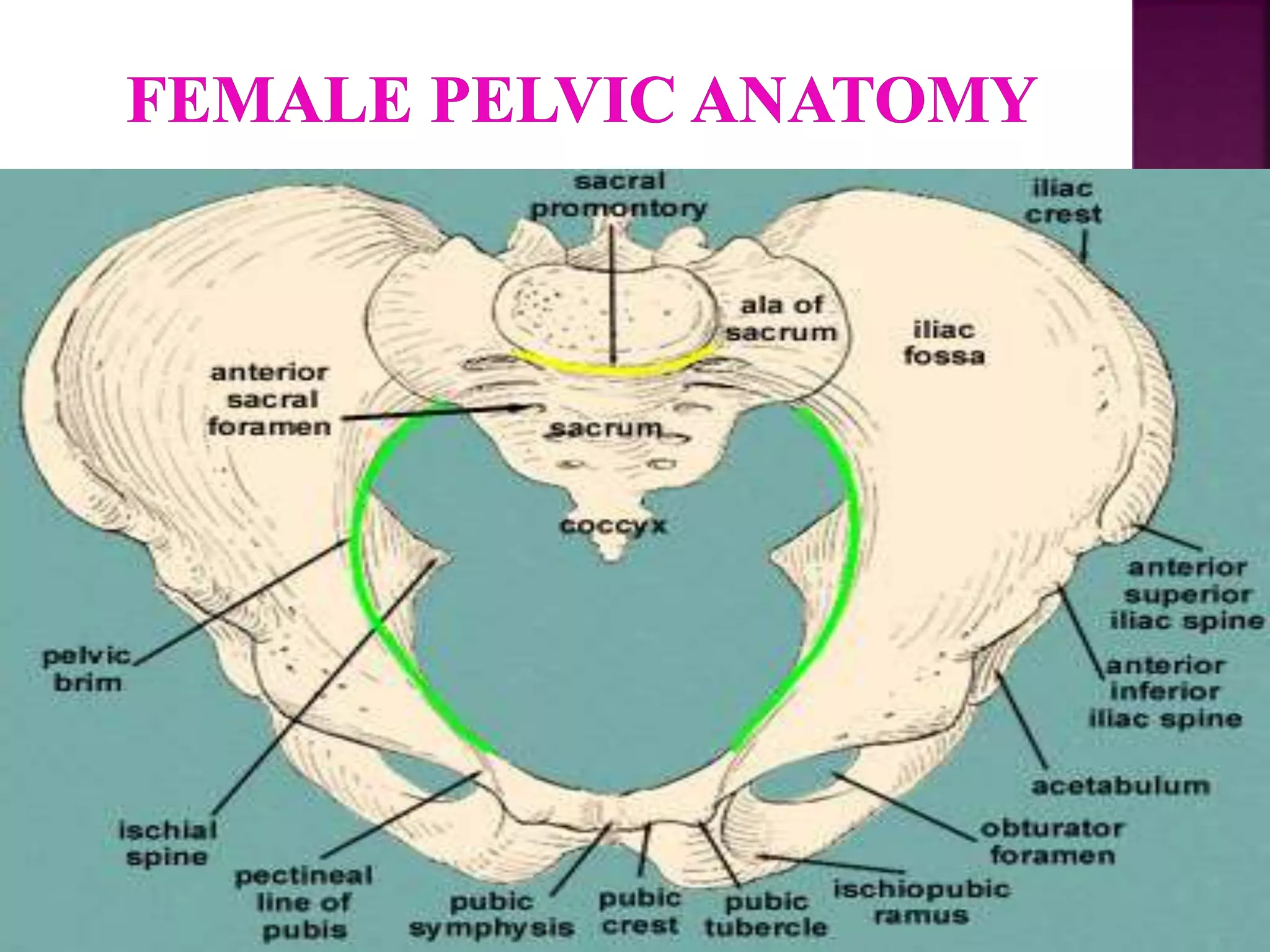
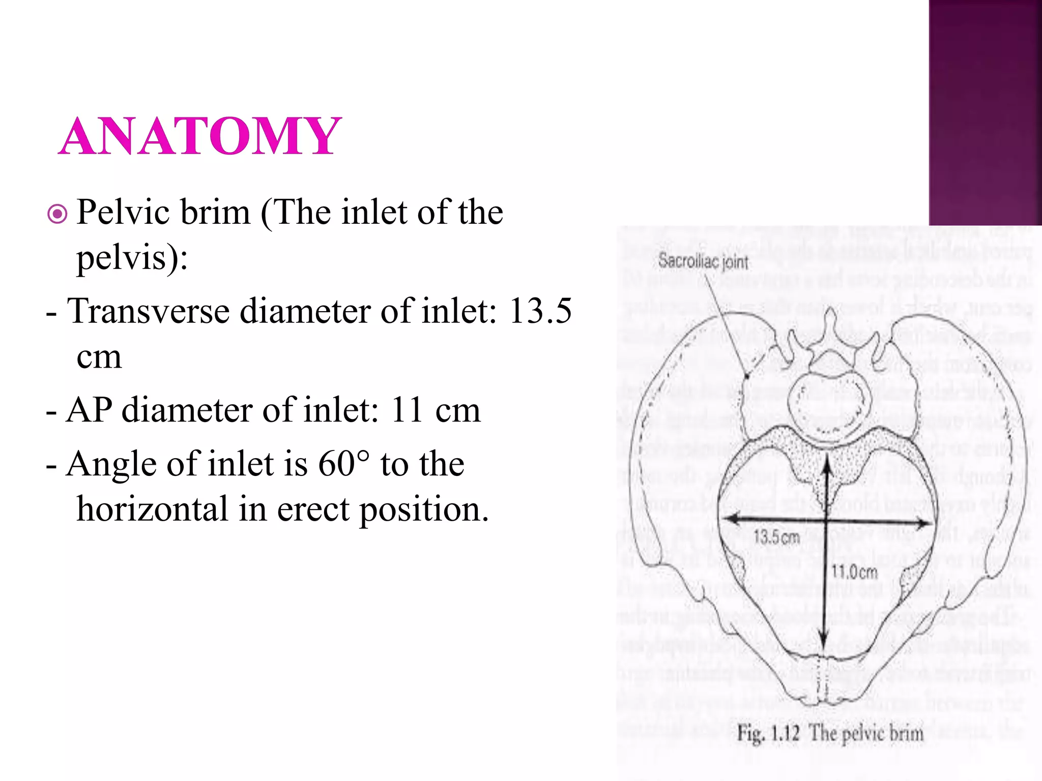
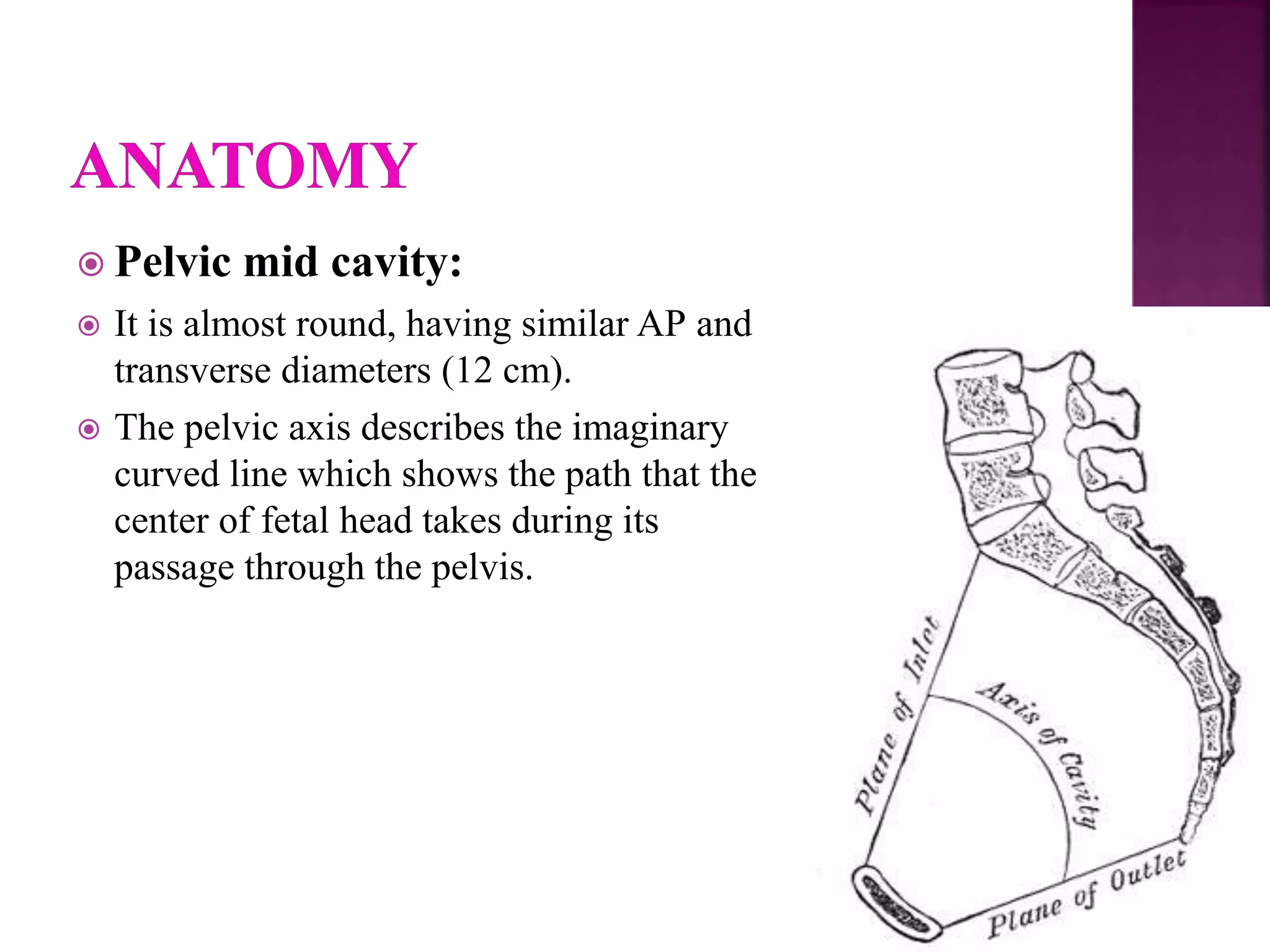
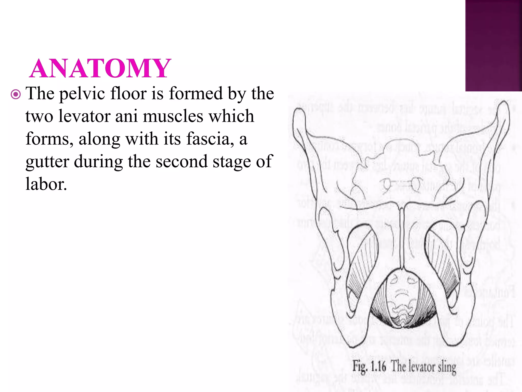
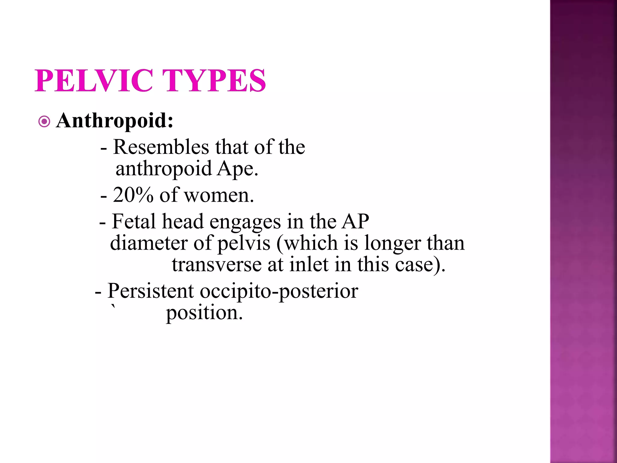
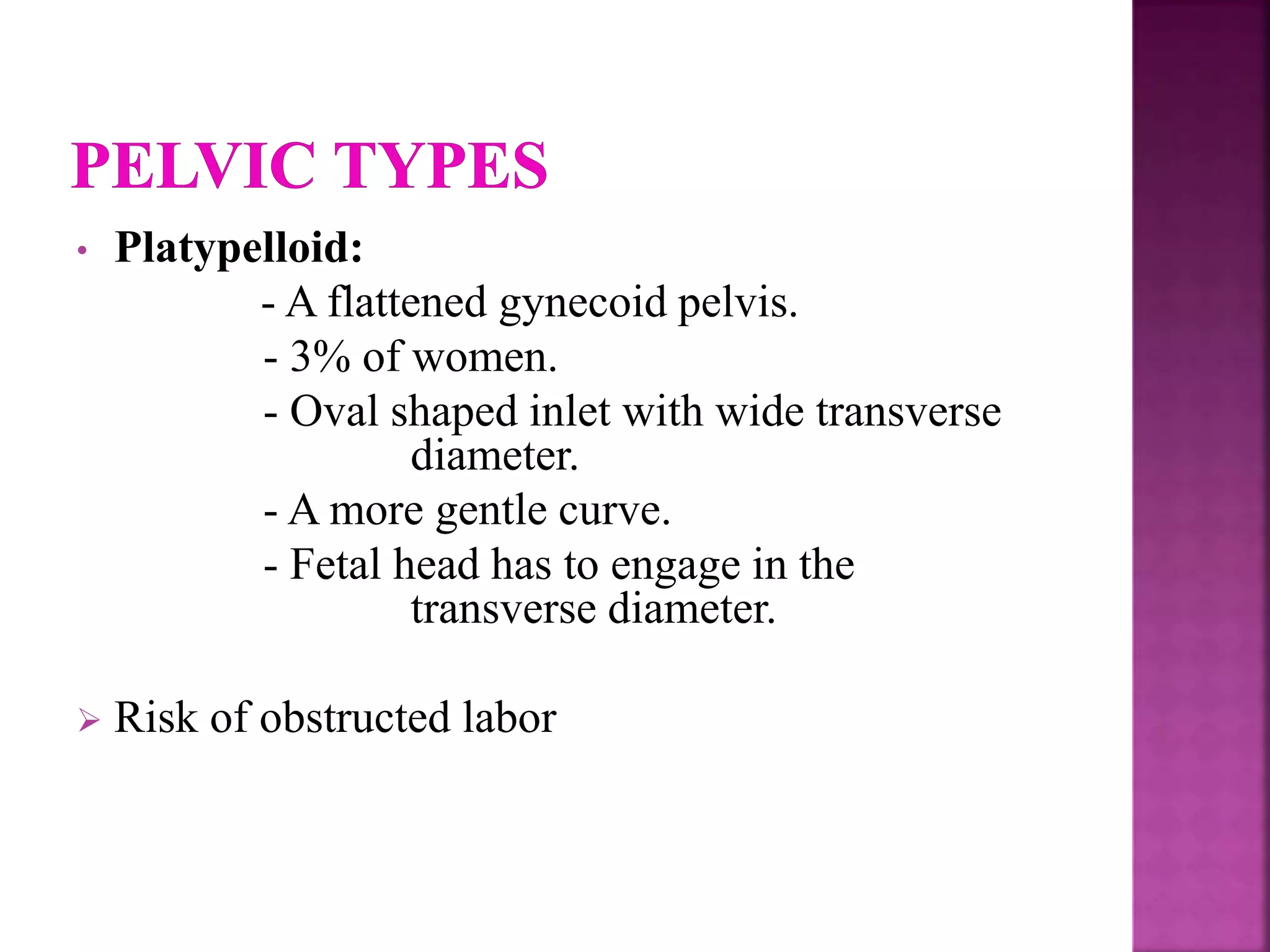
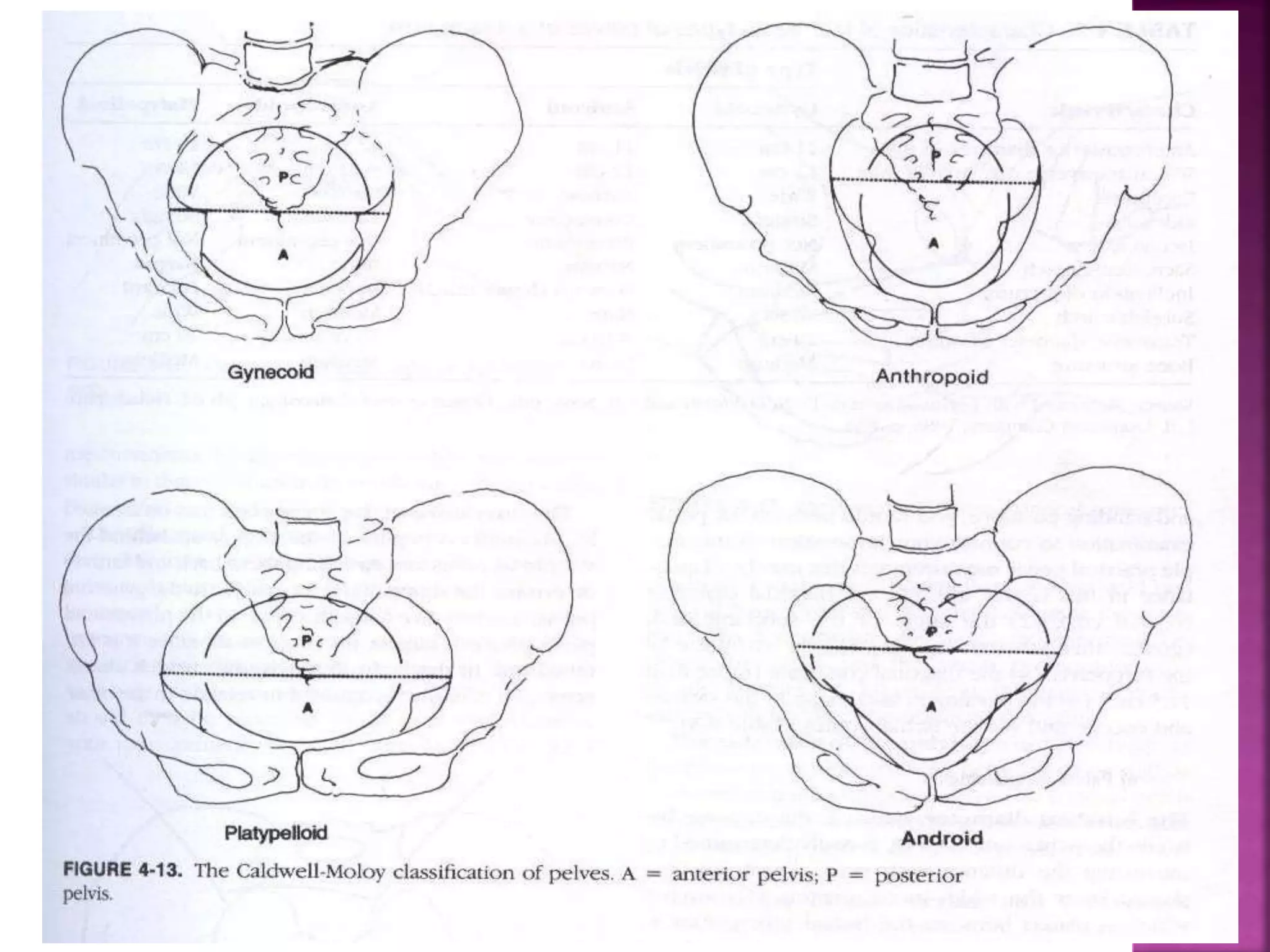
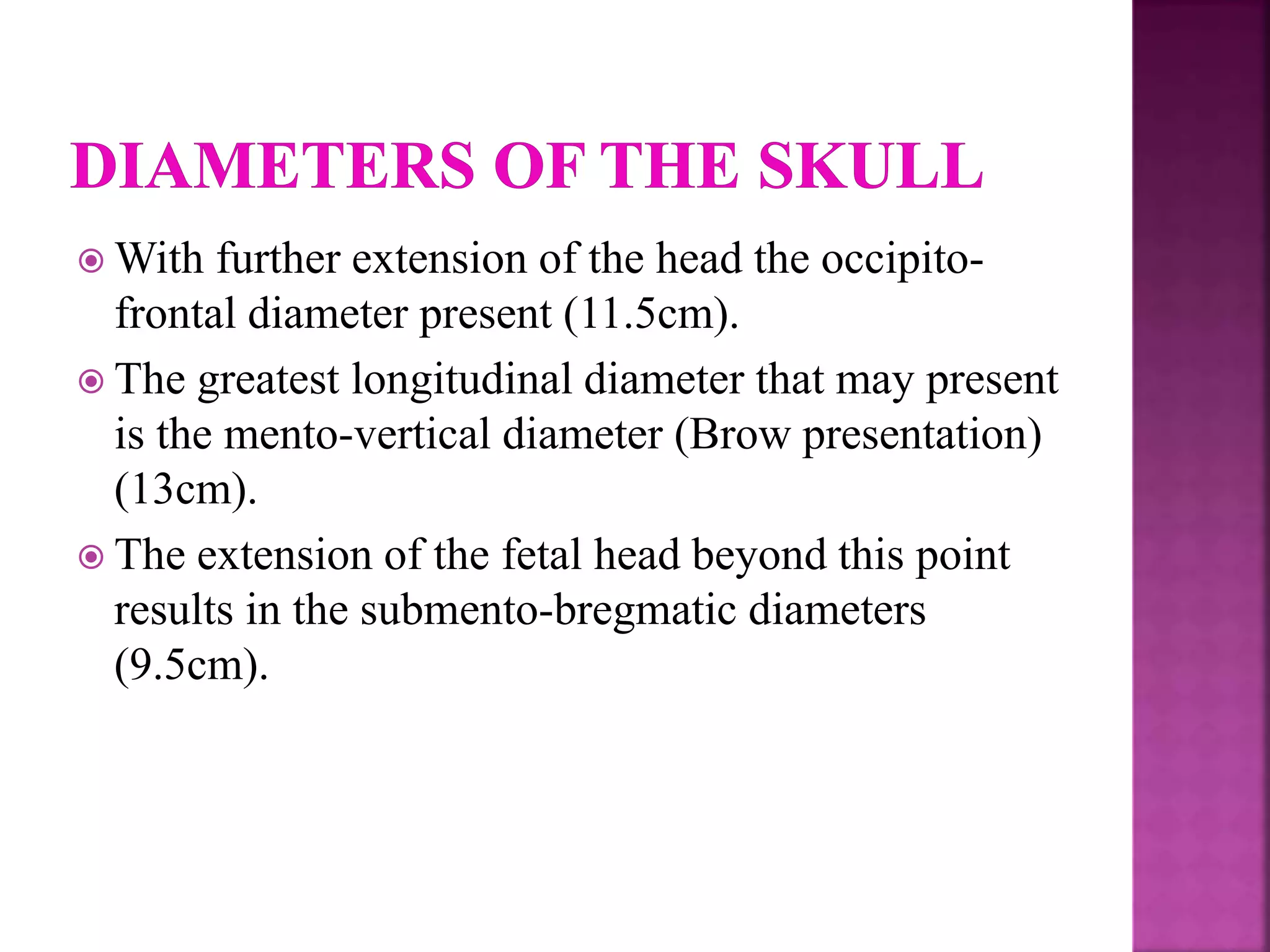
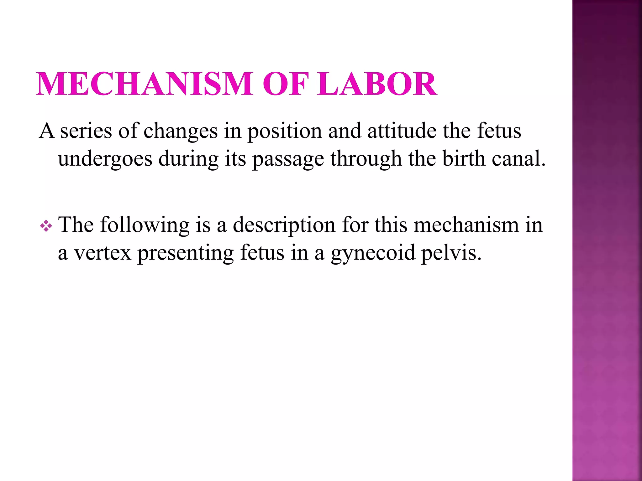
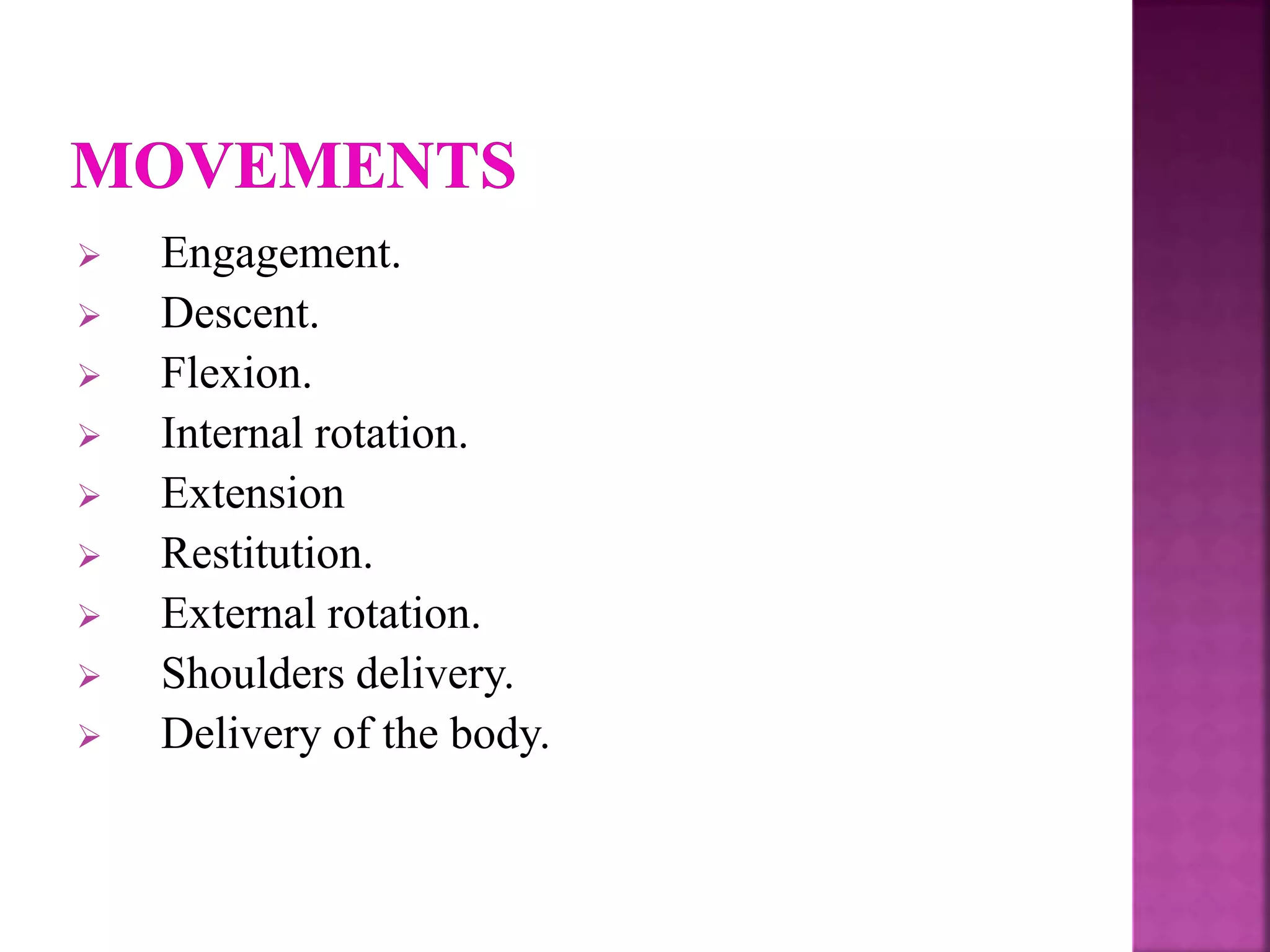
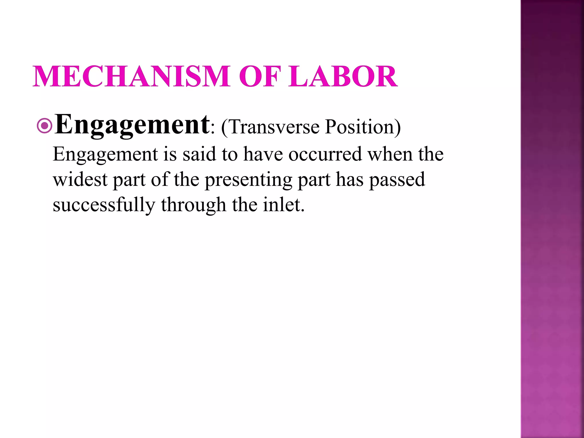
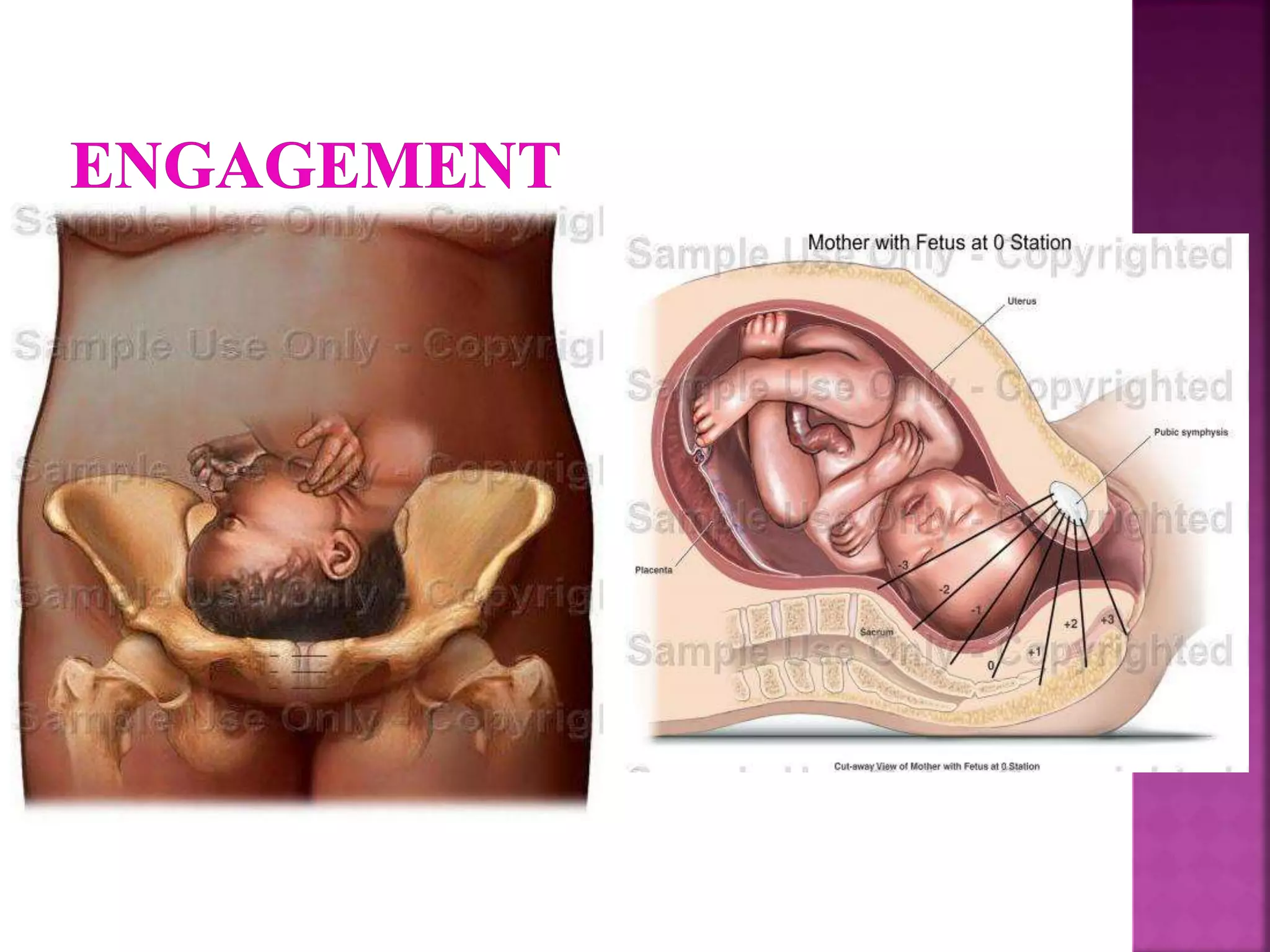
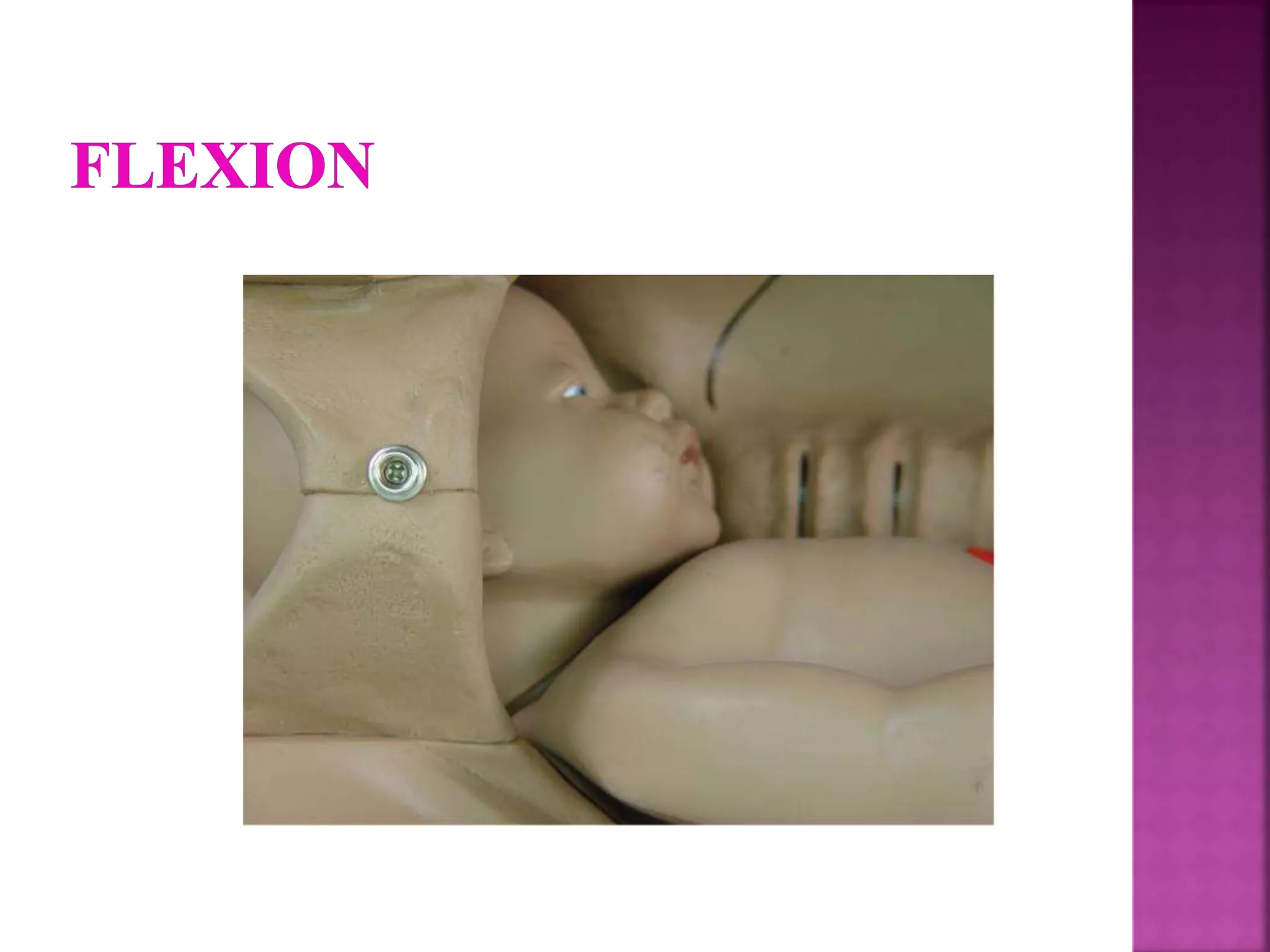
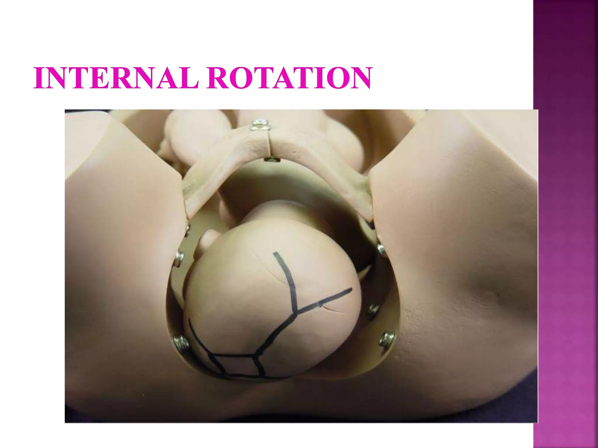
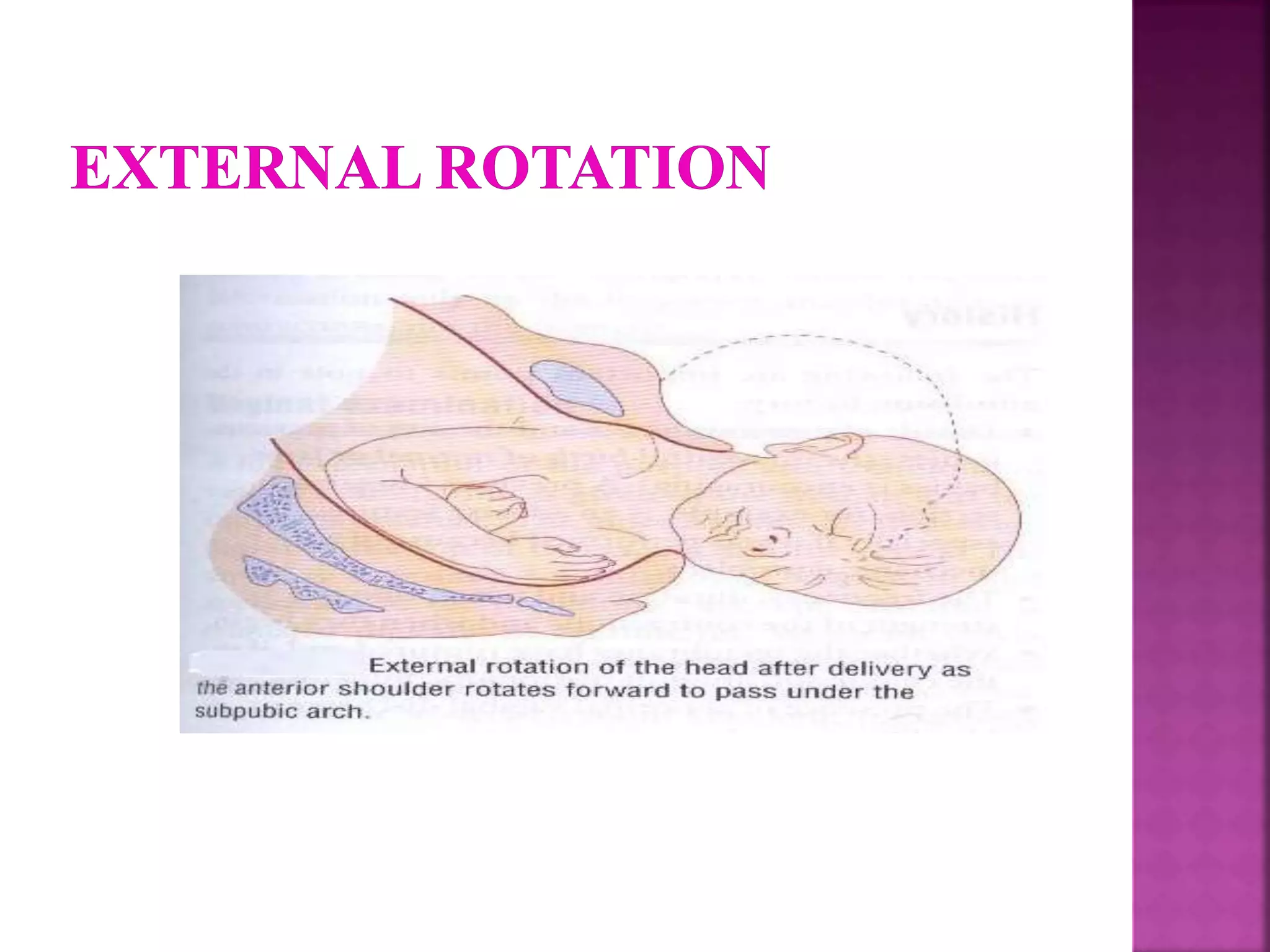
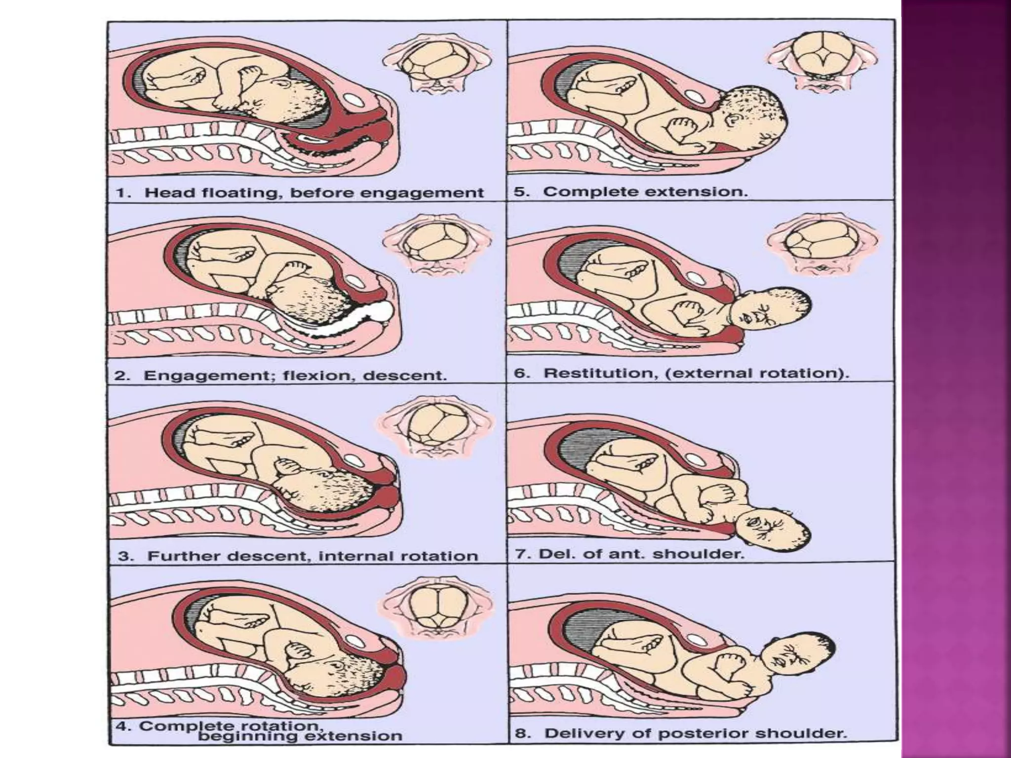
PRESENTED BY
ABHILASHA VERMA
LECTURER
OBG. GYNAE.
(JHALAWAR NURSING COLLEGE)
 Labour is the series of events (that is
rhythmic contraction and relaxation) take
place in uterus inorder to expel the products
of conception ( fetus, placenta and
membrane) from the womb through vagina to
out of the world.
 The powers
 Uterine contractions
 The passages
 bony pelvis, and soft tissues
 The passenger
 Fetus
2. Secondary powers:
# voluntary uterine contractions- abdominal muscles.
# leads to expulsion of the baby.
- Retraction: Progressive shortening of uterine smooth muscle cells in
the upper portion of the uterus as labor progresses.
1. Pelvic Anatomy:
# Bony pelvis &diameters.
# Pelvic Floor.
2. Pelvic Types.
 Pelvic brim (The inlet of the
pelvis):
- Transverse diameter of inlet: 13.5
cm
- AP diameter of inlet: 11 cm
- Angle of inlet is 60° to the
horizontal in erect position.
 Pelvic mid cavity:
 It is almost round, having similar AP and
transverse diameters (12 cm).
 The pelvic axis describes the imaginary
curved line which shows the path that the
center of fetal head takes during its
passage through the pelvis.
 Pelvic outlet:
Transverse Diameter: 11 cm.
AP diameter: 13.5 cm.
 Inlet: Transverse diameter is
wider.
 Outlet: AP diameter is wider.
 The pelvic floor is formed by the
two levator ani muscles which
forms, along with its fascia, a
gutter during the second stage of
labor.
 Gynecoid (Classic type):
- Found in 50% of women.
- It has a cylindrical shape.
- Is most favorable.
- Fetal head usually assumes an occipito- anterior
position in this type.
 Round at the inlet, with the widest transverse diameter only slightly
greater than the anteroposterior diameter
 Side walls straight
 Ischial spines of average prominence
 Well-curved sacrum
 Spacious subpubic arch, with an angle of approximately 90 degrees
• Android:
- Typical male pelvis.
- 30% of women.
- limited space at inlet and gets narrower as
you go down the pelvis (funneling).
- Fetal head is forced to be in occipito-
anterior position.
- The angle of the pubic arch is less than
90 degrees.
 Deep transverse arrest of descent is common at
midpelvis.
 Anthropoid:
- Resembles that of the
anthropoid Ape.
- 20% of women.
- Fetal head engages in the AP
diameter of pelvis (which is longer than
transverse at inlet in this case).
- Persistent occipito-posterior
` position.
• Platypelloid:
- A flattened gynecoid pelvis.
- 3% of women.
- Oval shaped inlet with wide transverse
diameter.
- A more gentle curve.
- Fetal head has to engage in the
transverse diameter.
 Risk of obstructed labor
 Note that all pelvic measurements relates to bony
pelvis. As the pelvic ligaments loosen in pregnancy.
 The pelvis is often more flexible than the
measurements would suggest which is why the
pelvic measurements may not be helpful.
• Fetal skull is the largest and the least compressible
part, thus it is the most important part of delivery
whatever the presentation is.
• The skull is made of: Vault, Face and Base.
• At the time of labor only unossified membranes at
the sutures join the bones of the vault, unlike the
face and the base which are firmly united.
• Between the bones there are four membranous
sutures: Sagittal, Frontal, Coronal and Lambdoidal.
• Fontanels are the junction of the various sutures:
Anterior fontanel (bregma) and Posterior fontanel.
• Vertex is the area of fetal skull bounded by two
parietal eminences laterally and the Ant. and Pos.
fontanels.
1. Coronal Suture
2. Anterior Fontanel (Bregma)
3. Anterolateral Fontanel
4. Squamosal Suture
5. Posterolateral Fontanel
6. Lamdoidal Suture
7. External Acoustic Meatus
8. Sagittal Suture
9. Posterior Fontanel
 The longitudinal diameters that presents in a well-
flexed head is the suboccipito-bregmatic diameter
(9.5cm).
 The longitudinal diameters that presents in a less
flexed head is the suboccipito-frontal diameter
(10cm).
 With further extension of the head the occipito-
frontal diameter present (11.5cm).
 The greatest longitudinal diameter that may present
is the mento-vertical diameter (Brow presentation)
(13cm).
 The extension of the fetal head beyond this point
results in the submento-bregmatic diameters
(9.5cm).
A series of changes in position and attitude the fetus
undergoes during its passage through the birth canal.
 The following is a description for this mechanism in
a vertex presenting fetus in a gynecoid pelvis.
 Engagement.
 Descent.
 Flexion.
 Internal rotation.
 Extension
 Restitution.
 External rotation.
 Shoulders delivery.
 Delivery of the body.
Engagement: (Transverse Position)
Engagement is said to have occurred when the
widest part of the presenting part has passed
successfully through the inlet.
Descent: during first stage and first phase of
second stage of labor descent occurs secondary to
uterine contractions. In the active phase of second
stage it is helped by voluntary use of abdominal
muscles and the Valsava maneuver (pushing).
 Flexion: it is a passive movement due to
resistance from the cervix, pelvic walls, and pelvic
floor. It minimizes the presenting diameter (from
occipitofrontal to suboccipitobregmatic).
Internal rotation:the occiput rotates
anteriorly, sagittal suture lies in AP diameter of
pelvic outlet (widest diameter), this probably
occurs as the fetal head meets the muscular
sling of the pelvic floor.
 Extension: occiput escapes from underneath
the symphysis pubis and distends through the
vulva.
 Episiotomy might be needed to reduce pernial
resistance.
 Crowning: When the largest diameter of fetal
head is encircled by the vulvar ring (vertex
reached a +5 station).
 Further extension, using the symphysis pubis as
a fulcrum, leads to appearance of the bregma,
followed by the face and the chin.
 Restitution: As soon as head escapes from the
vulva it aligns itself with the shoulders. The
occiput rotates one eighth of a circle (neck
untwists turning the head sideways).
 External rotation:
 Shoulders rotate to the AP plane.
 Occiput rotates a further one eighth of a circle to
the transverse position.
 Delivery of shoulders and body:
 Anterior shoulder delivers first followed by posterior
shoulder.
 Rest of fetal body is normally delivered easily.

 Thank you

Mechanism of labour

  • 1.
    PRESENTED BY ABHILASHA VERMA LECTURER OBG.GYNAE. (JHALAWAR NURSING COLLEGE)
  • 2.
     Labour isthe series of events (that is rhythmic contraction and relaxation) take place in uterus inorder to expel the products of conception ( fetus, placenta and membrane) from the womb through vagina to out of the world.
  • 3.
     The powers Uterine contractions  The passages  bony pelvis, and soft tissues  The passenger  Fetus
  • 4.
    2. Secondary powers: #voluntary uterine contractions- abdominal muscles. # leads to expulsion of the baby. - Retraction: Progressive shortening of uterine smooth muscle cells in the upper portion of the uterus as labor progresses.
  • 6.
    1. Pelvic Anatomy: #Bony pelvis &diameters. # Pelvic Floor. 2. Pelvic Types.
  • 8.
     Pelvic brim(The inlet of the pelvis): - Transverse diameter of inlet: 13.5 cm - AP diameter of inlet: 11 cm - Angle of inlet is 60° to the horizontal in erect position.
  • 9.
     Pelvic midcavity:  It is almost round, having similar AP and transverse diameters (12 cm).  The pelvic axis describes the imaginary curved line which shows the path that the center of fetal head takes during its passage through the pelvis.
  • 10.
     Pelvic outlet: TransverseDiameter: 11 cm. AP diameter: 13.5 cm.  Inlet: Transverse diameter is wider.  Outlet: AP diameter is wider.
  • 11.
     The pelvicfloor is formed by the two levator ani muscles which forms, along with its fascia, a gutter during the second stage of labor.
  • 12.
     Gynecoid (Classictype): - Found in 50% of women. - It has a cylindrical shape. - Is most favorable. - Fetal head usually assumes an occipito- anterior position in this type.  Round at the inlet, with the widest transverse diameter only slightly greater than the anteroposterior diameter  Side walls straight  Ischial spines of average prominence  Well-curved sacrum  Spacious subpubic arch, with an angle of approximately 90 degrees
  • 13.
    • Android: - Typicalmale pelvis. - 30% of women. - limited space at inlet and gets narrower as you go down the pelvis (funneling). - Fetal head is forced to be in occipito- anterior position. - The angle of the pubic arch is less than 90 degrees.  Deep transverse arrest of descent is common at midpelvis.
  • 14.
     Anthropoid: - Resemblesthat of the anthropoid Ape. - 20% of women. - Fetal head engages in the AP diameter of pelvis (which is longer than transverse at inlet in this case). - Persistent occipito-posterior ` position.
  • 15.
    • Platypelloid: - Aflattened gynecoid pelvis. - 3% of women. - Oval shaped inlet with wide transverse diameter. - A more gentle curve. - Fetal head has to engage in the transverse diameter.  Risk of obstructed labor
  • 16.
     Note thatall pelvic measurements relates to bony pelvis. As the pelvic ligaments loosen in pregnancy.  The pelvis is often more flexible than the measurements would suggest which is why the pelvic measurements may not be helpful.
  • 18.
    • Fetal skullis the largest and the least compressible part, thus it is the most important part of delivery whatever the presentation is. • The skull is made of: Vault, Face and Base. • At the time of labor only unossified membranes at the sutures join the bones of the vault, unlike the face and the base which are firmly united.
  • 19.
    • Between thebones there are four membranous sutures: Sagittal, Frontal, Coronal and Lambdoidal. • Fontanels are the junction of the various sutures: Anterior fontanel (bregma) and Posterior fontanel. • Vertex is the area of fetal skull bounded by two parietal eminences laterally and the Ant. and Pos. fontanels.
  • 20.
    1. Coronal Suture 2.Anterior Fontanel (Bregma) 3. Anterolateral Fontanel 4. Squamosal Suture 5. Posterolateral Fontanel 6. Lamdoidal Suture 7. External Acoustic Meatus 8. Sagittal Suture 9. Posterior Fontanel
  • 21.
     The longitudinaldiameters that presents in a well- flexed head is the suboccipito-bregmatic diameter (9.5cm).  The longitudinal diameters that presents in a less flexed head is the suboccipito-frontal diameter (10cm).
  • 22.
     With furtherextension of the head the occipito- frontal diameter present (11.5cm).  The greatest longitudinal diameter that may present is the mento-vertical diameter (Brow presentation) (13cm).  The extension of the fetal head beyond this point results in the submento-bregmatic diameters (9.5cm).
  • 24.
    A series ofchanges in position and attitude the fetus undergoes during its passage through the birth canal.  The following is a description for this mechanism in a vertex presenting fetus in a gynecoid pelvis.
  • 25.
     Engagement.  Descent. Flexion.  Internal rotation.  Extension  Restitution.  External rotation.  Shoulders delivery.  Delivery of the body.
  • 26.
    Engagement: (Transverse Position) Engagementis said to have occurred when the widest part of the presenting part has passed successfully through the inlet.
  • 28.
    Descent: during firststage and first phase of second stage of labor descent occurs secondary to uterine contractions. In the active phase of second stage it is helped by voluntary use of abdominal muscles and the Valsava maneuver (pushing).
  • 30.
     Flexion: itis a passive movement due to resistance from the cervix, pelvic walls, and pelvic floor. It minimizes the presenting diameter (from occipitofrontal to suboccipitobregmatic).
  • 32.
    Internal rotation:the occiputrotates anteriorly, sagittal suture lies in AP diameter of pelvic outlet (widest diameter), this probably occurs as the fetal head meets the muscular sling of the pelvic floor.
  • 34.
     Extension: occiputescapes from underneath the symphysis pubis and distends through the vulva.  Episiotomy might be needed to reduce pernial resistance.  Crowning: When the largest diameter of fetal head is encircled by the vulvar ring (vertex reached a +5 station).  Further extension, using the symphysis pubis as a fulcrum, leads to appearance of the bregma, followed by the face and the chin.
  • 36.
     Restitution: Assoon as head escapes from the vulva it aligns itself with the shoulders. The occiput rotates one eighth of a circle (neck untwists turning the head sideways).  External rotation:  Shoulders rotate to the AP plane.  Occiput rotates a further one eighth of a circle to the transverse position.
  • 38.
     Delivery ofshoulders and body:  Anterior shoulder delivers first followed by posterior shoulder.  Rest of fetal body is normally delivered easily.
  • 40.