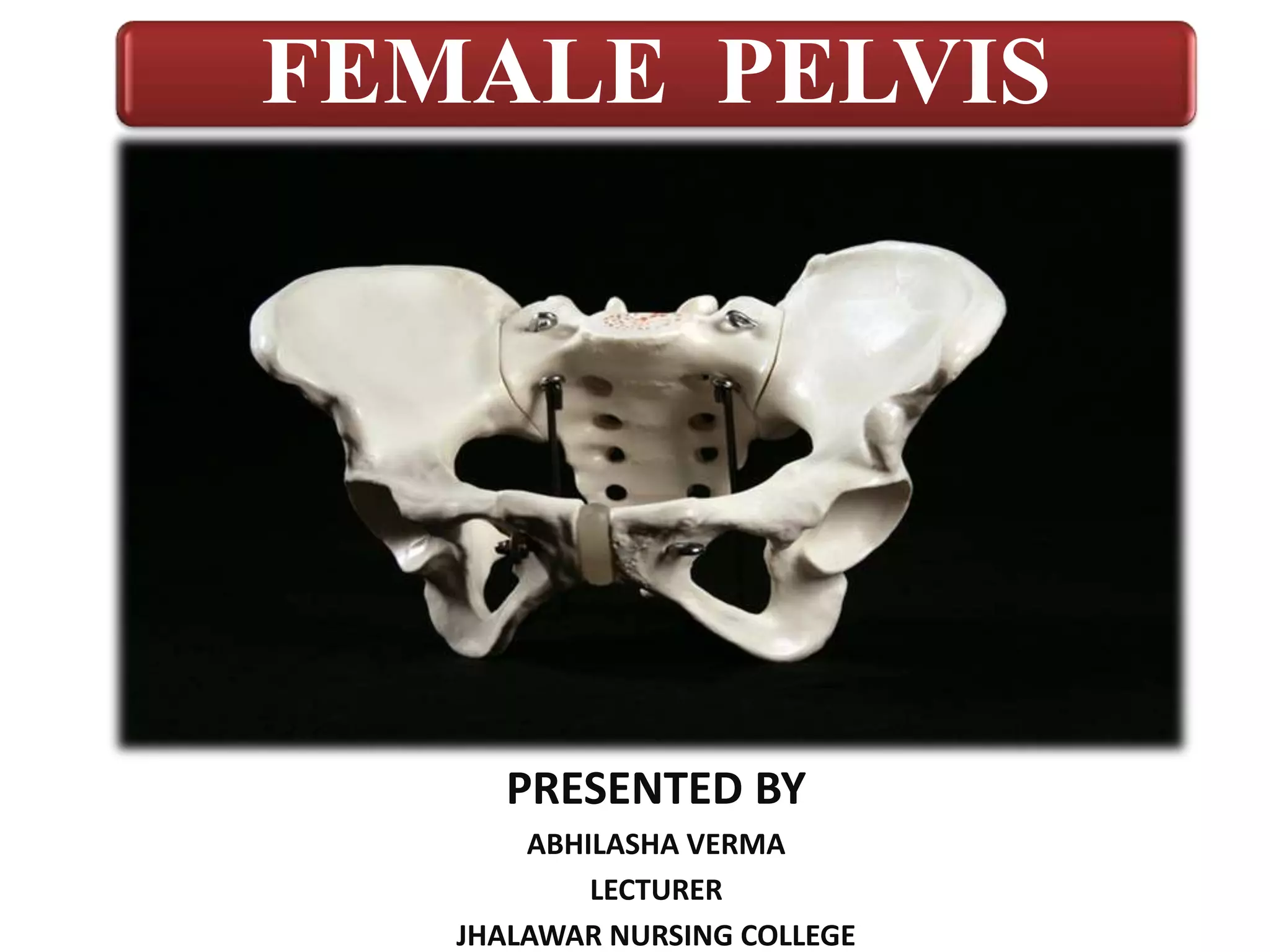
FEMALE PELVIS
PRESENTED BY
ABHILASHA VERMA
LECTURER
JHALAWAR NURSING COLLEGE
Difference b/w Male & Female Pelvis
FEMALE PELVIS
• Gynaecoid pelvis
• Larger and broader
• Larger inlet in oval shape.
• Angle between the inferior
pubic rami is 90° called as
pubic arch.
• Angle is more concave .
• Sacrum is shorter, wider,
more curved posteriorly.
MALE PELVIS
• Android pelvis
• Narrow and compact
• Heart shaped inlet because
of sacral promontory
projection.
• Angle between the inferior
pubic rami is 70°.
• Angle is more straight.
• Sacrum is longer, narrow,
straight.
2ABHILASHA VERMA
3
General Function-
1. Primary function to allow movement to body.
2. Helps in walking, sitting, running.
3. Pelvis contain and protect reproductive organs along with
bladder and rectum.
4. Sitting weight of body is taken up by the ischial tuberosity.
5. Helps in transmitting weight of body to legs.
 Obstetrical Function-
1. Adapted for child bearing in comparison to male pelvis, brim
is rounder and wider.
2. It supports gravid uterus.
3. It gives passage way to fetus.
Female pelvis is a skeletal ring, because of it’s characteristics, aids in child birth.
Function Of Female Pelvis
4
BONES OF PELVIS
 It consists of 4 bones-
• Inominate bones (2)
• Sacrum (1)
• Coccyx (1)
5ABHILASHA VERMA
Pelvis Demonstration Video
ABHILASHA VERMA 6
INNOMINATE BONE
• Each innominate bone madeup of 3 parts:
(Ilium, Ischium, pubic)
1. ILIUM BONE:
• Large flared out structure ( wide).
• Concave inner surface is iliac fossa.
• Curved upper border is iliac crest.
• The prominance at the front of iliac crest known as anterior
superior iliac spine, just below it, there is anterior inferior
iliac spine.
• At opposite end of iliac crest similar prominence are present
which are named posterior superior and posterior inferior
iliac spine.
7ABHILASHA VERMA
2. ISCHIUM BONE:
• It is thick lower part of innominate bone.
• Thick lower part has large prominance known as
ischial tuberosity. ( when you sit , sit on this)
• In sitting position body rest on ischial tuberosity.
• Slight projection are present are known as ischial
spine (2).
– In labour, station of fetal head estimated in relation to
ischial spine.
– Internal rotation of fetal head occur here.
– Greater and lessor sciatic notch are present.
8ABHILASHA VERMA
3. PUBIC BONE:
• They form anterior part of innominate bone.
• It has body and two oar like projection known as
superoir ramus and inferior ramus.
• The two pubic bone meet at symphysis pubis.
• Two inferior rami form pubic arch.
• Space enclosed by body of the pubic bone, ramus,
and ischium is called as obturator foramen.
• Two curves are found on lower border of pubic bone;
– Greater sciatic notch: Extends from post. Inferior iliac
spine to ischial spine, wide and rounded.
– Lessor sciatic notch: lies between ischial spine to ischial
tuberosity.
 The innominate has a deep cup to receive head of
femur, termed as acetabulum.
9ABHILASHA VERMA
SACRUM
• Lies between ilium.
• Wedge shaped bone consists of 5 fused
vertebrae.
• The prominent upper margin of first sacral
vertebra is called as sacral promontory.
• Anterior surface of sacrum is concave and
referred as hollow of sacrum. Four pairs of
holes or foramina are present which allows
passage of sacral nerves.
10ABHILASHA VERMA
COCCYX
• Small triangular bone with it’s base ( Upper most)
articulating with lower end of sacrum.
• It consists of four fused vertebras.
• It gives attachment to deep muscles, ligaments of
pelvic floor, and muscle fiber of anal sphincter.
• During labor coccyx moves backward to enlarge the
pelvic outlet allowing more space for passage of
fetus.
11ABHILASHA VERMA
PELVIC JOINTS
• Four pelvic joints-
1. SACROILIAC JOINTS (2)-
• Slightly movable joint.
• Formed where ilium join first 2 sacral vertebrae on either
side.
• Strongest joint of body.
• Connects spine to pelvis.
2. SYMPHYSIS PUBIS (1)-
• Cartilagenous fibrous joint between 2 pubic bones.
2. SACRO-COCCYGEAL (1)-
• Hinge joint between sacrum and coccyx.
• During pregnancy due to softening of ligament , provide
more space for fetal head to pass through pelvis. 12ABHILASHA VERMA
PELVIC LIGAMENTS
1. Sacro-iliac Ligaments:
 Pass infront of or behind each sacro iliac joints.
2. Pubic Ligaments:
 Superior pubic ligament connect pubic bones.
3. Sacro-tuberous Ligaments:
 Run from sacrum to ischial tuberosity.
4. Sacro-spinous Ligaments:
 Run on each side of sacrum and ischial spine.
5. Sacro-coccygeal Ligaments:
 Run on each from sacrum to coccyx.
13ABHILASHA VERMA
FALSE PELVIS & TRUE PELVIS
14
TRUE & FALSE PELVIS
15
 Linea terminalis divide pelvis into false and true pelvis.
 FALSE PELVIS OR GREATER PELVIS-
 False pelvis is portion above pelvic brim.
 It is formed by iliac portion of innominate bones and limited
above by iliac crests.
 It’s only obstetrical function is to support enlarged and gravid
uterus.
 It is bounded by-
Anterior-Anterior abdominal wall.
Posterior- Lumber Vertebrae
Laterally- Iliac fossa.
TRUE /LESSOR /MINOR PELVIS:
 Boney canal through which fetus must pass to be born
vaginally.
 It is situated below pelvic brim including brim.
 Boney wall complete then greater pelvis.
 It has brim( inlet), cavity and outlet.
 It is bounded by-
 Superior- Sacral promontory, linea terminalis, upper
margin of pubic bone.
 Inferior- Inferior margin of ischial tuberosity and tip of
coccyx.
 Anterior- Obturator foramen, post. Surface of symphysis
pubis, pubic bone, ascending ramus of pubic bone.
16ABHILASHA VERMA
DIAMETERS OF PELVIS (Brim/ Inlet)
1) Anterior-posterior Diameter of inlet-
a) Anatomical Conjugate(11cm)-
 Distance between mid point of sacral promontory to inner margin of
upper border of symphysis pubis.
 It measures about 11 cm.
b) Obsteric Conjugate (10 cm)-
 Distance between mid point of sacral promontory to prominent bony
projection in midline of inner surface of symphysis pubis.
 Shortest diameter of inlet, measures about 10 cm.
 Cann’t be clinicaly estimated, diagonal conjugate necessary.
Obstetric Conjugate = Diagonal conjugate - 2 cm
c) Diagonal Conjugate (12cm)-
 Distance between lower border of symphysis pubis to mid point of sacral
promontory.
 Measure by bimanual examination.
 It measures about 12 cm
17ABHILASHA VERMA
2) Transverse Diameter of inlet:
 Distance between 2 farthest point on pelvic brim over ilio-
pectineal lines.
 Measures about 13 cm.
 This divide brim into anterior and posterior segment.
3) Oblique Diameter of inlet:
 There are two oblique diameter right & left.
 Each extends from one sacro-iliac joint to opposite ilio-
pectineal eminance.
 It measures 12 cm
18ABHILASHA VERMA
DIAMETERS OF PELVIS (Cavity)
“Cavity is segment of pelvis bounded above by inlet and below
by outlet.”
 Anterior wall formed by pubic bone and symphysis pubis,
measure s 4 cm.
 Posterior wall formed by curve of sacrum and coccyx,
measures 12 cm.
 Shape; Almost round.
 It extands from midpoint of post. Surface of symphysis pubis
tojunction of 2nd and 3rd sacral vertebrae.
 Transverse and oblique diameter cannot measures exactly as
point is overlies soft tissues, so roughly we can say overall
diameter 12 cm.
ABHILASHA VERMA 19
DIAMETERS OF PELVIS (Outlet)
 Describes as anatomical and obstetrical outlet.
 Obstetrical outlet has practical value, through which fetus has to
pass outlet only.
 The oblique diameter cannot be measured exactly due to presence
of soft tissues, roughly measured about 12 cm
 Anatomical outlet-
• Anterio-Posterio diameter extends from Lower border of symphysis pubis to tip of
coccyx, measures about 13 cm.
• Transverse diameter is inter tuberous diameter , is the distance between inner border
of 2 ischial tuberosity, measures about 11 cm.
 Obstetrical outlet-
• Narrow through which fetus has to pass.
• Anterio-posterior diameter extends from the inferior border of sympysis pubis to tip of
sacrum, measures about 11 cm.
• Transverse diameter is bispinous diameter, it is distance between tip of two ischial
spine, measures about 10.5 cm.
• Both AP Diameter and transverse diameter form Diamond shaped structure collectively
called obstetric diameter.
ABHILASHA VERMA 20
1. Anterior Posterior Diameter-
• Extends from inferior border of symphysis pubis to
sacrococcygeal joint.
• Measures about 13 cm.
2. Transverse Diameter-
• Distance between two tip of ischial spine.
• Measures 11 cm.
3. Oblique Diameter-
• Between obturator foramen and sacrospinous ligament.
• Measures 12 cm
ABHILASHA VERMA 21
DIAMETERS OF PELVIS
ABHILASHA VERMA 22
LANDMARKS OF FEMALE PELVIS
23
• A bony fixed point where fetus comes in contact with
maternal pelvis at time of engagement.
• From anterior to posterior way in numerical sequence-
1. Symphysis pubis
2. Pubic crest
3. Pubic tubercle
4. Pectineal line
5. Ilio-pectineal eminance
6. ilio=-pectineal line
7. Sacroiliac joint
8. Anterior border of sacral ala
9. Sacral promontary.
THANK YOU
ABHILASHA VERMA 24

Female pelvis

  • 1.
    FEMALE PELVIS PRESENTED BY ABHILASHAVERMA LECTURER JHALAWAR NURSING COLLEGE
  • 2.
    Difference b/w Male& Female Pelvis FEMALE PELVIS • Gynaecoid pelvis • Larger and broader • Larger inlet in oval shape. • Angle between the inferior pubic rami is 90° called as pubic arch. • Angle is more concave . • Sacrum is shorter, wider, more curved posteriorly. MALE PELVIS • Android pelvis • Narrow and compact • Heart shaped inlet because of sacral promontory projection. • Angle between the inferior pubic rami is 70°. • Angle is more straight. • Sacrum is longer, narrow, straight. 2ABHILASHA VERMA
  • 3.
  • 4.
    General Function- 1. Primaryfunction to allow movement to body. 2. Helps in walking, sitting, running. 3. Pelvis contain and protect reproductive organs along with bladder and rectum. 4. Sitting weight of body is taken up by the ischial tuberosity. 5. Helps in transmitting weight of body to legs.  Obstetrical Function- 1. Adapted for child bearing in comparison to male pelvis, brim is rounder and wider. 2. It supports gravid uterus. 3. It gives passage way to fetus. Female pelvis is a skeletal ring, because of it’s characteristics, aids in child birth. Function Of Female Pelvis 4
  • 5.
    BONES OF PELVIS It consists of 4 bones- • Inominate bones (2) • Sacrum (1) • Coccyx (1) 5ABHILASHA VERMA
  • 6.
  • 7.
    INNOMINATE BONE • Eachinnominate bone madeup of 3 parts: (Ilium, Ischium, pubic) 1. ILIUM BONE: • Large flared out structure ( wide). • Concave inner surface is iliac fossa. • Curved upper border is iliac crest. • The prominance at the front of iliac crest known as anterior superior iliac spine, just below it, there is anterior inferior iliac spine. • At opposite end of iliac crest similar prominence are present which are named posterior superior and posterior inferior iliac spine. 7ABHILASHA VERMA
  • 8.
    2. ISCHIUM BONE: •It is thick lower part of innominate bone. • Thick lower part has large prominance known as ischial tuberosity. ( when you sit , sit on this) • In sitting position body rest on ischial tuberosity. • Slight projection are present are known as ischial spine (2). – In labour, station of fetal head estimated in relation to ischial spine. – Internal rotation of fetal head occur here. – Greater and lessor sciatic notch are present. 8ABHILASHA VERMA
  • 9.
    3. PUBIC BONE: •They form anterior part of innominate bone. • It has body and two oar like projection known as superoir ramus and inferior ramus. • The two pubic bone meet at symphysis pubis. • Two inferior rami form pubic arch. • Space enclosed by body of the pubic bone, ramus, and ischium is called as obturator foramen. • Two curves are found on lower border of pubic bone; – Greater sciatic notch: Extends from post. Inferior iliac spine to ischial spine, wide and rounded. – Lessor sciatic notch: lies between ischial spine to ischial tuberosity.  The innominate has a deep cup to receive head of femur, termed as acetabulum. 9ABHILASHA VERMA
  • 10.
    SACRUM • Lies betweenilium. • Wedge shaped bone consists of 5 fused vertebrae. • The prominent upper margin of first sacral vertebra is called as sacral promontory. • Anterior surface of sacrum is concave and referred as hollow of sacrum. Four pairs of holes or foramina are present which allows passage of sacral nerves. 10ABHILASHA VERMA
  • 11.
    COCCYX • Small triangularbone with it’s base ( Upper most) articulating with lower end of sacrum. • It consists of four fused vertebras. • It gives attachment to deep muscles, ligaments of pelvic floor, and muscle fiber of anal sphincter. • During labor coccyx moves backward to enlarge the pelvic outlet allowing more space for passage of fetus. 11ABHILASHA VERMA
  • 12.
    PELVIC JOINTS • Fourpelvic joints- 1. SACROILIAC JOINTS (2)- • Slightly movable joint. • Formed where ilium join first 2 sacral vertebrae on either side. • Strongest joint of body. • Connects spine to pelvis. 2. SYMPHYSIS PUBIS (1)- • Cartilagenous fibrous joint between 2 pubic bones. 2. SACRO-COCCYGEAL (1)- • Hinge joint between sacrum and coccyx. • During pregnancy due to softening of ligament , provide more space for fetal head to pass through pelvis. 12ABHILASHA VERMA
  • 13.
    PELVIC LIGAMENTS 1. Sacro-iliacLigaments:  Pass infront of or behind each sacro iliac joints. 2. Pubic Ligaments:  Superior pubic ligament connect pubic bones. 3. Sacro-tuberous Ligaments:  Run from sacrum to ischial tuberosity. 4. Sacro-spinous Ligaments:  Run on each side of sacrum and ischial spine. 5. Sacro-coccygeal Ligaments:  Run on each from sacrum to coccyx. 13ABHILASHA VERMA
  • 14.
    FALSE PELVIS &TRUE PELVIS 14
  • 15.
    TRUE & FALSEPELVIS 15  Linea terminalis divide pelvis into false and true pelvis.  FALSE PELVIS OR GREATER PELVIS-  False pelvis is portion above pelvic brim.  It is formed by iliac portion of innominate bones and limited above by iliac crests.  It’s only obstetrical function is to support enlarged and gravid uterus.  It is bounded by- Anterior-Anterior abdominal wall. Posterior- Lumber Vertebrae Laterally- Iliac fossa.
  • 16.
    TRUE /LESSOR /MINORPELVIS:  Boney canal through which fetus must pass to be born vaginally.  It is situated below pelvic brim including brim.  Boney wall complete then greater pelvis.  It has brim( inlet), cavity and outlet.  It is bounded by-  Superior- Sacral promontory, linea terminalis, upper margin of pubic bone.  Inferior- Inferior margin of ischial tuberosity and tip of coccyx.  Anterior- Obturator foramen, post. Surface of symphysis pubis, pubic bone, ascending ramus of pubic bone. 16ABHILASHA VERMA
  • 17.
    DIAMETERS OF PELVIS(Brim/ Inlet) 1) Anterior-posterior Diameter of inlet- a) Anatomical Conjugate(11cm)-  Distance between mid point of sacral promontory to inner margin of upper border of symphysis pubis.  It measures about 11 cm. b) Obsteric Conjugate (10 cm)-  Distance between mid point of sacral promontory to prominent bony projection in midline of inner surface of symphysis pubis.  Shortest diameter of inlet, measures about 10 cm.  Cann’t be clinicaly estimated, diagonal conjugate necessary. Obstetric Conjugate = Diagonal conjugate - 2 cm c) Diagonal Conjugate (12cm)-  Distance between lower border of symphysis pubis to mid point of sacral promontory.  Measure by bimanual examination.  It measures about 12 cm 17ABHILASHA VERMA
  • 18.
    2) Transverse Diameterof inlet:  Distance between 2 farthest point on pelvic brim over ilio- pectineal lines.  Measures about 13 cm.  This divide brim into anterior and posterior segment. 3) Oblique Diameter of inlet:  There are two oblique diameter right & left.  Each extends from one sacro-iliac joint to opposite ilio- pectineal eminance.  It measures 12 cm 18ABHILASHA VERMA
  • 19.
    DIAMETERS OF PELVIS(Cavity) “Cavity is segment of pelvis bounded above by inlet and below by outlet.”  Anterior wall formed by pubic bone and symphysis pubis, measure s 4 cm.  Posterior wall formed by curve of sacrum and coccyx, measures 12 cm.  Shape; Almost round.  It extands from midpoint of post. Surface of symphysis pubis tojunction of 2nd and 3rd sacral vertebrae.  Transverse and oblique diameter cannot measures exactly as point is overlies soft tissues, so roughly we can say overall diameter 12 cm. ABHILASHA VERMA 19
  • 20.
    DIAMETERS OF PELVIS(Outlet)  Describes as anatomical and obstetrical outlet.  Obstetrical outlet has practical value, through which fetus has to pass outlet only.  The oblique diameter cannot be measured exactly due to presence of soft tissues, roughly measured about 12 cm  Anatomical outlet- • Anterio-Posterio diameter extends from Lower border of symphysis pubis to tip of coccyx, measures about 13 cm. • Transverse diameter is inter tuberous diameter , is the distance between inner border of 2 ischial tuberosity, measures about 11 cm.  Obstetrical outlet- • Narrow through which fetus has to pass. • Anterio-posterior diameter extends from the inferior border of sympysis pubis to tip of sacrum, measures about 11 cm. • Transverse diameter is bispinous diameter, it is distance between tip of two ischial spine, measures about 10.5 cm. • Both AP Diameter and transverse diameter form Diamond shaped structure collectively called obstetric diameter. ABHILASHA VERMA 20
  • 21.
    1. Anterior PosteriorDiameter- • Extends from inferior border of symphysis pubis to sacrococcygeal joint. • Measures about 13 cm. 2. Transverse Diameter- • Distance between two tip of ischial spine. • Measures 11 cm. 3. Oblique Diameter- • Between obturator foramen and sacrospinous ligament. • Measures 12 cm ABHILASHA VERMA 21
  • 22.
  • 23.
    LANDMARKS OF FEMALEPELVIS 23 • A bony fixed point where fetus comes in contact with maternal pelvis at time of engagement. • From anterior to posterior way in numerical sequence- 1. Symphysis pubis 2. Pubic crest 3. Pubic tubercle 4. Pectineal line 5. Ilio-pectineal eminance 6. ilio=-pectineal line 7. Sacroiliac joint 8. Anterior border of sacral ala 9. Sacral promontary.
  • 24.