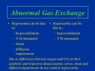
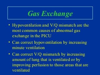
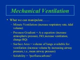
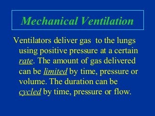
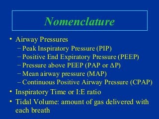
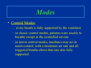
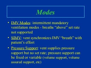
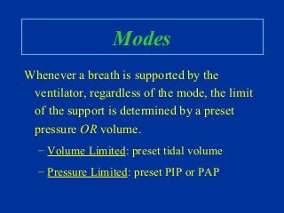
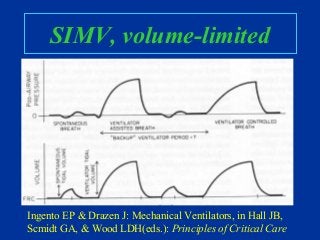
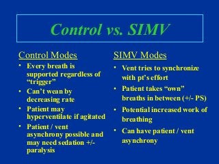
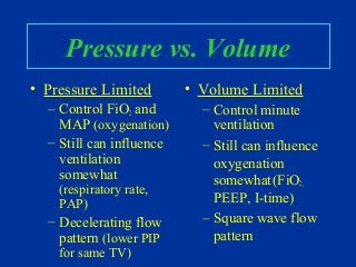
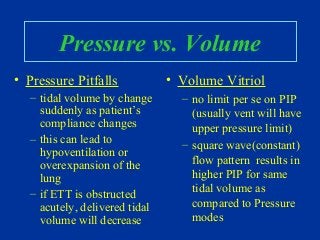
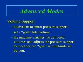
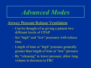
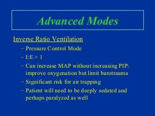
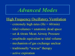
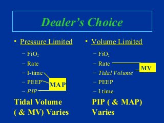
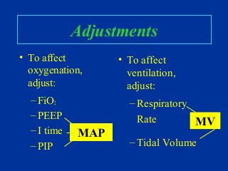
This document provides an overview of mechanical ventilation including:
- Indications for mechanical ventilation including respiratory and cardiac failure.
- Basic anatomy and physiology of ventilation including the roles of airways, alveoli, and pressures.
- Common modes of ventilation like assist-control, IMV, SIMV and their characteristics.
- Factors to consider when selecting initial settings like rate, pressures, and tidal volumes.
- How to adjust settings to impact oxygenation and ventilation.
- Potential problems that can arise with mechanical ventilation.
This document provides an overview of mechanical ventilation settings, modes, advantages and disadvantages of different modes, guidelines for initiation, and examples of troubleshooting. It discusses settings like trigger sensitivity, tidal volume, PEEP, and rates. Modes covered include assist-control, pressure support, and SIMV. Guidelines recommend starting with low tidal volumes and optimizing PEEP and FiO2. Troubleshooting examines causes of high pressures, coping with COPD patients, improving synchrony, and managing ARDS.
This document discusses breathing systems used during anesthesia. It begins with definitions and a brief history of breathing systems. It then classifies different breathing systems and describes the working principles and components of various systems, including Mapleson systems and the circle system. Key points covered include how fresh gas flow rates impact carbon dioxide levels, components of the circle system like the reservoir bag and carbon dioxide absorbers, and factors that influence the absorptive capacity of different carbon dioxide absorbents.
Prof. Mridul Panditrao wants to share his much acclaimed CME lecture in ISACON 2014, Madurai, India and many other places, on one of the very very important but often ununderstood and neglected essential topics in Anesthesia..... Vaporizers!!
Anesthetic equipment and breathing systems are used to deliver precise concentrations of oxygen, anesthetic gases, and ventilation to patients during procedures requiring anesthesia. Key components include:
1) An anesthetic machine with flow meters to control gas delivery and a vaporizer to add anesthetic gas to the carrier gas.
2) An anesthetic breathing system, such as a semi-closed circle system, to transport gases between the machine and patient while preventing excessive rebreathing and absorbing carbon dioxide using a reservoir bag and absorbent canister.
3) Monitoring devices to ensure proper gas delivery and ventilation are provided to the patient.
1) Vaporizers are devices that convert liquid anesthetic agents into vapor and add a controlled amount of vapor to the breathing system.
2) There are various types of vaporizers that use different mechanisms for vaporization and temperature compensation. Common vaporizers include the TEC, Goldman, and Copper Kettle vaporizers.
3) Special desflurane vaporizers are required due to desflurane's high vapor pressure, as standard vaporizers could result in dangerously high concentrations being delivered to the patient.
This document provides information on various types of non-rebreathing valves used in anesthesia and resuscitation equipment. It describes the components, mechanics, and classifications of common valves such as the AMBU, Fink, Frumin, Laerdal, Lewis-Leigh, and Ruben valves. The valves are classified based on their mechanical characteristics and whether they are designed for spontaneous, controlled, or both types of ventilation. Their functions, evaluations in terms of resistance, dead space, and back flow, and care instructions are also outlined.
This document provides information about vaporizers used in anesthesia. It defines key terms like vapor, gas, and vaporizer. It then discusses the history of vaporizers, describing important developments from the 1500s to modern electronic vaporizers. The rest of the document covers the physics principles behind vaporizers like vapor pressure and boiling point. It also explains different vaporizer designs, mechanisms for regulating output concentrations, and methods of vaporization and temperature compensation.
This document discusses ventilator settings and modes. It begins by defining a ventilator and listing some key settings such as respiratory rate, tidal volume, minute ventilation, fraction of inspired oxygen, and positive end expiratory pressure. It then discusses the different types of ventilator modes: controlled modes (e.g. volume control, pressure control), supported modes (e.g. pressure support), and combination modes (e.g. SIMV with pressure support). The document concludes by outlining the steps for assessing a patient's readiness for weaning from the ventilator and describing methods for weaning such as a spontaneous breathing trial.
This document provides an overview of respiratory physiology. It discusses the processes of external and internal respiration, ventilation, lung volumes and capacities, pressures and gradients, spontaneous and positive pressure ventilation, lung characteristics, compliance, elastic forces, surface tension, surfactant, ventilation, perfusion, gas tensions, the oxygen cascade, oxygen transport via dissolved oxygen and hemoglobin, and factors affecting hemoglobin dissociation.
This document provides information on breathing systems used in anesthesia. It defines breathing systems and discusses their key components and classifications. The Dr. discusses the ideal characteristics of breathing systems and describes various Mapleson systems (A-F) - how they function during spontaneous and controlled ventilation. Highlights include that Mapleson A is efficient for spontaneous breathing but not controlled ventilation, while Bain's modification of Mapleson D is well-suited for both. The document also covers testing and advantages/disadvantages of different breathing circuits.
This document describes various components and types of breathing circuits used in anesthesia. It discusses the basic principles of delivering oxygen/gases and eliminating carbon dioxide. The key components described include the reservoir bag, breathing tubes, adjustable pressure limiting valve, and filters. Circuits are classified based on gas flow and include open, semi-open, closed, and semi-closed types. Specific circuits discussed in detail include the Mapleson A-F circuits, Bain's circuit, and the circle breathing system. Advantages and disadvantages of each system are provided.
Vaporizers are used to convert liquid anesthetic agents into vapor and add a controlled amount of this vapor to the fresh gas flow. The output concentration of a vaporizer can be regulated by adjusting the splitting ratio between the gas flowing through the vaporizing chamber and bypass chamber. Temperature compensation mechanisms like a bimetallic strip are used to maintain a constant output despite temperature fluctuations. Factors like gas flow rate, carrier gas composition, ambient pressure, and vaporizer design can influence the vaporizer's output concentration.
Mechanical ventilation provides oxygen and removes carbon dioxide when a patient is unable to breathe adequately on their own. It requires an understanding of pulmonary physiology and close collaboration between nurses, doctors, and respiratory therapists to set ventilation goals and monitor the patient's response. Positive outcomes depend on tailoring care to individual patient needs and ensuring open communication within the healthcare team.
This document discusses different types of breathing systems used in anaesthesia. It describes the components and ideal properties of breathing systems. Open, semi-open, and semi-closed systems are defined. Several specific semi-open systems are outlined, including the Mapleson A, D, F systems. Semi-closed systems require CO2 absorbents and lower fresh gas flows than open systems. Types of semi-closed systems are also defined. The document emphasizes the importance of ensuring tight connections between breathing system components.
The document discusses mechanical ventilation and the mechanics of breathing. It covers topics like spontaneous breathing, respiration, ventilation, gas flow and pressure gradients in the lungs during breathing, compliance, resistance, time constants, and different types of ventilators including conventional and high frequency ventilators.
Capnography measures ventilation by detecting exhaled carbon dioxide (CO2) and provides a graphical waveform that can be interpreted. Pulse oximetry measures oxygenation by detecting oxygen levels in the blood. Capnography is useful for confirming endotracheal tube placement, detecting tube displacement, assessing chest compressions during CPR, and detecting return of spontaneous circulation. It also helps evaluate and monitor respiratory conditions, hypoventilation states, and low perfusion states in intubated and non-intubated patients.
The document discusses different types of breathing circuits used in anesthesia. It begins by describing the basic components and functions of a breathing circuit, which delivers oxygen and anesthetic gases to patients while removing carbon dioxide. Circuits are classified as open, semi-open, semi-closed, or closed based on how exhaust gases are handled. Several specific circuit types are then outlined in detail, including the Mapleson A, Bain, Ayres T-piece, and Jackson-Rees systems. Key features and uses of each system are provided. Semi-closed circuits are explained as using a carbon dioxide absorber to remove carbon dioxide from exhaled gases so they can be rebreathed, allowing for lower fresh gas flow rates than open systems
This document discusses various types of breathing systems used in anesthesia including open, semi-open, semi-closed and closed systems. It provides details on common breathing systems such as the circle system, Mapleson classifications A-F, Bain system and Jackson-Rees modification. The ideal properties of a breathing system are also listed.
Mechanical ventilation graphics provide important information to interpret patient response, disease status, and ventilator function. Scalars plot pressure, volume, or flow over time, while loops plot pressure versus volume or flow versus volume with no time component. Common waveforms include square, ramp, and sine waves. Pressure modes result in square pressure waves while volume modes produce ramp waves. Loops can indicate breath type and assess issues like air trapping, resistance, compliance, and asynchrony. Graphical analysis is a critical tool for ventilator management and optimization.
Vaporizers are devices that change liquid anesthetic agents into vapor and add a controlled amount of vapor to the gas flow or breathing system. They do this by utilizing concepts like vapor pressure, boiling point, and partial pressure. There are several types of vaporizers including concentration calibrated vaporizers, measured flow vaporizers, and electronic vaporizers. Key factors that affect vaporization include temperature, flow rate, volatility of the agent, and carrier gas composition. Ambient pressure changes from high altitude, hyperbaric conditions, or back pressure can impact the vaporizer's output.
The document provides information on breathing systems used in anesthesia. It discusses the components and classifications of breathing systems. The key types discussed are the Mapleson systems (A, B, C, D, E), which are bidirectional flow systems classified by the placement of the reservoir bag. The Mapleson systems are analyzed in terms of their efficiency for spontaneous and controlled ventilation. The Bain modification of the Mapleson D system is also described.
This document discusses breathing systems used in anesthesia. It defines a breathing system and lists its main components. The key requirements of an effective breathing system are to deliver accurate gas concentrations, eliminate carbon dioxide, minimize dead space, and have low resistance. Various configurations are described, including open, semi-open, semi-closed and closed systems. Popular breathing circuits like Mapleson A, B, C, D, E and F are explained along with the Ayre's T-piece and reservoir bag. The document provides details on how different breathing systems function during spontaneous and controlled ventilation.
This document provides an overview of mechanical ventilation, including:
1) How mechanical ventilation helps reduce the work of breathing and restore gas exchange through invasive and noninvasive positive pressure ventilation.
2) The basics of monitoring pressure, volume, flow, and pressure-time curves at the bedside.
3) Important considerations for mechanical ventilation including potential adverse effects on hemodynamics, lungs, and gas exchange, and how to address issues like auto-PEEP.
The document contains a series of slides related to mechanical ventilation. It includes questions about adjusting ventilator settings to increase oxygenation or wash out carbon dioxide. It also discusses concepts like compliance, plateau pressure, modes of ventilation including CPAP, PS, CMV, SIMV, and SIMV + PS. Pressure, flow, and volume waves are shown. Potential complications and functions of mechanical ventilation are listed.
Respiratory changes during anesthesia and ippvImran Sheikh
Anesthesia causes impairment of respiratory function through several mechanisms. It decreases functional residual capacity and lung compliance while increasing respiratory resistance. This leads to atelectasis in 15-20% of the lung and ventilation/perfusion mismatching. Maintaining muscle tone, applying positive end-expiratory pressure, recruitment maneuvers using sustained high inspiratory pressures, and limiting oxygen concentrations can help prevent atelectasis formation. Anesthesia also redistributes ventilation away from dependent lung regions and inhibits hypoxic pulmonary vasoconstriction.
This document summarizes the history and components of breathing systems used in anesthesiology. It discusses the evolution of breathing circuits from early simple open systems to more advanced closed and semi-closed systems incorporating reservoirs, valves, filters and CO2 absorbers. Key systems are described, including Mapleson classifications and the Magill circuit. The essential criteria of an ideal breathing system and desirable secondary criteria are also outlined.
This document discusses hypertensive crisis management in hypoxemic ICU patients. It first presents a case report where administration of a vasodilator to treat hypertension worsened the patient's hypoxia by inhibiting hypoxic pulmonary vasoconstriction. It then reviews the physiological role and mechanisms of hypoxic pulmonary vasoconstriction, including how it redistributes blood flow to optimally ventilated lung regions. Finally, it addresses understanding and managing hypertensive crisis in hypoxic patients, noting how conditions like pain, awakening, and rebound effects can provoke sympathetic hyperactivity and rise in blood pressure.
Neo Lucratia Montja is a South African female with a National Diploma in Credit Management from the University of Johannesburg from 2003 to 2005. She is currently studying for a BCompt Accounting degree from the University of South Africa. She has over 10 years of experience in banking and credit roles at First National Bank including teller, debt collector, credit analyst, relationship consultant, and currently a customer portfolio analyst. She has a driver's license and is proficient in Microsoft Office.
Acuity is a top-ranked property and casualty insurance firm that attributes its success and competitive differentiation to low voluntary staff turnover of less than 2% and benefits/perks. The company gains loyalty through long-term staff retention and diversity initiatives like cross-training employees and protecting customer privacy. Acuity also fosters social responsibility through community events and uses integrity, creativity, and simplicity to maintain its high ranking as the top insurance provider in the United States.
Digital Marketing Setup and Strategy WorkshopDan Stratford
Digital Marketing workshop taught by Author of SEO for Dummies Peter Kent, industry experts Dan Stratford, Dave Carlson and Laura Pence. Includes teaching on SEO, paid search, website development, social media and more.
Thomas Lewis is a senior at Rock Ridge High School who has volunteered with his church's youth programs and for charitable causes in Africa. He has served as captain of his high school varsity soccer team and club soccer team, and is a member of the Educators Rising program. Thomas has leadership skills and proficiency with Microsoft Office, and is working to learn American Sign Language.
TIPO DE CAMBIO DEL DÓLAR DE ESTADOS UNIDOS 20 DE ENERO DE 2016 Tipo de cambio publicado por el Banco de México en el Diario Oficial de la Federación para solventar obligaciones denominadas en moneda extranjera pagaderas en la República Mexicana.
The document discusses the benefits of exercise for mental health. Regular physical activity can help reduce anxiety and depression and improve mood and cognitive functioning. Exercise causes chemical changes in the brain that may help protect against mental illness and improve symptoms.
Thank you for the detailed presentation on mechanical ventilation in pediatrics. I appreciate you taking the time to explain the key concepts and parameters.
This document provides information on basic mechanical ventilation. It discusses various indications for mechanical ventilation including conditions like pneumonia, ARDS, pulmonary edema, and neuromuscular disorders. It then describes the basic components and functions of a mechanical ventilator including volume change, time, gas flow, and pressure difference. Key parameters like compliance, PEEP, and I:E ratio that are important for mechanical ventilation are explained. Different ventilator modes are outlined including pressure control, volume control, SIMV, and PSV. Settings like tidal volume, pressure, and respiratory rate that should be optimized are also reviewed.
This document discusses various modes of mechanical ventilation. It begins by defining what a ventilation mode is, noting that a mode describes the control, phase, and conditional variables in mandatory, spontaneous, or combined breaths. It then discusses different control variables like pressure, volume, and flow. It explains phase variables that initiate, sustain, and end inspiration. Limit and cycle variables that determine the magnitude and end of inspiration are also covered. Common modes like pressure control, volume control, and their advantages and disadvantages are summarized. The document provides details on interpreting pressure waveforms and calculating plateau pressure.
This document provides an overview of various ventilator modes and concepts. It begins by defining what a ventilator mode is and describes the key components of a ventilator breath, including control variables, phase variables, and conditional variables. It then explains different types of breaths that can be delivered and various modes, focusing on pressure control, volume control, SIMV, pressure support, and their advantages and disadvantages. Key concepts like auto-PEEP, rise time, and cycling are discussed in the context of pressure support ventilation. Overall, the document aims to educate on the fundamental principles of mechanical ventilation.
Newer modes of ventilation aim to improve on conventional modes by being "closed loop" and adapting to changes in the patient's lung mechanics and respiratory effort. PRVC is one such mode that maintains a target tidal volume with automatic adjustment of pressure support. Other modes like APRV, PAV, and NAVA aim to improve patient-ventilator synchrony and reduce the work of breathing. Modes like VAPS and ASV use both pressure and volume control to guarantee a minimum tidal volume. Neurally adjusted modes like NAVA base support on neural respiratory drive rather than pressures or flows. Overall, newer modes try to prevent lung injury, asynchrony, and promote faster weaning through closed-loop feedback and adaptation
The document discusses the different modes, parameters, and variables of mechanical ventilation, providing definitions and examples of various modes like volume control, pressure control, PRVC, SIMV, and pressure support and discussing parameters like tidal volume, respiratory rate, PEEP, and I:E ratio that must be set and monitored to effectively ventilate patients using these different modes.
This document provides information on mechanical ventilation, including indications, criteria, principles, terminology, modes, pressures, and settings. The key points are:
1. Mechanical ventilation is indicated for respiratory failure (type I or II) or to provide airway protection. Criteria include clinical assessment, ABGs, and physiological parameters.
2. Ventilation aims to facilitate CO2 release while maintaining normal PaCO2. Oxygenation aims to maximize O2 delivery by improving V/Q matching.
3. Common modes include controlled mandatory ventilation (CMV), intermittent mandatory ventilation (IMV), and synchronized IMV (SIMV). Settings must be tailored to the individual patient.
Mechanical ventilation in neonates by dr naved akhterDr Naved Akhter
Mechanical ventilation is used to support gas exchange and clinical status in neonates. The goals are to maintain sufficient oxygenation and ventilation until the underlying disease resolves, while protecting the lungs from damage. Modes of ventilation include mandatory, SIMV, assist/control, and pressure support. Parameters like tidal volume, PIP, PEEP, and FiO2 are adjusted based on blood gas levels to optimize oxygenation and ventilation. Ventilator graphics and pulmonary monitoring are used to assess patient-ventilator interaction and guide management.
This document provides an overview of basic concepts and applications of mechanical ventilation. It discusses various ventilation modes including controlled, assisted, assist-control, IMV, and SIMV modes. It also covers settings such as tidal volume, respiratory rate, I:E ratio, and FIO2. Key aspects of setting up and monitoring mechanical ventilation are summarized, including how to initially set parameters based on patient size and desired minute ventilation. Factors that affect oxygenation and ventilation are outlined. Waveforms and pressure-volume loops are presented to illustrate lung mechanics under different conditions. The importance of monitoring airway pressures and compliance is emphasized to optimize ventilation.
The document provides information on various modes of mechanical ventilation and strategies for weaning patients off ventilators. It discusses negative pressure ventilation techniques like iron lungs as well as modern positive pressure modes like pressure control ventilation, synchronized intermittent mandatory ventilation (SIMV), and proportional assist ventilation (PAV). The goals of mechanical ventilation are to maintain ventilation and tissue oxygenation while decreasing the work of breathing. Modes are selected based on the level of support needed and to facilitate eventual weaning from the ventilator.
Mechanical ventilation uses positive pressure to deliver gas to the lungs. There are several modes that have evolved over time including negative pressure ventilation and newer microprocessor controlled positive pressure systems. The basic function is to deliver gas to the lungs while parameters like tidal volume, respiratory rate, pressures and timing are adjusted based on the patient's condition and response. Common modes include controlled mandatory ventilation which provides all breaths from the ventilator, assist control which provides mandatory breaths plus additional breaths if patient triggers, and synchronized intermittent mandatory ventilation which aims to prevent breath stacking by synchronizing mandatory breaths with patient effort.
Mechanical ventilation & Pulmonary Rehabilitation -1.pdfAdamu Mohammad
Mechanical ventilation is used to support patients with respiratory failure by controlling parameters like tidal volume, respiratory rate, and pressure. It requires careful setting and monitoring to prevent complications. Modes include controlled, assisted, and combined settings. Pulmonary rehabilitation uses exercise, education, and breathing techniques to improve symptoms and quality of life for patients with chronic lung disease.
This document discusses different modes of mechanical ventilation. It begins by introducing mechanical ventilation and its purpose of providing respiratory support. It then describes the basic components of a ventilator and ventilator circuit. The document outlines several modes of mechanical ventilation including controlled mechanical ventilation, assist-control ventilation, intermittent mandatory ventilation, and synchronized intermittent mandatory ventilation. It provides details on the characteristics, advantages, and disadvantages of each mode.
Andreas Vesalius in 1555 suggested opening the trachea and inserting a tube to allow the lung to reinflate and strengthen the heart, representing one of the earliest descriptions of mechanical ventilation.
Dr. Nikhil Yadav's document discusses various modes of mechanical ventilation including controlled modes like volume control and pressure control ventilation, assisted modes like assist-control and synchronized intermittent mandatory ventilation, and spontaneous breathing modes like pressure support ventilation and proportional assist ventilation. The summary provides a high-level overview of the key topics and historical context covered in the document.
1. Ventilation involves delivering gas to the lungs at an appropriate tidal volume, minute ventilation, and oxygen concentration to ensure adequate oxygenation and carbon dioxide elimination.
2. Various modes of ventilation include pressure-triggered modes like SIMV and pressure support ventilation as well as newer hybrid modes that combine features. Parameters like PIP, PEEP, rates and triggers must be optimized.
3. Weaning from ventilation requires gradually reducing support by lowering PIP, PEEP and FiO2 while monitoring blood gases to ensure adequate oxygen and carbon dioxide levels are maintained before considering extubation.
This document provides an overview of mechanical ventilation including:
- The basic components and goals of mechanical ventilators.
- Different modes of ventilation such as controlled, assisted, and pressure support ventilation.
- Parameters for setting up and monitoring ventilation like tidal volume, PEEP, and blood gases.
- Indications for initiating and weaning from ventilation.
- Potential complications and ways to troubleshoot issues with the ventilator or patient ventilation.
Does Over-Masturbation Contribute to Chronic Prostatitis.pptxwalterHu5
In some case, your chronic prostatitis may be related to over-masturbation. Generally, natural medicine Diuretic and Anti-inflammatory Pill can help mee get a cure.
Travel vaccination in Manchester offers comprehensive immunization services for individuals planning international trips. Expert healthcare providers administer vaccines tailored to your destination, ensuring you stay protected against various diseases. Conveniently located clinics and flexible appointment options make it easy to get the necessary shots before your journey. Stay healthy and travel with confidence by getting vaccinated in Manchester. Visit us: www.nxhealthcare.co.uk
Clinic ^%[+27633867063*Abortion Pills For Sale In Tembisa Central19various
Clinic ^%[+27633867063*Abortion Pills For Sale In Tembisa Central Clinic ^%[+27633867063*Abortion Pills For Sale In Tembisa CentralClinic ^%[+27633867063*Abortion Pills For Sale In Tembisa CentralClinic ^%[+27633867063*Abortion Pills For Sale In Tembisa CentralClinic ^%[+27633867063*Abortion Pills For Sale In Tembisa Central
These lecture slides, by Dr Sidra Arshad, offer a simplified look into the mechanisms involved in the regulation of respiration:
Learning objectives:
1. Describe the organisation of respiratory center
2. Describe the nervous control of inspiration and respiratory rhythm
3. Describe the functions of the dorsal and respiratory groups of neurons
4. Describe the influences of the Pneumotaxic and Apneustic centers
5. Explain the role of Hering-Breur inflation reflex in regulation of inspiration
6. Explain the role of central chemoreceptors in regulation of respiration
7. Explain the role of peripheral chemoreceptors in regulation of respiration
8. Explain the regulation of respiration during exercise
9. Integrate the respiratory regulatory mechanisms
10. Describe the Cheyne-Stokes breathing
Study Resources:
1. Chapter 42, Guyton and Hall Textbook of Medical Physiology, 14th edition
2. Chapter 36, Ganong’s Review of Medical Physiology, 26th edition
3. Chapter 13, Human Physiology by Lauralee Sherwood, 9th edition
Histololgy of Female Reproductive System.pptxAyeshaZaid1
Dive into an in-depth exploration of the histological structure of female reproductive system with this comprehensive lecture. Presented by Dr. Ayesha Irfan, Assistant Professor of Anatomy, this presentation covers the Gross anatomy and functional histology of the female reproductive organs. Ideal for students, educators, and anyone interested in medical science, this lecture provides clear explanations, detailed diagrams, and valuable insights into female reproductive system. Enhance your knowledge and understanding of this essential aspect of human biology.
10 Benefits an EPCR Software should Bring to EMS Organizations Traumasoft LLC
The benefits of an ePCR solution should extend to the whole EMS organization, not just certain groups of people or certain departments. It should provide more than just a form for entering and a database for storing information. It should also include a workflow of how information is communicated, used and stored across the entire organization.
- Video recording of this lecture in English language: https://youtu.be/Pt1nA32sdHQ
- Video recording of this lecture in Arabic language: https://youtu.be/uFdc9F0rlP0
- Link to download the book free: https://nephrotube.blogspot.com/p/nephrotube-nephrology-books.html
- Link to NephroTube website: www.NephroTube.com
- Link to NephroTube social media accounts: https://nephrotube.blogspot.com/p/join-nephrotube-on-social-media.html
Cell Therapy Expansion and Challenges in Autoimmune DiseaseHealth Advances
There is increasing confidence that cell therapies will soon play a role in the treatment of autoimmune disorders, but the extent of this impact remains to be seen. Early readouts on autologous CAR-Ts in lupus are encouraging, but manufacturing and cost limitations are likely to restrict access to highly refractory patients. Allogeneic CAR-Ts have the potential to broaden access to earlier lines of treatment due to their inherent cost benefits, however they will need to demonstrate comparable or improved efficacy to established modalities.
In addition to infrastructure and capacity constraints, CAR-Ts face a very different risk-benefit dynamic in autoimmune compared to oncology, highlighting the need for tolerable therapies with low adverse event risk. CAR-NK and Treg-based therapies are also being developed in certain autoimmune disorders and may demonstrate favorable safety profiles. Several novel non-cell therapies such as bispecific antibodies, nanobodies, and RNAi drugs, may also offer future alternative competitive solutions with variable value propositions.
Widespread adoption of cell therapies will not only require strong efficacy and safety data, but also adapted pricing and access strategies. At oncology-based price points, CAR-Ts are unlikely to achieve broad market access in autoimmune disorders, with eligible patient populations that are potentially orders of magnitude greater than the number of currently addressable cancer patients. Developers have made strides towards reducing cell therapy COGS while improving manufacturing efficiency, but payors will inevitably restrict access until more sustainable pricing is achieved.
Despite these headwinds, industry leaders and investors remain confident that cell therapies are poised to address significant unmet need in patients suffering from autoimmune disorders. However, the extent of this impact on the treatment landscape remains to be seen, as the industry rapidly approaches an inflection point.
Travel Clinic Cardiff: Health Advice for International TravelersNX Healthcare
Travel Clinic Cardiff offers comprehensive travel health services, including vaccinations, travel advice, and preventive care for international travelers. Our expert team ensures you are well-prepared and protected for your journey, providing personalized consultations tailored to your destination. Conveniently located in Cardiff, we help you travel with confidence and peace of mind. Visit us: www.nxhealthcare.co.uk
2. Introduction
• Indications
• Basic anatomy and physiology
• Modes of ventilation
• Selection of mode and settings
• Common problems
• Complications
• Weaning and extubation
4. Indications
• Cardiac Insufficiency
– eliminate work of breathing
– reduce oxygen consumption
• Neurologic dysfunction
– central hypoventilation/ frequent apnea
– patient comatose, GCS < 8
– inability to protect airway
5. Basic Anatomy
• Upper Airway
– humidifies inhaled gases
– site of most resistance to airflow
• Lower Airway
– conducting airways (anatomic dead space)
– respiratory bronchioles and alveoli (gas
exchange)
6. Basic Physiology
• Negative pressure circuit
– Gradient between mouth and pleural
space is the driving pressure
– need to overcome resistance
– maintain alveolus open
• overcome elastic recoil forces
– Balance between elastic recoil of chest
wall and the lung
9. Ventilation
• Carbon Dioxide
PaCO2= k * metabolic production
alveolar minute ventilation
Alveolar MV = resp. rate * effective tidal vol.
Effective TV = TV - dead space
Dead Space = anatomic + physiologic
10. Oxygenation
• Oxygen:
– Minute ventilation is the amount of fresh gas
delivered to the alveolus
– Partial pressure of oxygen in alveolus (PAO2) is the
driving pressure for gas exchange across the
alveolar-capillary barrier
– PAO2 = ({Atmospheric pressure - water
vapor}*FiO2) - PaCO2 / RQ
– Match perfusion to alveoli that are well ventilated
– Hemoglobin is fully saturated 1/3 of the way thru
the capillary
13. Abnormal Gas Exchange
• Hypoxemia can be due
to:
– hypoventilation
– V/Q mismatch
– shunt
– diffusion
impairments
• Hypercarbia can be
due to:
– hypoventilation
– V/Q mismatch
Due to differences between oxygen and CO2 in their
solubility and respective disassociation curves, shunt and
diffusion impairments do not result in hypercarbia
14. Gas Exchange
• Hypoventilation and V/Q mismatch are the
most common causes of abnormal gas
exchange in the PICU
• Can correct hypoventilation by increasing
minute ventilation
• Can correct V/Q mismatch by increasing
amount of lung that is ventilated or by
improving perfusion to those areas that are
ventilated
15. Mechanical Ventilation
• What we can manipulate……
– Minute Ventilation (increase respiratory rate, tidal
volume)
– Pressure Gradient = A-a equation (increase
atmospheric pressure, FiO2,increase ventilation,
change RQ)
– Surface Area = volume of lungs available for
ventilation (increase volume by increasing airway
pressure, i.e., mean airway pressure)
– Solubility = ?perflurocarbons?
16. Mechanical Ventilation
Ventilators deliver gas to the lungs
using positive pressure at a certain
rate. The amount of gas delivered
can be limited by time, pressure or
volume. The duration can be
cycled by time, pressure or flow.
17. Nomenclature
• Airway Pressures
– Peak Inspiratory Pressure (PIP)
– Positive End Expiratory Pressure (PEEP)
– Pressure above PEEP (PAP or ΔP)
– Mean airway pressure (MAP)
– Continuous Positive Airway Pressure (CPAP)
• Inspiratory Time or I:E ratio
• Tidal Volume: amount of gas delivered with
each breath
18. Modes
• Control Modes:
– every breath is fully supported by the ventilator
– in classic control modes, patients were unable to
breathe except at the controlled set rate
– in newer control modes, machines may act in
assist-control, with a minimum set rate and all
triggered breaths above that rate also fully
supported.
19. Modes
• IMV Modes: intermittent mandatory
ventilation modes - breaths “above” set rate
not supported
• SIMV: vent synchronizes IMV “breath” with
patient’s effort
• Pressure Support: vent supplies pressure
support but no set rate; pressure support can
be fixed or variable (volume support, volume
assured support, etc)
20. Modes
Whenever a breath is supported by the
ventilator, regardless of the mode, the limit
of the support is determined by a preset
pressure OR volume.
– Volume Limited: preset tidal volume
– Pressure Limited: preset PIP or PAP
21. Mechanical Ventilation
If volume is set, pressure varies…..if
pressure is set, volume varies…..
….according to the compliance…...
COMPLIANCE =
∆ Volume / ∆ Pressure
22. Compliance
Burton SL & Hubmayr RD: Determinants of Patient-Ventilator Interactions:
Bedside Waveform Analysis, in Tobin MJ (ed): Principles & Practice of Intensive
Care Monitoring
23. Assist-control, volume
Ingento EP & Drazen J: Mechanical Ventilators, in Hall JB,
Scmidt GA, & Wood LDH(eds.): Principles of Critical Care
24. IMV, volume-limited
Ingento EP & Drazen J: Mechanical Ventilators, in Hall JB,
Scmidt GA, & Wood LDH(eds.): Principles of Critical Care
25. SIMV, volume-limited
Ingento EP & Drazen J: Mechanical Ventilators, in Hall JB,
Scmidt GA, & Wood LDH(eds.): Principles of Critical Care
26. Control vs. SIMV
Control Modes
• Every breath is
supported regardless of
“trigger”
• Can’t wean by
decreasing rate
• Patient may
hyperventilate if agitated
• Patient / vent
asynchrony possible and
may need sedation +/-
paralysis
SIMV Modes
• Vent tries to synchronize
with pt’s effort
• Patient takes “own”
breaths in between (+/- PS)
• Potential increased work of
breathing
• Can have patient / vent
asynchrony
27. Pressure vs. Volume
• Pressure Limited
– Control FiO2 and
MAP (oxygenation)
– Still can influence
ventilation
somewhat
(respiratory rate,
PAP)
– Decelerating flow
pattern (lower PIP
for same TV)
• Volume Limited
– Control minute
ventilation
– Still can influence
oxygenation
somewhat(FiO2,
PEEP, I-time)
– Square wave flow
pattern
28. Pressure vs. Volume
• Pressure Pitfalls
– tidal volume by change
suddenly as patient’s
compliance changes
– this can lead to
hypoventilation or
overexpansion of the
lung
– if ETT is obstructed
acutely, delivered tidal
volume will decrease
• Volume Vitriol
– no limit per se on PIP
(usually vent will have
upper pressure limit)
– square wave(constant)
flow pattern results in
higher PIP for same
tidal volume as
compared to Pressure
modes
29. Trigger
• How does the vent know when to give a
breath? - “Trigger”
– patient effort
– elapsed time
• The patient’s effort can be “sensed” as a
change in pressure or a change in flow
(in the circuit)
30. Need a hand??
Pressure Support
• “Triggering” vent requires certain amount of
work by patient
• Can decrease work of breathing by providing
flow during inspiration for patient triggered
breaths
• Can be given with spontaneous breaths in IMV
modes or as stand alone mode without set rate
• Flow-cycled
31. Advanced Modes
• Pressure-regulated volume control
(PRVC)
• Volume support
• Inverse ratio (IRV) or airway-pressure
release ventilation (APRV)
• Bilevel
• High-frequency
32. Advanced Modes
PRVC
A control mode, which delivers a set
tidal volume with each breath at the
lowest possible peak pressure. Delivers
the breath with a decelerating flow
pattern that is thought to be less injurious
to the lung…… “the guided hand”.
33. Advanced Modes
Volume Support
– equivalent to smart pressure support
– set a “goal” tidal volume
– the machine watches the delivered
volumes and adjusts the pressure support
to meet desired “goal” within limits set
by you.
34. Advanced Modes
Airway Pressure Release Ventilation
– Can be thought of as giving a patient two
different levels of CPAP
– Set “high” and “low” pressures with release
time
– Length of time at “high” pressure generally
greater than length of time at “low” pressure
– By “releasing” to lower pressure, allow lung
volume to decrease to FRC
35. Advanced Modes
Inverse Ratio Ventilation
– Pressure Control Mode
– I:E > 1
– Can increase MAP without increasing PIP:
improve oxygenation but limit barotrauma
– Significant risk for air trapping
– Patient will need to be deeply sedated and
perhaps paralyzed as well
36. Advanced Modes
High Frequency Oscillatory Ventilation
– extremely high rates (Hz = 60/min)
– tidal volumes < anatomic dead space
– set & titrate Mean Airway Pressure
– amplitude equivalent to tidal volume
– mechanism of gas exchange unclear
– traditionally “rescue” therapy
– active expiration
37. Advanced Modes
High Frequency Oscillatory Ventilation
– patient must be paralyzed
– cannot suction frequently as disconnecting the
patient from the oscillator can result in volume
loss in the lung
– likewise, patient cannot be turned frequently so
decubiti can be an issue
– turn and suction patient 1-2x/day if they can
tolerate it
38. Advanced Modes
Non Invasive Positive Pressure Ventilation
– Deliver PS and CPAP via tight fitting mask
(BiPAP: bi-level positive airway pressure)
– Can set “back up” rate
– May still need sedation
39. Initial Settings
• Pressure Limited
– FiO2
– Rate
– I-time or I:E ratio
– PEEP
– PIP or PAP
• Volume Limited
– FiO2
– Rate
– I-time or I:E ratio
– PEEP
– Tidal Volume
These choices are with time - cycled ventilators.
Flow cycled vents are available but not commonly
used in pediatrics.
40. Initial Settings
• Settings
– Rate: start with a rate that is somewhat
normal; i.e., 15 for adolescent/child, 20-30
for infant/small child
– FiO2: 100% and wean down
– PEEP: 3-5
– Control every breath (A/C) or some (SIMV)
– Mode ?
43. Adjustments
• PEEP
Can be used to help prevent alveolar
collapse at end inspiration; it can also
be used to recruit collapsed lung spaces
or to stent open floppy airways
44. Except...
• Is it really that simple ?
– Increasing PEEP can increase dead space,
decrease cardiac output, increase V/Q
mismatch
– Increasing the respiratory rate can lead to
dynamic hyperinflation (aka auto-PEEP),
resulting in worsening oxygenation and
ventilation
45. Troubleshooting
• Is it working ?
–Look at the patient !!
–Listen to the patient !!
– Pulse Ox, ABG, EtCO2
– Chest X ray
– Look at the vent (PIP; expired TV;
alarms)
46. Troubleshooting
• When in doubt, DISCONNECT THE
PATIENT FROM THE VENT, and begin
bag ventilation.
• Ensure you are bagging with 100% O2.
• This eliminates the vent circuit as the
source of the problem.
• Bagging by hand can also help you gauge
patient’s compliance
47. Troubleshooting
• Airway first: is the tube still in? (may need
DL/EtCO2 to confirm) Is it patent? Is it in the
right position?
• Breathing next: is the chest rising? Breath
sounds present and equal? Changes in exam?
Atelectasis, bronchospasm, pneumothorax,
pneumonia? (Consider needle thoracentesis)
• Circulation: shock? Sepsis?
48. Troubleshooting
• Well, it isn’t working…..
– Right settings ? Right Mode ?
– Does the vent need to do more work ?
• Patient unable to do so
• Underlying process worsening (or new
problem?)
– Air leaks?
– Does the patient need to be more sedated ?
– Does the patient need to be extubated ?
– Vent is only human…..(is it working ?)
49. Troubleshooting
• Patient - Ventilator Interaction
– Vent must recognize patient’s
respiratory efforts (trigger)
– Vent must be able to meet patient’s
demands (response)
– Vent must not interfere with patient’s
efforts (synchrony)
50. Troubleshooting
• Improving Ventilation and/or Oxygenation
– can increase respiratory rate (or decrease rate if
air trapping is an issue)
– can increase tidal volume/PAP to increase tidal
volume
– can increase PEEP to help recruit collapsed
areas
– can increase pressure support and/or decrease
sedation to improve patient’s spontaneous
effort
51. Lowered Expectations
• Permissive Hypercapnia
– accept higher PaCO2s in exchange for limiting
peak airway pressures
– can titrate pH as desired with sodium
bicarbonate or other buffer
• Permissive Hypoxemia
– accept PaO2 of 55-65; SaO2 88-90% in
exchange for limiting FiO2 (<.60) and PEEP
– can maintain oxygen content by keeping
hematocrit > 30%
52. Adjunctive Therapies
• Proning
– re-expand collapsed dorsal areas of the lung
– chest wall has more favorable compliance curve
in prone position
– heart moves away from the lungs
– net result is usually improved oxygenation
– care of patient (suctioning, lines, decubiti)
trickier but not impossible
– not everyone maintains their response or even
responds in the first place
53. Adjunctive Therapies
• Inhaled Nitric Oxide
– vasodilator with very short half life that can be
delivered via ETT
– vasodilate blood vessels that supply ventilated
alveoli and thus improve V/Q
– no systemic effects due to rapid inactivation by
binding to hemoglobin
– improves oxygenation but does not improve
outcome
55. Complications
• Cardiovascular Complications
– Impaired venous return to RH
– Bowing of the Interventricular Septum
– Decreased left sided afterload (good)
– Altered right sided afterload
• Sum Effect…..decreased cardiac output
(usually, not always and often we don’t
even notice)
57. Extubation
• Weaning
– Is the cause of respiratory failure gone or
getting better ?
– Is the patient well oxygenated and
ventilated ?
– Can the heart tolerate the increased work
of breathing ?
58. Extubation
• Weaning (cont.)
– decrease the PEEP (4-5)
– decrease the rate
– decrease the PIP (as needed)
• What you want to do is decrease what
the vent does and see if the patient can
make up the difference….
59. Extubation
• Extubation
– Control of airway reflexes
– Patent upper airway (air leak around tube?)
– Minimal oxygen requirement
– Minimal rate
– Minimize pressure support (0-10)
– “Awake ” patient
Editor's Notes
This presentation will review the basics of mechanical ventilation. First there will be a review of the indications for mechanical ventilation, followed by a brief review of basic anatomy and a more extensive discussion of the physiology of gas exchange in the lungs. Different modes and settings on the ventilator will be examined as well as some problems and potential solutions. After a section on complications related to mechanical ventilation, the process of weaning and extubation will be reviewed.
Patients are often (ideally) intubated before they reach the point of respiratory failure. Respiratory distress can be due to inadequate ventilation, oxygenation or a combination thereof. The process can be either intrinsic to the lungs (pneumonia, for example) or to the chest wall (“pump failure”, as in muscular dystrophies). For some patients, the work of breathing may be such that they are unable to gain weight even in the face of adequate ventilation and oxygenation.
Not every patient who is intubated has a primary pulmonary pathology. For patients in cardiogenic shock or with CHF, the demands of the respiratory system may precipitate cardiovascular collapse. Supporting the patient with mechanical ventilation can reduce the demands on the heart, allowing it to recover.
Intubation can also serve to protect the airway for those who cannot do it themselves. Mechanical ventilation offers the option of hyperventilation for patients with intracranial hypertension.
Remember that the upper airway is a significant site of resistance and this can be added to by iatrogenic devices (nasogastric tubes) or relieved by other devices (nasal trumpets).
When delivering a gas to any patient, give it the way nature does - give it humidified.
The lower airways can be broken down into dead space and the sites where gas exchange occurs. Anything that increases dead space (e.g., PEEP) will impact upon ventilation unless it also increases the area where gas exchange occurs.
During inspiration the gradient becomes more negative as you approach the alveolus. This is an active process; part of the energy used for inspiration is stored in the tissues. This is used in exhalation, which is effectively a passive process.
Recall that work = volume x pressure. For infants with their more compliant chest wall, they will have a proportionally greater work of breathing which helps predispose them to respiratory distress.
The point at which the recoil between the alveolus and the chest wall is balanced is equivalent to functional residual capacity.
I wanted to try to give some background to this slide without getting too weighed down in the physiology of breathing and respiratory failure. Besides, it always confuses me.
http://www.biology.eku.edu/RITCHISO/301notes6.htm
http://physioweb.med.uvm.edu/pulmonary_physiology/
Note that the compliance characteristics of the system (T) is the sum of the compliance curve for the lung (L) and of that for the chest wall (W). At FRC the total pressure in the system is zero - the outward recoil of the chest wall balances the tendency for the alveolus to collapse.
The partial pressure of carbon dioxide in the arterial blood is directly related to metabolic production and indirectly related to minute ventilation. To be accurate, it is alveolar minute ventilation that matters. When a child is tachypneic, the minute ventilation may not change since the increase in rate is balanced by a decrease in tidal volume. However, the amount of dead space has not changed so the effective tidal volume will decrease and hence the effective minute ventilation and thus PaCO2 will increase despite the increased respiratory rate. Likewise, any process that increases dead space without changing minute ventilation will result in an increase in PaCO2 .
To be simplistic, oxygenation involves getting enough oxygen to an alveolus that is perfused. The more volume of gas that can be delivered for exchange, the better. The higher the driving pressure for that gas exchange, the better. Ideally, ventilation (V) and perfusion (Q) are matched so that oxygen is where the blood is (V/Q = 1).
When gas exchange does take place, it is so rapid that a hemoglobin molecule passing through an alveolar capillary is fully saturated before it is one third of the way across. The rest of the capillary represents a reserve for when transit time is increased (e.g., tachycardia) or for when diffusion is slowed (e.g., pulmonary edema, fibrosis) so that hemoglobin still may be fully saturated when it exits the capillary.
[I made this slide to replace the old one that listed factors that affect the rate of gas exchange. I agree that V/Q mismatch will be hard to do well but it is important. I can’t believe that I never realized it wasn’t in my original set of slides. I tell residents that saying “V/Q mismatch” in response to any respiratory question in the ICU will usually be right………What I would like to try to do is to link the physiology of normal gas exchange to abnormal gas exchange and then link that to what we do with the vent…..I.e., increase PEEP to increase area for gas exchange and improve oxygenation, etc etc. This will be tough to do without discussing V/Q mismatch. For what it is worth, here is my attempt at this….]
http://www.biology.eku.edu/RITCHISO/301notes6.html
The sigmoidal shape of the oxygen disassociation curve is of critical importance. Hemoglobin can only carry so much oxygen (1.34 ml per gram of hemoglobin) regardless of high the PaO2 may be. Furthermore, dissolved oxygen contributes very little to oxygen content (0.003 ml oxygen/dL/mmHg PaO2 ). As a result, increasing the PaO2 oxygen in oxygenated blood cannot overcome the effect of shunted blood that is deoxygenated and the patient will be desaturated to a degree proportional to the magnitude of the shunt.
I am going to see if I can scan the curve for carbon dioxide and put it after this slide…..This would be the text for that slide…...
The disassociation curve for carbon dioxide is far more linear and is steeper. As a result, there is much less of a limit on how much carbon dioxide can be carried by hemoglobin and exchanged at the alveolar-capillary barrier. Since carbon dioxide is also far more soluble than oxygen (by a factor of 20), dissolved CO2 contributes significantly -about 10% - to the amount that is exchanged at the alveolus. For these reasons, PaCO2 is not affected by shunt or diffusion barriers as is oxygen (and explains why a child with cyanotic heart disease is hypoxemic but not hypercarbic).
As outlined in the preceding slides, hypoxemia (not hypoxia) can be the result of hypoventilation (not enough delivered) or not matching the delivery to the loading sites (V/Q mismatch). Shunt, whether intracardiac or intrapulmonary, is the ultimate form of V/Q mismatch (V/Q = ). Diffusion impairments must be significant to result in hypoxemia and are rarely of clinical relevance in pediatrics. Hypoventilation is the primary cause of hypercarbia. V/Q mismatch must be profound before hypercarbia results for the reasons discussed in the previous slide.
Maneuvers designed to improve V/Q are beyond the scope of this introductory lecture. Suffice to say, it is hard to selectively manipulate one area of the lung without affecting the entire system. For example, inhaled bronchodilators improve ventilation by relieving bronchospasm; they can also improve perfusion by producing vasodilatation in the alveolar capillary and thereby improve V/Q mismatch. However, there can be systemic absorption of these bronchodilators; this can lead to vasodilatation of vessels that were vasoconstricted secondary to hypoxia. The result is worsening V/Q mismatch and hypoxemia.
The last sentence is awkward on the slide …..as you said, V/Q will be tough to do……I would also like to try and get the topic of proning in briefly later on…the whole idea of “good lung down” or is that up? I think would be hard to do if we want to keep this at 50-60 slides…..should we just mention it but not discuss it ?
We can manipulate the same things a patient does - increase the respiratory rate and tidal volume for each breath. We can also try to keep alveoli from collapsing (as a baby does when they grunt)with positive end expiratory pressure. Unlike patients, we can manipulate the FiO2 or even the atmospheric pressure.
We can also try to open up collapsed areas by applying increased positive pressure. Remember that an alveolus that has collapsed will require a greater amount of pressure for a given change in volume than an alveolus that starts at FRC (see the section on compliance). Increasing the area available for gas exchange improves oxygenation and ventilation.
Perflurocarbons are experimental drugs; they are liquids through which oxygenation and ventilation can occur; one of their advantages is that oxygen is more soluble in perflurocarbons than it is in air. A full discussion of perflurocarbons is beyond the scope of this lecture.
Use this slide to bride pathophsy and vents…..
Most ventilators used in pediatrics are time cycled and operate either in a pressure limited or volume limited mode. Flow cycled ventilators are available but not commonly used.
As almost all of my experience is with servo’s I do not know of any time-limited vents….what would be the difference between a flow-limited and time-cycled vent and a time-limited flow cycled vent? Should we clean up this slide and just say you can limit by pressure or volume and cycle by flow or time?
These are some of the basic terms used with ventilators. CPAP is equivalent to PEEP except the term is usually used when referring to patients who are not intubated (i.e., on nasal CPAP).
In a control mode, the ventilator will guarantee that the patient receives the set tidal volume or PAP with every breath. The patient can breathe “above” the set rate but will receive full support regardless of their effort.
IMV modes support breaths only at the set rate and interval. If the set rate is 10, then every six seconds the patient will receive a machine triggered breath (synchronized to their own effort if the mode is SIMV). In between those 10 breaths, the patient is free to breathe but those breaths are not supported. These breaths can be supported with pressure support (see more below). Lastly, the vent may not give any breaths at all but support the patient’s spontaneous efforts - this is pressure support/CPAP. Newer machines allow for the pressure support to be fixed (e.g., 10 mmHg) or variable (enough support so that the patient receives a tidal volume of 200cc).
As stated before, the limit of support can be volume or pressure.
If one parameter is set, then the other will vary as dictated by the patient’s compliance. The parameter that varies can be followed as an index of the patient’s compliance.
Ref: Burton SL and Hubmayr RD: Determinants of Patient-Ventilator Interactions: Bedside Waveform Analysis,s in Tobin MJ (ed): Principles and Practice of Intensive Care Monitoring. New York, McGraw-Hill, Inc, 1998, p. 656.
Note that the FRC rests on a favorable part of the compliance curve. Small changes in pressure result in large changes in volume. If a patient’s compliance is on this part of the curve, then a given tidal volume will result in low peak pressures. If the compliance worsens (i.e., moves to the left or far right) then the pressure needed to deliver that same tidal volume will increase and the PIP will increase. The same is true if a set pressure is delivered - as compliance improves, the tidal volume will increase. If compliance worsens then a smaller tidal volume will result for the same PAP. Ideally, you want the alveolus to be at the bottom inflection point (FRC) at the beginning of each breath (end of each breath).
Ref: Ingento EP and Drazen J: Mechanical ventilators, in Hall JB, Scmidt GA, and Wood LDH(eds.): Principles of Critical Care. New York, McGraw-Hill, Inc., 1992, p.144.
In assist control modes (pressure control, volume control), the machine will deliver a full breath whether it is triggered by patient effort (note the negative deflection in the uppermost graph indicating patient effort) or triggered by the machine (the machine will act if a set amount of time (T) elapses without discernible patient effort).
Ref: Ingento EP and Drazen J: Mechanical ventilators, in Hall JB, Scmidt GA, and Wood LDH(eds.): Principles of Critical Care. New York, McGraw-Hill, Inc., 1992, p.145.
“Positive pressure, volume-cycled breaths are delivered at a preset rate similar to control mode ventilation, except that between breaths, the inspiratory valve to the patient is open, allowing for spontaneous breathing.”
Ref: Ingento EP and Drazen J: Mechanical ventilators, in Hall JB, Scmidt GA, and Wood LDH(eds.): Principles of Critical Care. New York, McGraw-Hill, Inc., 1992, p.146.
(Would we also want to put slides with pressure mode graphs as well to show the different flow characteristics ??)
During SIMV, the ventilator divides time by the set rate to determine cycle-length. During the early part of this cycle, the patient may breath spontaneously without support. During the terminal phase of this cycle (% varies by manufacturer) the ventilator will synchronize a full breath with detected effort by the patient.
Control modes are used when complete control over the patient’s ventilation and/or oxygenation is desired. This is usually because the patient’s lung disease is significant enough that you that you wish to give maximal support. Another scenario may be one in which you want to precisely control the PaCO2, as in hyperventilation for increased intracranial pressure. Patients placed on control modes are often deeply sedated and may be given neuromuscular blockers.
SIMV modes are chosen when you want the patient to do as much work as they can tolerate and try to minimize the support from the ventilator. SIMV modes are used to wean patients; as you decrease the set rate, the patient will need to do more on their own to maintain normal blood gases. In control modes, if you decrease the rate, the patient’s spontaneous efforts will be fully supported so you will not know how much of that particular tidal volume they are generating on their own.
Note that for the paralyzed patient there is no significant difference between assist control and SIMV.
PRESSURE-LIMITED
I would not say that I have limited ability to affect ventilation in PC, though I may choose to increase the PAP recognizing that I accept the potential for increased baro/volutrauma at the same time
I also accept that I may suffer a decrease in ventilation with changes in compliance.
VOLUME-LIMITED
Accept that changes in compliance may lead to increases in peak airway pressures and associated baro/volutrauma.
Whichever mode one chooses, one needs to be aware of the limitations of that mode. In pressure modes, the tidal volume can drop resulting in hypoventilation or it can increase, leading to overdistention. With volume modes, the peak pressure can increase, resulting in barotrauma if the pulmonary compliance worsens. Regardless of the parameter that is controlled, the other must be monitored as it is a reflection of the compliance and hence the patient’s pulmonary function. Increasing peak pressures on volume mode (or decreasing tidal volumes in pressure modes) can also be a sign that the ETT is obstructed or of another problem with the ventilator circuit.
Ventilators deliver breaths when they are told to do so. This occurs when a certain amount of time has elapsed (e.g., 5 seconds if the rate is 12 [60 sec/12 b/m = 5 sec]) or when the patient makes an effort. A patient’s effort may be sensed as a change in pressure in the circuit (negative deflection) or as a change in flow (also a negative deflection). Flow sensors tend to have a more rapid response time. The amount of support delivered with a patient triggered breath will depend on the mode (assist control vs. IMV vs. SIMV) and the amount of pressure support that is set.
A patient needs to generate a certain amount of work in order to trigger it. Additionally, a patient has to breathe through an ETT that is almost always narrower than their own airway and ventilate the increased dead space imposed by the vent circuit. A patient may not be able to generate adequate tidal volumes for these reasons. To compensate for this increase in the work of breathing, pressure support is given. The ventilator generates pressure support by adding flow to the circuit during patient-triggered breaths in IMV or SIMV modes. This does not make it easier for the patient to trigger the ventilator but it does help the patient generate larger tidal volumes. Pressure support usually terminates when the flow in the circuit is 25% of the peak flow.
As ventilator technology has advanced, newer modes have been developed. Some are variations of volume or pressure modes and some are completely unrelated to conventional mechanical ventilation. It is important to recognize that none of these modes have been shown to be better than another or to reduce mortality for any disease.
This mode combines the benefit of a volume mode (guaranteed minute ventilation) with the benefits of a pressure mode (decelerating flow pattern and a lower PIP for the same tidal volume as compared to volume control).
This mode is designed for spontaneously breathing patients. The machine adjusts the pressure support given with each breath so that a minimal tidal volume and minute ventilation will be achieved. As the patient improves, the amount of pressure support given will decrease. If the patient worsens, pressure support will be increased - smart pressure support. Other modes are available in which the amount of pressure support given is adjusted to the patient’s effort as compared to the actual tidal volume generated. Overall, the principle is the same - the amount of support varies in relation to the patient’s efforts.
I am not sure how clear this is and if we should even include a discussion of it…as far as I know it is only available with the Drager and I have never used it (have contemplated its use)…..
A somewhat complex mode. Two different levels of CPAP are given. The patient can breathe spontaneously at either level and at their own rate. The CPAP is “released” to a lower pressure at set intervals to avoid hyperinflation of the lungs.
By increasing I-time, the MAP will move closer to the PIP although the PIP has not changed. This is one way to increase MAP (and thereby oxygenation) without increasing the PIP (and thereby limiting barotrauma).
I call it a “rescue” therapy but I am not sure if this is accurate…..could we use a second slide on this ?….
A mode completely unrelated to conventional mechanical ventilation. Breaths are delivered at a rate of 3-10 Hz (180-600 breaths/min) with tidal volumes that are less than anatomic dead space. For reasons that remain unclear, it is believed that there is less barotrauma with this mode. Proposed mechanisms of gas exchange include bulk flow, molecular diffusion and pendelluft effect (lung units that fill rapidly empty into lung units that fill slowly and vice versa).
Although the oscillator is used aggressively in some institutions, it has not been conclusively shown to be more effective than conventional mechanical ventilation. In addition, the care of patient’s on the oscillator demands skill and experience. Having said that, the oscillator is thought to be less injurious to the lungs. By oscillating around the MAP, cyclic overdistention of the lungs can be avoided and lung recruitment is easier to achieve.
Positive pressure can be delivered to a patient either through a face mask or nasal prongs, not just an ETT. Pressure modes are used; a back up rate can be set. Benefits of NIPPV include a decreased need for sedation and the ability to avoid intubation. NIPPV can be used to support patients with obstructive sleep apnea at night, as a bridge from mechanical ventilation or in patients with ARDS as a primary mode of support.
The initial settings for both volume and pressure are similar with one exception - you set a tidal volume or a peak pressure (or PAP). A starting tidal volume is usually 10-12 cc/kg. The starting PAP is what is needed to adequately move the patient’s chest and to generate breath sounds. This number can be between 15-20 mmH2O above PEEP.
The starting breath rate is usually one that would be physiologically appropriate for the patient. The starting number may be increased or decreased as dictated by the clinical situation.
Immediately after intubation, patients are placed on an FiO2 of 100% (1.00 to be accurate). This can be weaned down as long as the oxygen saturation remains acceptable.
PEEP usually is set at 5 mmH2O and then increased as needed to achieve acceptable oxygen saturation with a FiO2 &lt;0.6. In some cases (asthma, head trauma), the PEEP may be set at 3 mmH2O to start.
Most patients are started off in an SIMV mode. If their clinical situation worsens, the mode may be changed to assist control to decrease their work of breathing and give the clinician more precise control over ventilatory function.
The decision to choose volume or pressure as a mode is based on what the clinician is more interested in directly affecting. Volume modes offer a guaranteed minute ventilation while pressure modes allow one to directly manipulate the MAP. In infants less than 5-10 kgs., pressure modes are usually chosen due to the inability of the ventilator to give small volumes (&lt;50 cc) accurately.
Clinical situations may not always allow for such a tidy paradigm. If a patient is hypoventilating as a result of sedation, oxygenation will improve if minute ventilation is increased. Needless to say, increasing the FiO2 in this scenario would also improve oxygenation. Likewise, the patient with tracheomalacia will have improved ventilation if PEEP is applied to stent open the airway.
PEEP has many uses. It can be used to help recruit alveoli that have collapsed or prevent alveolar collapse. By maintaining lung volume (or recruiting lung volume) PEEP can improve oxygenation and ventilation. It also can stent open areas of malacia and thereby improve ventilation and oxygenation even if these areas in of themselves do not participate in gas exchange.
No good deed ever goes unpunished. Lung function is a dynamic process; improving one thing might worsen another. PEEP can be used to prevent alveolar collapse in cases of pulmonary edema. However this same distending pressure is applied to anatomic dead space, which is then increased. Hence, ventilation may worsen. Usually it won’t because the increase in alveolar tidal volumes outweighs the increase in dead space. PEEP can also impair cardiac output (see below) and in severe cases, this can result in worsening hypoxemia.
A difficult circumstance to face is the patient with asthma and respiratory failure. These patients often require long inspiratory and expiratory times for effective gas exchange. Increasing the rate when the patient is hypercarbic may in fact worsen the PaCO2. This is because at higher rates, the lung has less time to empty completely back to FRC. Air is trapped in the alveolus and the alveolus becomes overdistended. Ventilation in neighboring alveoli can become impaired and overall, ventilation worsens even though the rate has been increased. This is an example of dynamic hyperinflation.
Both these scenarios are examples of how changing one parameter on the ventilator can have a number of effects, not all of which are desirable.
Nothing ever replaces the clinical exam. Look at the patient and listen to judge for yourself how the patient is doing. Does the patient appear pink and well perfused? Has the respiratory rate of the patient decreased? Is the chest moving? Are breath sounds present and equal? Now look at the pulse oximeter; send an ABG and maybe check an end-tidal CO2. These tests are used more often than not to confirm your clinical impression. They certainly have a role when the clinical exam may be indeterminate and they can be more sensitive in detecting small changes.
A chest x-ray can be used to confirm that the tube is in good position and can be used to follow the progression of the patient’s disease process. Chest x rays may also be helpful if there are acute changes, such as a pneumothorax or right upper lobe collapse.
The ventilator can also be a source of information. Is it delivering what it is set for? Has the PIP increased suddenly? Has the expired tidal volume dropped? A significant difference between the inhaled tidal volume and exhaled tidal volume may be indicative of a leak within the circuit.
Just as examining the patient yourself is vital so is ventilating the patient yourself. Doing this allows you to decide if it is the patient or the ventilator that is the source of the problem. It is easy or hard to bag the patient? How long does it take for the lungs to exhale? Is it easy to bag after the patient has been suctioned or repositioned? This is a good example of one of the prime rules of critical care - always check your equipment.
When trying to decide where the problem is, remember the ABC’s. ETTs can become obstructed with secretions or be dislodged if the patient moves (or is moved). Leaks can develop around the ETT, resulting in hypoventilation. Mucus plugs can block lower airways as easily as they can obstruct ETTs.
Bronchospasm or a pneumothorax may cause more of an acute change as compared to pneumonia or atelectasis. If evidence of a tension pneumothorax is present (tachycardia, hypotension, absent breath sounds on one side, deviated trachea and distended jugular veins) needle thoracentesis is indicated emergently.
Cardiovascular compromise can worsen pulmonary function. Cardiogenic shock may lead to pulmonary edema with worsening compliance. Tachypnea can be an early sign of sepsis; patients with impaired respiratory function may not be able to handle this burden and progress (or regress as the case may be) to respiratory failure. Just as an adequate amount of gas must be brought to the alveolus for exchange, so must an adequate blood supply be present. This is a rare cause of respiratory dysfunction due to the low resistance in the pulmonary bed. An extreme example is cardiac arrest - without any forward flow, there is no blood to oxygenate regardless of the alveolar PO2.
If the patient is not responding in the way that you expect them to and you have ruled out the previously mentioned causes, you have to re-assess your original decisions. Does the patient need more support? Are they weaker than you expected? Are they still sleeping off the 20 mg of Ativan that the ER gave them for status epilecticus? Have you controlled the bronchospasm?
Are there air leaks in the circuit? How about in the patient (pneumothorax, etc.) ? Can the patient work with the ventilator(see more below) ? Perhaps the patient is not working with the ventilator because they are ready to be extubated and would prefer to breathe without a tube in their throat. The work of breathing on a ventilator may be greater than the work of breathing imposed by the disease process. This can be seen especially with small infants. Lastly, is the vent working properly - check that equipment!
In order for a patient to breathe comfortably on the ventilator, their demands must be recognized and quickly met. The delay between recognition of a patient’s effort and the opening of the inspiratory valve has decreased markedly with newer generation ventilators. The less work the patient needs to do to trigger the vent, the more work they can spend on generating a tidal volume. Also, the ventilator and patient should not conflict. That is to say that the ventilator should not try to give a breath when a patient is exhaling. You do not want a situation in which both the ventilator and the patient are working hard - one should be asked to work less. If the patient is ready to do more, than decrease the vent settings. If the patient is not ready, then increase the settings in order to decrease the patient’s distress.
Okay, you have now dismissed the vent as the source of the problem; there doesn’t appear to be any patient-ventilator dysnchrony and any acute problems have been addressed. You are left with a patient that has unacceptable ventilation and/or oxygenation. What can you do?
Recall the basics - you can increase the FiO2 or PEEP to improve oxygenation; you can increase the rate or tidal volume to increase ventilation. Before deciding what to do, you must first decide what the problem is. Is hypercarbia due to inadequate tidal volumes? Is there increased dead space? Has the metabolic production of carbon dioxide changed? Is the worsening oxygenation due to increased V/Q mismatch? Is this due to decreased ventilation? Is there increased dead space? Increasing the PEEP may improve alveolar ventilation to a greater extent than it increases dead space. Increasing the rate may improve alveolar ventilation such that both oxygenation and ventilation improve. Increasing the FiO2 may be all that is needed to bring the SaO2 back to an acceptable level.
What is an acceptable level of PaO2 or PaCO2 ? Like so many things in life, an acceptable level is often determined by what you are willing to do to get there. Ideally, a patient should have a SaO2 greater than 95%. If this can be accomplished with a PEEP of 7 and an FiO2 of &lt;0.60, that would be acceptable. If achieving a SaO2 greater than 95% means that the PEEP is 12 and the FiO2 is 1.0, this is not acceptable. Along these same lines, if achieving a PaCO2 of 40 mmHg results in a peak pressure of 30 mmH2O, this is acceptable. Peak pressures greater than 35-40 mmH2O are not as acceptable. In general, the upper limit for an acceptable FiO2 is 0.60; peak pressures ideally would be less than 35-40 mmH2O; PEEP should probably be kept at 15 mmH2O or less. Recognize that these limits are arbitrary in nature and no study has ever demonstrated what a safe PIP or FiO2 is. Furthermore, these “limits” must be made in the context of the patient’s clinical situation and expected clinical course.
When you reach your limits with the ventilator, you must decide to either exceed these limits or change your goals. PaCO2 is somewhat easier to deal with. Hypercarbia in of itself does not pose much danger (CO2 narcosis should not be of concern for a patient on a ventilator). It does result in a respiratory acidosis. One must decide what pH they will accept. Some may accept a pH of 7.20, other 7.30, still others in between or even lower than 7.20. You can buffer the pH with bicarbonate or let the kidneys do it for you and accept the metabolic alkalosis. Again, the clinical situation must be taken into account - you will not accept the same PaCO2 for the child with ARDS as you would for the child with head trauma. You may also want a normal pH if the patient is receiving vasopressor support.
Oxygen can be somewhat trickier. The body has a significant ability to tolerate decreases in oxygen delivery without a change in oxygen consumption. When this changes however can be difficult to predict, especially in critically ill patients, who may have altered delivery-consumption relationships. Recall that contributes very little to oxygen content. Also, increasing the SaO2 from 90% to 95% increases oxygen content by 6% but may require significant increases in ventilatory support. If you increase the hemoglobin from 9 mg/dL to 12 then you have increased oxygen content by 33%. For this reason a SaO2, of 88-90% may be tolerated if an acceptable hematocrit is maintained. Again these numbers are arbitrary and this strategy has never been proven to decrease mortality. For the patient in shock, you may want a greater than 93%. For the patient with cyanotic heart disease, a of 70% may be acceptable. Decide what you want and what you will pay (or what the patient can pay).
There are options beyond adjusting the ventilator. We will briefly touch on the concept of proning. It is well established that atelectasis develops in the dorsal areas of the lung when patients are supine for any extended period of time. “Flipping” a patient can help re-expand these collapsed areas and improve alveolar ventilation and hence gas exchange. Additionally, the chest wall has a more favorable compliance curve in the prone position. Most patients will usually have improved oxygenation when prone and can tolerate being prone for 20 hours at a time. It has not been established that proning improves mortality but it can be useful in the patient that is difficult to oxygenate.
Nitric oxide was originally thought to be the “magic bullet” for ARDS and other forms of severe respiratory failure. Given its ability to vasodilate blood vessels, be delivered as a gas (act on blood vessels that perfuse ventilated alveoli - selective pulmonary vasodilatation) and its short half-life (no systemic hypotension), it was studied with great interest. iNO does improve oxygenation in ARDS, it just doesn’t improve outcome. It does have more of a beneficial effect in patients with pulmonary hypertension, especially neonates with persistent pulmonary hypertension of the newborn or post-operative cardiac patients.
Positive pressure ventilation can be injurious to the lungs, which were designed as a negative pressure circuit. What exactly qualifies as a dangerous pressure or even what is the best pressure to follow is not exactly known. Recently, a plateau pressure of &gt;35 mmH2O has been proposed as a limit. Plateau pressures are measured during an inspiratory “pause” - the lungs are kept inflated but there is no gas flow occurring. Also, recent studies have demonstrated that cyclic overdistention/collapse of an alveolus can contribute to lung injury by causing the release of inflammatory mediators. You want to keep the alveolus on the steep part of the compliance curve - not ride between the bottom plateau and the upper plateau (refer to slide #22). See below for the side effects of PEEP.
What constitutes a safe FiO2 is also not exactly known. 0.60 is an acceptable limit to some, albeit arbitrary. Oxygen toxicity results in the production of free radicals with resulting cell damage and death.
Increasing the intrathoracic pressure, as mechanical ventilation does, has several effects on the heart. First and foremost, it decreases venous return to the right side of the heart and subsequently to the left side of the heart. Patients may need a higher CVP to maintain cardiac output. Positive pressure also causes the interventricular septum to bow to the left, which decreases the filling capacity of the left ventricle and thus its output. Positive pressure does decrease the afterload the LV faces. This can be of use in patients with left heart failure. The effects on right sided afterload depend on the lung volumes. If positive pressure increases the total lung volume to FRC then pulmonary vascular resistance will decrease (as hypoxic vasoconstriction is relieved) and right sided afterload will decrease. If positive pressure increases lung volumes beyond FRC, then overdistended alveoli will compress blood vessels, increasing PVR and right sided afterload.
More often than not, a patient will have decreased cardiac output when on positive pressure. This can often be ameliorated by fluid administration. The degree of cardiac compromise may often limit the amount of PEEP a patient can tolerate and hence what you are willing to use.
Other complications from mechanical ventilation are as listed.
[Should we discuss post-extubation stridor and laryngeal edema or leave that for the folks doing the airway talk?]
In truth, you are always weaning a patient in the sense that you are always trying to minimize the ventilator settings. “True” weaning implies a different expectation - that the patient is improving and will soon not need mechanical ventilation. This usually happens when the disease process is improving or resolved and the patient has acceptable parameters. It is important to assess the ability of the heart to handle the increased demands that extubation may place upon it (e.g., pneumonia/ARDS has resolved but significant septic shock with cardiovascular collapse is present).
Weaning is really the transfer of demands from the ventilator to the patient. By decreasing the rate, the FiO2 and the PEEP, you are asking the patient to do more. The rate at these parameters are decreased will often depend on the acuity of the disease process. The patient who was intubated because of sedation secondary to a drug overdose may wean rapidly when they are awake as compared to the child recovering from ARDS who may take weeks to completely wean from mechanical ventilatory support. The rate can be decreased in increments of 2-5 breaths/minute (or more) as dictated by the clinical situation. An arterial blood gas or end tidal CO2 monitor can be used to assess the PaCO2 after these changes. PEEP is generally lowered in increments of 1-2 mmH2O per change. As changes in oxygenation or ventilation may not be immediately apparent after a decrease in PEEP, these changes are not made more often than every 6-8 hours. [fair statement?]
When is a patient ready to be extubated? First, they must be able to protect their airway. They should have an acceptable SaO2 on an FiO2 of no more than .30-.35. They should be breathing at a comfortable rate with a set ventilator rate of 5-8. Patients may be trialed on just pressure support/CPAP to make sure they are generating an adequate spontaneous minute ventilation. The amount of pressure support should be just enough to compensate for the added work of breathing imposed by the vent and ETT. The PEEP should be at 5 mmH2O.
If these are the circumstances, then the patient is ready for an attempt at extubation and their time on mechanical ventilation (and this presentation) has come to an end.