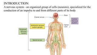
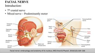
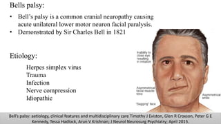
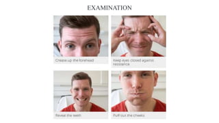
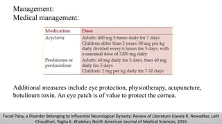
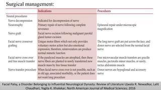
SlideShare a Scribd company logo
FACIAL NERVE
Dr. Kathirvel Gopalakrishnan
M.D.S (OMFS)
Contents
Nerve - Introduction
Cranial nerve
Facial nerve – Introduction
Embryology
Components and functions
Nuclei
Course
Ganglion
Branches and distribution
Applied aspects
INTRODUCTION:
A nervous system - an organized group of cells (neurons), specialized for the
conduction of an impulse to and from different parts of its body
NEURON – PARTS AND FUNCTIONS
CRANIAL NERVES
FACIAL NERVE
Introduction:
• 7th cranial nerve
• Mixed nerve – Predominantly motor
Facial nerve: embryology and anatomy of its nucleus; MOJ Anat & Physiol. 2018;5(3):164‒166
Embryology:
Develops from the second branchial arch
and supply all the elements that derive from it
Facial nerve: embryology and anatomy of its nucleus; MOJ Anat & Physiol. 2018;5(3):164‒166
3rd week
of IUL
Facio-acoustic primordium
appears
4th week
of IUL
1st branch appears in rostral
aspect of embryo
5th week
of IUL
Nervus intermedius becomes
evident
4th week
7th
week
Facial nerve: embryology and anatomy of its nucleus; MOJ Anat & Physiol. 2018;5(3):164‒166
7th week of
IUL
Chorda tympani derived
Joins lingual nerve
Middle of 7th week, trunk
bifurcates into
temporofacial and
cervicofacial branches
End of 7th week, temporal,
zygomatic, buccal, marginal
mandibular and cervical
regions are recognized
clearly
7 ½ week7th week end
8th week
of IUL
The cartilaginous capsule
forms a groove around
• Facial nerve
• Stapedial artery and
• Stapes muscle.
This groove will become the
facial canal
12th week
of IUL
All the muscles of the face
are formed and all are
innervated by one of the
branches of the facial nerve
Facial nerve: embryology and anatomy of its nucleus; MOJ Anat & Physiol. 2018;5(3):164‒166
21st week of IUL The ossification of the facial canal begins,
which will only end when the collateral
branches of the facial nerve are found in their
definitive location
End of pregnancy The tympanic bone and the mastoid process are
not fully developed, so the petrous portion of
the facial nerve does not exist and will not be
formed until 4 years of age
Facial nerve: embryology and anatomy of its nucleus; MOJ Anat & Physiol. 2018;5(3):164‒166
Component Function
General visceral efferent Preganglionic parasympathetic innervation of
sublingual/submandibular gland, lacrimal gland,
nasal mucosa/mucous membrane
Special visceral efferent Motor innervation of muscles of facial
expression, stylohyoid, stapedius, posterior belly
of digastric
General somatic afferent Provides sensory input from the auricular concha,
portions of external auditory canal and tympanic
membrane
Special visceral afferent Provides taste from the anterior two-thirds of the
tongue via the chorda tympani nerve
Facial Nerve and Parotid Gland Anatomy Amit Kochhar, MDa , Babak Larian, MDa,b , Babak Azizzadeh, MDa,b, ;
Otolaryngol Clin N Am 49 (2016) 273–284
Facial nerve: embryology and anatomy of its nucleus; MOJ Anat & Physiol. 2018;5(3):164‒166
NUCLEI
Facial nerve
 Motor
Nervus Intermedius
 General sensory
 Special sensory
 Superior salivatory
 Lacrimal
Origin
The facial nerve arises from pons and exits the
brain stem from its ventrolateral surface
Consists of two parts:
• Facial nerve
• Nervus Intermedius
The Facial nerve - motor component and very
small general somatic afferent component
The nervus intermedius- sensory and
parasympathetic visceromotor components.
Neuroanatomy, Cranial Nerve 7 (Facial) ; Dominika Dulak; Imama A. Naqvi. ; Stat pearls publishing, 2020 Jan
Course
Divided based on its relation to the cranium and the temporal bone into
• Intracranial
• Intratemporal
• Extratemporal
Neuroanatomy, Cranial Nerve 7 (Facial) ; Dominika Dulak; Imama A. Naqvi. ; Stat pearls publishing, 2020 Jan
Intracranial part:
Two roots, Facial nerve motor root
and nervus intermedius arises from the
pons at a lateral aspect
It runs along with the
vestibulocochlear nerve and enters
internal acoustic meatus
Intratemporal part:
The Facial nerve and nervus
intermedius travel through the internal
acoustic meatus, a 1cm long opening in
the petrous part of the temporal bone.
Enters into the facial canal, a ‘Z’
shaped structure which consists of
 Meatal segment
 Labyrinthine segment
 Tympanic segment
 Mastoid segment
Facial Nerve Anatomy and Clinical Applications; Sinali O. Seneviratne; Bhupendra C. Patel; StatPearls Publishing LLC; 2020
Meatal segment:
The superior quadrant of the internal
acoustic meatus.
Labyrinthine segment:
•Facial nerve and nervus intermedius
fuse to form the geniculate ganglion
•Greater petrosal nerve –
parasympathetic fibres to mucous
glands and lacrimal gland.
Facial Nerve Anatomy and Clinical Applications; Sinali O. Seneviratne; Bhupendra C. Patel; StatPearls Publishing LLC; 2020
Tympanic segment:
Runs in the medial wall of the
middle ear cavity
Mastoid segment:
•Nerve to stapedius – motor fibres to
stapedius muscle of the middle ear.
•Chorda tympani – special sensory
fibres to the anterior 2/3 tongue and
parasympathetic fibres to the
submandibular and sublingual glands.
Facial Nerve Anatomy and Clinical Applications; Sinali O. Seneviratne; Bhupendra C. Patel; StatPearls Publishing LLC; 2020
Extratemporal parts:
Leaves the cranium through the
stylomastoid foramen, located just
posterior to the styloid process of the
temporal bone.
As it exits, it gives the first branch
posterior auricular supplying to the
pinna of the ear and external auditory
meatus
Then it gives branches to the posterior
belly of digastric, stylohyoid
Neuroanatomy, Cranial Nerve 7 (Facial) ; Dominika Dulak; Imama A. Naqvi. ; Stat pearls publishing, 2020 Jan
Divides at the end of the posterior
edge of the parotid gland into the
terminal branches into two trunks:
• Temporofacial trunk
• Cervicofacial trunk
•Temporal branch – innervating the
frontalis and orbicularis oculi muscles
and the muscles in the upper part of
the face
•Zygomatic branch – innervating the
middle part of the face
•Buccal branch – innervating the
cheek muscles, including the
buccinator muscle
•Marginal mandibular branch –
innervating muscles of the lower
part of the face
•Cervical branch – innervating the
muscles below the chin and the
platysma muscle
Neuroanatomy, Cranial Nerve 7 (Facial) ; Dominika Dulak; Imama A. Naqvi. ; Stat pearls publishing, 2020 Jan
Classification of Terminal branching pattern of facial nerve by Davis et al (1956)
Classification of Terminal branching pattern of facial nerve by Katz et al (1987)
TYPE 1a TYPE 1b TYPE 2 TYPE 3a TYPE 3b
TYPE 3cTYPE 5TYPE 4bTYPE 4a TYPE 4bTYPE 4a
Classification of Terminal branching pattern of facial nerve by Kopuz et al (1994)
Branch of facial nerve Distribution
Within facial canal
Greater Petrosal Nerve Enters foramen lacerum and joins deep
petrosal nerve to form Nerve of Pterygoid
canal.
It then relays with Pterygopalatine ganglion
and supply lacrimal gland and mucosal
gland of nose and palate
Chorda tympani Nerve Passes through Pterygopalatine fissure and
joins with Lingual nerve
Taste sensation from anterior 2/3rd of the
tongue
Nerve to stapedius Stapedius muscle
After exiting Stylomastoid foramen
Posterior Auricular nerve Posterior auricular, Superior auricular,
Occipital belly of occipitofrontalis
Nerve to digastric Posterior belly of digastric muscle
Nerve to stylohyoid Stylohyoid
Terminal Branches
Temporal Anterior auricular, Superior auricular, Part of
frontalis, upper part of Orbicularis oculi
Zygomatic Lower part of Orbicularis oculi
Buccal Buccinator, Orbicularis oris, Muscles of upper lip
Marginal mandibular Mentalis, Orbicularis oris, Muscles of lower lip
Cervical Platysma
Ganglia:
Geniculate Ganglion:
• Sensory ganglion
• Located on the first bend of
facial nerve in relation to
medial wall of ear in facial
canal
• Taste sensation to anterior
2/3rd of tongue
B.D CHAURASIA’S HUMAN ANATOMY VOLUME 3
Submandibular Ganglion:
• Parasympathetic ganglion
• Secretomotor fibres for submandibular and sublingual gland relay with
this ganglion
• Preganglionic fibres from chorda tympani nerve
B.D CHAURASIA’S HUMAN ANATOMY VOLUME 3
Pterygopalatine Ganglion
• Largest Parasympathetic ganglion
• Secretomotor fibres for lacrimal gland relay with this ganglion
B.D CHAURASIA’S HUMAN ANATOMY VOLUME 3
Neuroanatomy, Cranial Nerve 7 (Facial) ; Dominika Dulak; Imama A. Naqvi. ; Stat pearls publishing, 2020 Jan
BRANCHES AND DISTRIBUTION
Neuroanatomy, Cranial Nerve 7 (Facial) ; Dominika Dulak; Imama A. Naqvi. ; Stat pearls publishing, 2020 Jan
BRANCHES AND DISTRIBUTION
Applied aspects
Lesion:
• Upper motor neuron lesion
• Lower motor neuron lesion
The neurons to the upper face receive
bilateral UMN innervation
The neurons to the lower face receive
UMN from the contralateral motor cortex
Facial Palsy, a Disorder Belonging to Influential Neurological Dynasty: Review of Literature Ujwala R. Newadkar, Lalit
Chaudhari, Yogita K. Khalekar; North American Journal of Medical Sciences; 2016
Upper motor lesion:
Causes unilateral facial palsy with some sparing of the frontalis and orbicularis
oculi muscles because of the bilateral cortical representation.
Furthermore, although voluntary facial movements are impaired, the face may still
move with emotional responses, for example on laughing.
Paresis of the ipsilateral arm (monoparesis) or arm and leg (hemiparesis), or
dysphasia may be associated because of more extensive cerebrocortical damage.
Facial Palsy, a Disorder Belonging to Influential Neurological Dynasty: Review of Literature Ujwala R. Newadkar, Lalit
Chaudhari, Yogita K. Khalekar; North American Journal of Medical Sciences; 2016
Lower motor neuron
Characterized by unilateral paralysis of all muscles of facial expression for both
voluntary and emotional responses.
The forehead is unfurrowed and the patient is unable to close the eye on that side.
Facial Palsy, a Disorder Belonging to Influential Neurological Dynasty: Review of Literature Ujwala R. Newadkar, Lalit
Chaudhari, Yogita K. Khalekar; North American Journal of Medical Sciences; 2016
Bells palsy:
• Bell’s palsy is a common cranial neuropathy causing
acute unilateral lower motor neuron facial paralysis.
• Demonstrated by Sir Charles Bell in 1821
Etiology:
Herpes simplex virus
Trauma
Infection
Nerve compression
Idiopathic
Bell’s palsy: aetiology, clinical features and multidisciplinary care Timothy J Eviston, Glen R Croxson, Peter G E
Kennedy, Tessa Hadlock, Arun V Krishnan; J Neurol Neurosurg Psychiatry; April 2015.
Bell’s palsy: aetiology, clinical features and multidisciplinary care Timothy J Eviston, Glen R Croxson, Peter G E
Kennedy, Tessa Hadlock, Arun V Krishnan; J Neurol Neurosurg Psychiatry; April 2015.
Clinical features:
1. Unilateral involvement
2. Absence of wrinkles in forehead
3. Loss of blinking reflex
4. Inability to close the eye, wink or smile
5. Impossible to whistle
6. Obliteration of nasolabial fold
7. Drooping of corner of the mouth
6. Mask like appearance of face
7. Bell’s Sign: In an attempt to close the eyelid, the eyeball turns
upward so that the pupil is covered and only the white sclera is visible.
BELL’S SIGN
EXAMINATION
•Blink test (corneal reflex):
When tapping on the patient’s glabella, a
suspension in blinking will occur on the affected side
•Schirmer test (assessing lacrimation of the
lacrimal gland):
Lacrimation will be decreased by 75%
compared to the normal side using a folded strip of
blotting paper in the lower conjunctival fornix.
Facial nerve palsy; Nathan R. Walker; Rakesh K. Mistry; Thomas Mazzoni; StatPearls Publishing; Jan 2020.
•Stapedial test:
This involuntary reaction in response to high-intensity sound stimuli causes
contraction of the stapedius muscle and gets mediated by the facial nerve.
Testing of the stapedius reflex can be performed using tympanometry.
•Salivary Test:
Salivation rate is assessed from a submandibular
duct following stimulation with a 6% citric acid
solution.
If positive, there will be a reduction in salivation
by 25% at the affected side and indicate a lesion at the
chorda tympani.
•Taste test:
Salt, sweet, sour and bitter tastes applied along
the lateral aspects of the anterior two-thirds of the
tongue.
A positive result will indicate a lesion at the
chorda tympani.
Facial nerve palsy; Nathan R. Walker; Rakesh K. Mistry; Thomas Mazzoni; StatPearls Publishing; Jan 2020.
Approaches to Grading Facial Nerve Function Michael J. Brenner, M.D and J. Gail Neely, M.D., F.A.C.S
Management:
Medical management:
Additional measures include eye protection, physiotherapy, acupuncture,
botulinum toxin. An eye patch is of value to protect the cornea.
Facial Palsy, a Disorder Belonging to Influential Neurological Dynasty: Review of Literature Ujwala R. Newadkar, Lalit
Chaudhari, Yogita K. Khalekar; North American Journal of Medical Sciences; 2016
Facial Palsy, a Disorder Belonging to Influential Neurological Dynasty: Review of Literature Ujwala R. Newadkar, Lalit
Chaudhari, Yogita K. Khalekar; North American Journal of Medical Sciences; 2016
Surgical management:
Facial Palsy, a Disorder Belonging to Influential Neurological Dynasty: Review of Literature Ujwala R. Newadkar, Lalit
Chaudhari, Yogita K. Khalekar; North American Journal of Medical Sciences; 2016
Surgical management:
Ramsay hunt syndrome:
Peripheral facial nerve palsy
accompanied by an erythematous vesicular
rash on the ear or in the mouth.
Varicella zoster virus (VZV) causes
Ramsay Hunt syndrome.
Bell’s palsy is significantly associated
with herpes simplex virus (HSV)
Ramsay Hunt syndrome C J Sweeney, D H Gilden; Jounal of Neurology, Neurosurgery and Psychiatry 2001
Signs and Symptoms:
Facial paralysis
Rash – Ear, Palate and tongue
Tinnitus
Hearing loss
Nausea
Vomiting
Vertigo
Nystagmus.
Due to close proximity of the geniculate
ganglion to the vestibulocochlear nerve
within the bony facial canal.
Management:
Ramsay Hunt syndrome C J Sweeney, D H Gilden; Jounal of Neurology, Neurosurgery and Psychiatry 2001
Medication Dose
Famciclovir 500 mg, three times daily for 7–10 days
Acyclovir 800 mg, five times daily for 7-10 days
Oral prednisone 60 mg daily for 3–5 days
Melkersson Rosenthal syndrome:
•Also known as Orofacial Granulomatosis
• Triad:
Relapsing facial paralysis
Persistent or recurrent orofacial edema
Fissured tongue
Rare to observe all aspects of the triad at the same time.
Melkersson–Rosenthal syndrome: a case report of a rare disease with overlapping features; Mauro Cancian, Stefano Giovannini, Annalisa
Angelini, Marny Fedrigo, Raffaele Bendo, Riccardo Senter & Stefano Sivolella; Allergy, Asthma and clinical immunology; 2019
Anatomical considerations:
Safe dissection:
Frontal Branch:
When using a lateral or superior approach,
staying right on or deep to the superficial leaf of
the deep temporal fascia is a safe plane of
dissection.
Anatomical Considerations to Prevent Facial Nerve Injury; Jason Roostaeian, Rod J. Rohrich, James M. Stuzin; The
American Society of Plastic Surgeons; 2015
Identification of facial nerve:
THANK YOU

More Related Content

What's hot

Facial nerve
Facial nerveFacial nerve
Facial nerve
NeurologyKota
 
Facial nerve anatomy
Facial nerve anatomyFacial nerve anatomy
Facial nerve anatomy
Meghna Bagalkotkar
 
Trigeminal nerve
Trigeminal nerveTrigeminal nerve
Trigeminal nerve
Anchal Mehra
 
Pterygopalatine fossa
Pterygopalatine fossaPterygopalatine fossa
Pterygopalatine fossa
Chitransha03
 
Dr Nirav patel seminar on trigeminal nerve
Dr Nirav patel seminar on trigeminal nerveDr Nirav patel seminar on trigeminal nerve
Dr Nirav patel seminar on trigeminal nerve
Nirav Patel
 
Anatomy of Facial Nerve
Anatomy of Facial NerveAnatomy of Facial Nerve
Anatomy of Facial Nerve
Diptiman Baliarsingh
 
Facial nerve
Facial nerveFacial nerve
Facial nerve
Ram Yadav
 
PTERYGOPALATINE FOSSA.pptx
PTERYGOPALATINE FOSSA.pptxPTERYGOPALATINE FOSSA.pptx
PTERYGOPALATINE FOSSA.pptx
Dr. sana yaseen
 
SURICAL ANATOMY OF FACIAL NERVE
SURICAL ANATOMY OF FACIAL NERVESURICAL ANATOMY OF FACIAL NERVE
SURICAL ANATOMY OF FACIAL NERVE
Amar Shinde
 
Infratemporal fossa
Infratemporal fossaInfratemporal fossa
Infratemporal fossa
ddert
 
Pterygopalatine fossa and approaches by Dr.Ashwin Menon
Pterygopalatine fossa and approaches by Dr.Ashwin MenonPterygopalatine fossa and approaches by Dr.Ashwin Menon
Pterygopalatine fossa and approaches by Dr.Ashwin Menon
Dr.Ashwin Menon
 
Trigeminal nerve dr. gosai
Trigeminal nerve dr. gosaiTrigeminal nerve dr. gosai
Trigeminal nerve dr. gosaiDr.B.B. Gosai
 
The mylohyoid muscle
The mylohyoid muscleThe mylohyoid muscle
The mylohyoid muscle
Idris Siddiqui
 
Facial nerve
Facial nerveFacial nerve
Facial nerve
Dr Himanshu Soni
 
Trigeminal nerve
Trigeminal nerve  Trigeminal nerve
Trigeminal nerve
DrKamini Dadsena
 
Mandibular nerve
Mandibular nerveMandibular nerve
Mandibular nerve
Thaslim Fathima
 
Anatomy of skull base
Anatomy of skull baseAnatomy of skull base
Anatomy of skull base
Dr. Shilpa M J
 
Trigeminal nerve
Trigeminal nerveTrigeminal nerve
Trigeminal nerve
ashish25200
 

What's hot (20)

Trigeminal nerve
Trigeminal nerveTrigeminal nerve
Trigeminal nerve
 
Facial nerve
Facial nerveFacial nerve
Facial nerve
 
Facial nerve anatomy
Facial nerve anatomyFacial nerve anatomy
Facial nerve anatomy
 
Trigeminal nerve
Trigeminal nerveTrigeminal nerve
Trigeminal nerve
 
Pterygopalatine fossa
Pterygopalatine fossaPterygopalatine fossa
Pterygopalatine fossa
 
Dr Nirav patel seminar on trigeminal nerve
Dr Nirav patel seminar on trigeminal nerveDr Nirav patel seminar on trigeminal nerve
Dr Nirav patel seminar on trigeminal nerve
 
Anatomy of Facial Nerve
Anatomy of Facial NerveAnatomy of Facial Nerve
Anatomy of Facial Nerve
 
Facial nerve
Facial nerveFacial nerve
Facial nerve
 
PTERYGOPALATINE FOSSA.pptx
PTERYGOPALATINE FOSSA.pptxPTERYGOPALATINE FOSSA.pptx
PTERYGOPALATINE FOSSA.pptx
 
SURICAL ANATOMY OF FACIAL NERVE
SURICAL ANATOMY OF FACIAL NERVESURICAL ANATOMY OF FACIAL NERVE
SURICAL ANATOMY OF FACIAL NERVE
 
Infratemporal fossa
Infratemporal fossaInfratemporal fossa
Infratemporal fossa
 
Pterygopalatine fossa and approaches by Dr.Ashwin Menon
Pterygopalatine fossa and approaches by Dr.Ashwin MenonPterygopalatine fossa and approaches by Dr.Ashwin Menon
Pterygopalatine fossa and approaches by Dr.Ashwin Menon
 
Trigeminal nerve dr. gosai
Trigeminal nerve dr. gosaiTrigeminal nerve dr. gosai
Trigeminal nerve dr. gosai
 
The mylohyoid muscle
The mylohyoid muscleThe mylohyoid muscle
The mylohyoid muscle
 
Facial nerve
Facial nerveFacial nerve
Facial nerve
 
Trigeminal nerve
Trigeminal nerve  Trigeminal nerve
Trigeminal nerve
 
Mandibular nerve
Mandibular nerveMandibular nerve
Mandibular nerve
 
Table 2 Foramina Of Skull
Table 2 Foramina Of SkullTable 2 Foramina Of Skull
Table 2 Foramina Of Skull
 
Anatomy of skull base
Anatomy of skull baseAnatomy of skull base
Anatomy of skull base
 
Trigeminal nerve
Trigeminal nerveTrigeminal nerve
Trigeminal nerve
 

Similar to Facial nerve

Facialnerve
FacialnerveFacialnerve
Facial nerve by Dr. Apoorv
Facial nerve by Dr. ApoorvFacial nerve by Dr. Apoorv
Facial nerve by Dr. Apoorv
Apoorv Pandey
 
Facial nerve and its extracranial and intracranial rots
Facial nerve and its extracranial and intracranial rotsFacial nerve and its extracranial and intracranial rots
Facial nerve and its extracranial and intracranial rots
sonambohra2
 
Facial nerve done
Facial nerve doneFacial nerve done
Facial nerve done
DrRakashree Chakraborty
 
Facial nerve
Facial nerveFacial nerve
Facial nerve
SanghamitraGhosh12
 
FACIAL NERVE.pptx
FACIAL NERVE.pptxFACIAL NERVE.pptx
FACIAL NERVE.pptx
malti19
 
Facial nerve and it's applied aspect
Facial nerve and it's applied aspectFacial nerve and it's applied aspect
Facial nerve and it's applied aspect
Prachi Jha
 
Trigeminal nerve ppt
Trigeminal nerve ppt  Trigeminal nerve ppt
Trigeminal nerve ppt
Dr Fariya Ashraf
 
Final facial nerve palsy dr. zaimal
Final facial nerve palsy dr. zaimalFinal facial nerve palsy dr. zaimal
Facial nerve and its applied aspects
Facial nerve and its applied aspectsFacial nerve and its applied aspects
Facial nerve and its applied aspects
sadaf syed
 
Facial Nerve Anatomy
Facial Nerve AnatomyFacial Nerve Anatomy
Facial Nerve Anatomy
DIVYA BANU
 
Facial nerve and its applied aspect - seminar 3 [Autosaved].pptx
Facial nerve and its applied aspect - seminar 3 [Autosaved].pptxFacial nerve and its applied aspect - seminar 3 [Autosaved].pptx
Facial nerve and its applied aspect - seminar 3 [Autosaved].pptx
drpriyanka8
 
3.TRIGEMINAL NERVE.pptx
3.TRIGEMINAL NERVE.pptx3.TRIGEMINAL NERVE.pptx
3.TRIGEMINAL NERVE.pptx
SusovanGiri6
 
Anatomy facial nerve
Anatomy facial nerveAnatomy facial nerve
Anatomy facial nerve
محمد حسين خميس
 
Facial nerve
Facial nerveFacial nerve
Facial nerve
Chitransha03
 
Anatomy of Cranial Nerves 5,7,8
Anatomy of Cranial Nerves 5,7,8Anatomy of Cranial Nerves 5,7,8
Anatomy of Cranial Nerves 5,7,8
RushdanZakariah
 
Innervation,okkkkkkkkkkkkkkk.docx
Innervation,okkkkkkkkkkkkkkk.docxInnervation,okkkkkkkkkkkkkkk.docx
Innervation,okkkkkkkkkkkkkkk.docx
ibraheem yahia
 
FACIAL NERVE.pptx
FACIAL NERVE.pptxFACIAL NERVE.pptx
FACIAL NERVE.pptx
Bipin Koirala
 
Prof. laila kAU Trigeminal Nerve .
Prof. laila kAU  Trigeminal  Nerve .Prof. laila kAU  Trigeminal  Nerve .
Prof. laila kAU Trigeminal Nerve .
Laila M. Aboul Mahasen Morsy
 
cranial nerve 5 detailed decription and presentation
cranial nerve 5 detailed decription and presentationcranial nerve 5 detailed decription and presentation
cranial nerve 5 detailed decription and presentation
MuhammadTouseefKhan2
 

Similar to Facial nerve (20)

Facialnerve
FacialnerveFacialnerve
Facialnerve
 
Facial nerve by Dr. Apoorv
Facial nerve by Dr. ApoorvFacial nerve by Dr. Apoorv
Facial nerve by Dr. Apoorv
 
Facial nerve and its extracranial and intracranial rots
Facial nerve and its extracranial and intracranial rotsFacial nerve and its extracranial and intracranial rots
Facial nerve and its extracranial and intracranial rots
 
Facial nerve done
Facial nerve doneFacial nerve done
Facial nerve done
 
Facial nerve
Facial nerveFacial nerve
Facial nerve
 
FACIAL NERVE.pptx
FACIAL NERVE.pptxFACIAL NERVE.pptx
FACIAL NERVE.pptx
 
Facial nerve and it's applied aspect
Facial nerve and it's applied aspectFacial nerve and it's applied aspect
Facial nerve and it's applied aspect
 
Trigeminal nerve ppt
Trigeminal nerve ppt  Trigeminal nerve ppt
Trigeminal nerve ppt
 
Final facial nerve palsy dr. zaimal
Final facial nerve palsy dr. zaimalFinal facial nerve palsy dr. zaimal
Final facial nerve palsy dr. zaimal
 
Facial nerve and its applied aspects
Facial nerve and its applied aspectsFacial nerve and its applied aspects
Facial nerve and its applied aspects
 
Facial Nerve Anatomy
Facial Nerve AnatomyFacial Nerve Anatomy
Facial Nerve Anatomy
 
Facial nerve and its applied aspect - seminar 3 [Autosaved].pptx
Facial nerve and its applied aspect - seminar 3 [Autosaved].pptxFacial nerve and its applied aspect - seminar 3 [Autosaved].pptx
Facial nerve and its applied aspect - seminar 3 [Autosaved].pptx
 
3.TRIGEMINAL NERVE.pptx
3.TRIGEMINAL NERVE.pptx3.TRIGEMINAL NERVE.pptx
3.TRIGEMINAL NERVE.pptx
 
Anatomy facial nerve
Anatomy facial nerveAnatomy facial nerve
Anatomy facial nerve
 
Facial nerve
Facial nerveFacial nerve
Facial nerve
 
Anatomy of Cranial Nerves 5,7,8
Anatomy of Cranial Nerves 5,7,8Anatomy of Cranial Nerves 5,7,8
Anatomy of Cranial Nerves 5,7,8
 
Innervation,okkkkkkkkkkkkkkk.docx
Innervation,okkkkkkkkkkkkkkk.docxInnervation,okkkkkkkkkkkkkkk.docx
Innervation,okkkkkkkkkkkkkkk.docx
 
FACIAL NERVE.pptx
FACIAL NERVE.pptxFACIAL NERVE.pptx
FACIAL NERVE.pptx
 
Prof. laila kAU Trigeminal Nerve .
Prof. laila kAU  Trigeminal  Nerve .Prof. laila kAU  Trigeminal  Nerve .
Prof. laila kAU Trigeminal Nerve .
 
cranial nerve 5 detailed decription and presentation
cranial nerve 5 detailed decription and presentationcranial nerve 5 detailed decription and presentation
cranial nerve 5 detailed decription and presentation
 

More from KathirvelGopalakrish

Condylar fracture.pptx
Condylar fracture.pptxCondylar fracture.pptx
Condylar fracture.pptx
KathirvelGopalakrish
 
Mandibular osteotomies.pptx
Mandibular osteotomies.pptxMandibular osteotomies.pptx
Mandibular osteotomies.pptx
KathirvelGopalakrish
 
Midface fracture.pptx
Midface fracture.pptxMidface fracture.pptx
Midface fracture.pptx
KathirvelGopalakrish
 
Primary assessment in maxillofacial trauma.pptx
Primary assessment in maxillofacial trauma.pptxPrimary assessment in maxillofacial trauma.pptx
Primary assessment in maxillofacial trauma.pptx
KathirvelGopalakrish
 
Rhinoplasty.pptx
Rhinoplasty.pptxRhinoplasty.pptx
Rhinoplasty.pptx
KathirvelGopalakrish
 
TMJ ankylosis and reconstruction.pptx
TMJ ankylosis and reconstruction.pptxTMJ ankylosis and reconstruction.pptx
TMJ ankylosis and reconstruction.pptx
KathirvelGopalakrish
 
Preop assessment and operation theatre protocols.pptx
Preop assessment and operation theatre protocols.pptxPreop assessment and operation theatre protocols.pptx
Preop assessment and operation theatre protocols.pptx
KathirvelGopalakrish
 
Osteomyelitis of jaw
Osteomyelitis of jawOsteomyelitis of jaw
Osteomyelitis of jaw
KathirvelGopalakrish
 
Maxillary canine and molar impaction
Maxillary canine and molar impactionMaxillary canine and molar impaction
Maxillary canine and molar impaction
KathirvelGopalakrish
 
Maxillary nerve block
Maxillary nerve blockMaxillary nerve block
Maxillary nerve block
KathirvelGopalakrish
 
Anatomy of orbit
Anatomy of orbitAnatomy of orbit
Anatomy of orbit
KathirvelGopalakrish
 
General anesthesia
General anesthesiaGeneral anesthesia
General anesthesia
KathirvelGopalakrish
 
Unconsciousness and syncope - Management
Unconsciousness and syncope - ManagementUnconsciousness and syncope - Management
Unconsciousness and syncope - Management
KathirvelGopalakrish
 
Anterior triangles of neck
Anterior triangles of neckAnterior triangles of neck
Anterior triangles of neck
KathirvelGopalakrish
 
Vascular supply of face and neck
Vascular supply of face and neckVascular supply of face and neck
Vascular supply of face and neck
KathirvelGopalakrish
 

More from KathirvelGopalakrish (15)

Condylar fracture.pptx
Condylar fracture.pptxCondylar fracture.pptx
Condylar fracture.pptx
 
Mandibular osteotomies.pptx
Mandibular osteotomies.pptxMandibular osteotomies.pptx
Mandibular osteotomies.pptx
 
Midface fracture.pptx
Midface fracture.pptxMidface fracture.pptx
Midface fracture.pptx
 
Primary assessment in maxillofacial trauma.pptx
Primary assessment in maxillofacial trauma.pptxPrimary assessment in maxillofacial trauma.pptx
Primary assessment in maxillofacial trauma.pptx
 
Rhinoplasty.pptx
Rhinoplasty.pptxRhinoplasty.pptx
Rhinoplasty.pptx
 
TMJ ankylosis and reconstruction.pptx
TMJ ankylosis and reconstruction.pptxTMJ ankylosis and reconstruction.pptx
TMJ ankylosis and reconstruction.pptx
 
Preop assessment and operation theatre protocols.pptx
Preop assessment and operation theatre protocols.pptxPreop assessment and operation theatre protocols.pptx
Preop assessment and operation theatre protocols.pptx
 
Osteomyelitis of jaw
Osteomyelitis of jawOsteomyelitis of jaw
Osteomyelitis of jaw
 
Maxillary canine and molar impaction
Maxillary canine and molar impactionMaxillary canine and molar impaction
Maxillary canine and molar impaction
 
Maxillary nerve block
Maxillary nerve blockMaxillary nerve block
Maxillary nerve block
 
Anatomy of orbit
Anatomy of orbitAnatomy of orbit
Anatomy of orbit
 
General anesthesia
General anesthesiaGeneral anesthesia
General anesthesia
 
Unconsciousness and syncope - Management
Unconsciousness and syncope - ManagementUnconsciousness and syncope - Management
Unconsciousness and syncope - Management
 
Anterior triangles of neck
Anterior triangles of neckAnterior triangles of neck
Anterior triangles of neck
 
Vascular supply of face and neck
Vascular supply of face and neckVascular supply of face and neck
Vascular supply of face and neck
 

Recently uploaded

ARTIFICIAL INTELLIGENCE IN HEALTHCARE.pdf
ARTIFICIAL INTELLIGENCE IN  HEALTHCARE.pdfARTIFICIAL INTELLIGENCE IN  HEALTHCARE.pdf
ARTIFICIAL INTELLIGENCE IN HEALTHCARE.pdf
Anujkumaranit
 
Superficial & Deep Fascia of the NECK.pptx
Superficial & Deep Fascia of the NECK.pptxSuperficial & Deep Fascia of the NECK.pptx
Superficial & Deep Fascia of the NECK.pptx
Dr. Rabia Inam Gandapore
 
ARTHROLOGY PPT NCISM SYLLABUS AYURVEDA STUDENTS
ARTHROLOGY PPT NCISM SYLLABUS AYURVEDA STUDENTSARTHROLOGY PPT NCISM SYLLABUS AYURVEDA STUDENTS
ARTHROLOGY PPT NCISM SYLLABUS AYURVEDA STUDENTS
Dr. Vinay Pareek
 
New Drug Discovery and Development .....
New Drug Discovery and Development .....New Drug Discovery and Development .....
New Drug Discovery and Development .....
NEHA GUPTA
 
BENIGN PROSTATIC HYPERPLASIA.BPH. BPHpdf
BENIGN PROSTATIC HYPERPLASIA.BPH. BPHpdfBENIGN PROSTATIC HYPERPLASIA.BPH. BPHpdf
BENIGN PROSTATIC HYPERPLASIA.BPH. BPHpdf
DR SETH JOTHAM
 
NVBDCP.pptx Nation vector borne disease control program
NVBDCP.pptx Nation vector borne disease control programNVBDCP.pptx Nation vector borne disease control program
NVBDCP.pptx Nation vector borne disease control program
Sapna Thakur
 
Couples presenting to the infertility clinic- Do they really have infertility...
Couples presenting to the infertility clinic- Do they really have infertility...Couples presenting to the infertility clinic- Do they really have infertility...
Couples presenting to the infertility clinic- Do they really have infertility...
Sujoy Dasgupta
 
Flu Vaccine Alert in Bangalore Karnataka
Flu Vaccine Alert in Bangalore KarnatakaFlu Vaccine Alert in Bangalore Karnataka
Flu Vaccine Alert in Bangalore Karnataka
addon Scans
 
micro teaching on communication m.sc nursing.pdf
micro teaching on communication m.sc nursing.pdfmicro teaching on communication m.sc nursing.pdf
micro teaching on communication m.sc nursing.pdf
Anurag Sharma
 
Triangles of Neck and Clinical Correlation by Dr. RIG.pptx
Triangles of Neck and Clinical Correlation by Dr. RIG.pptxTriangles of Neck and Clinical Correlation by Dr. RIG.pptx
Triangles of Neck and Clinical Correlation by Dr. RIG.pptx
Dr. Rabia Inam Gandapore
 
263778731218 Abortion Clinic /Pills In Harare ,
263778731218 Abortion Clinic /Pills In Harare ,263778731218 Abortion Clinic /Pills In Harare ,
263778731218 Abortion Clinic /Pills In Harare ,
sisternakatoto
 
Non-respiratory Functions of the Lungs.pdf
Non-respiratory Functions of the Lungs.pdfNon-respiratory Functions of the Lungs.pdf
Non-respiratory Functions of the Lungs.pdf
MedicoseAcademics
 
Lung Cancer: Artificial Intelligence, Synergetics, Complex System Analysis, S...
Lung Cancer: Artificial Intelligence, Synergetics, Complex System Analysis, S...Lung Cancer: Artificial Intelligence, Synergetics, Complex System Analysis, S...
Lung Cancer: Artificial Intelligence, Synergetics, Complex System Analysis, S...
Oleg Kshivets
 
Physiology of Special Chemical Sensation of Taste
Physiology of Special Chemical Sensation of TastePhysiology of Special Chemical Sensation of Taste
Physiology of Special Chemical Sensation of Taste
MedicoseAcademics
 
The Normal Electrocardiogram - Part I of II
The Normal Electrocardiogram - Part I of IIThe Normal Electrocardiogram - Part I of II
The Normal Electrocardiogram - Part I of II
MedicoseAcademics
 
Maxilla, Mandible & Hyoid Bone & Clinical Correlations by Dr. RIG.pptx
Maxilla, Mandible & Hyoid Bone & Clinical Correlations by Dr. RIG.pptxMaxilla, Mandible & Hyoid Bone & Clinical Correlations by Dr. RIG.pptx
Maxilla, Mandible & Hyoid Bone & Clinical Correlations by Dr. RIG.pptx
Dr. Rabia Inam Gandapore
 
Surat @ℂall @Girls ꧁❤8527049040❤꧂@ℂall @Girls Service Vip Top Model Safe
Surat @ℂall @Girls ꧁❤8527049040❤꧂@ℂall @Girls Service Vip Top Model SafeSurat @ℂall @Girls ꧁❤8527049040❤꧂@ℂall @Girls Service Vip Top Model Safe
Surat @ℂall @Girls ꧁❤8527049040❤꧂@ℂall @Girls Service Vip Top Model Safe
Savita Shen $i11
 
How to Give Better Lectures: Some Tips for Doctors
How to Give Better Lectures: Some Tips for DoctorsHow to Give Better Lectures: Some Tips for Doctors
How to Give Better Lectures: Some Tips for Doctors
LanceCatedral
 
Ocular injury ppt Upendra pal optometrist upums saifai etawah
Ocular injury  ppt  Upendra pal  optometrist upums saifai etawahOcular injury  ppt  Upendra pal  optometrist upums saifai etawah
Ocular injury ppt Upendra pal optometrist upums saifai etawah
pal078100
 
For Better Surat #ℂall #Girl Service ❤85270-49040❤ Surat #ℂall #Girls
For Better Surat #ℂall #Girl Service ❤85270-49040❤ Surat #ℂall #GirlsFor Better Surat #ℂall #Girl Service ❤85270-49040❤ Surat #ℂall #Girls
For Better Surat #ℂall #Girl Service ❤85270-49040❤ Surat #ℂall #Girls
Savita Shen $i11
 

Recently uploaded (20)

ARTIFICIAL INTELLIGENCE IN HEALTHCARE.pdf
ARTIFICIAL INTELLIGENCE IN  HEALTHCARE.pdfARTIFICIAL INTELLIGENCE IN  HEALTHCARE.pdf
ARTIFICIAL INTELLIGENCE IN HEALTHCARE.pdf
 
Superficial & Deep Fascia of the NECK.pptx
Superficial & Deep Fascia of the NECK.pptxSuperficial & Deep Fascia of the NECK.pptx
Superficial & Deep Fascia of the NECK.pptx
 
ARTHROLOGY PPT NCISM SYLLABUS AYURVEDA STUDENTS
ARTHROLOGY PPT NCISM SYLLABUS AYURVEDA STUDENTSARTHROLOGY PPT NCISM SYLLABUS AYURVEDA STUDENTS
ARTHROLOGY PPT NCISM SYLLABUS AYURVEDA STUDENTS
 
New Drug Discovery and Development .....
New Drug Discovery and Development .....New Drug Discovery and Development .....
New Drug Discovery and Development .....
 
BENIGN PROSTATIC HYPERPLASIA.BPH. BPHpdf
BENIGN PROSTATIC HYPERPLASIA.BPH. BPHpdfBENIGN PROSTATIC HYPERPLASIA.BPH. BPHpdf
BENIGN PROSTATIC HYPERPLASIA.BPH. BPHpdf
 
NVBDCP.pptx Nation vector borne disease control program
NVBDCP.pptx Nation vector borne disease control programNVBDCP.pptx Nation vector borne disease control program
NVBDCP.pptx Nation vector borne disease control program
 
Couples presenting to the infertility clinic- Do they really have infertility...
Couples presenting to the infertility clinic- Do they really have infertility...Couples presenting to the infertility clinic- Do they really have infertility...
Couples presenting to the infertility clinic- Do they really have infertility...
 
Flu Vaccine Alert in Bangalore Karnataka
Flu Vaccine Alert in Bangalore KarnatakaFlu Vaccine Alert in Bangalore Karnataka
Flu Vaccine Alert in Bangalore Karnataka
 
micro teaching on communication m.sc nursing.pdf
micro teaching on communication m.sc nursing.pdfmicro teaching on communication m.sc nursing.pdf
micro teaching on communication m.sc nursing.pdf
 
Triangles of Neck and Clinical Correlation by Dr. RIG.pptx
Triangles of Neck and Clinical Correlation by Dr. RIG.pptxTriangles of Neck and Clinical Correlation by Dr. RIG.pptx
Triangles of Neck and Clinical Correlation by Dr. RIG.pptx
 
263778731218 Abortion Clinic /Pills In Harare ,
263778731218 Abortion Clinic /Pills In Harare ,263778731218 Abortion Clinic /Pills In Harare ,
263778731218 Abortion Clinic /Pills In Harare ,
 
Non-respiratory Functions of the Lungs.pdf
Non-respiratory Functions of the Lungs.pdfNon-respiratory Functions of the Lungs.pdf
Non-respiratory Functions of the Lungs.pdf
 
Lung Cancer: Artificial Intelligence, Synergetics, Complex System Analysis, S...
Lung Cancer: Artificial Intelligence, Synergetics, Complex System Analysis, S...Lung Cancer: Artificial Intelligence, Synergetics, Complex System Analysis, S...
Lung Cancer: Artificial Intelligence, Synergetics, Complex System Analysis, S...
 
Physiology of Special Chemical Sensation of Taste
Physiology of Special Chemical Sensation of TastePhysiology of Special Chemical Sensation of Taste
Physiology of Special Chemical Sensation of Taste
 
The Normal Electrocardiogram - Part I of II
The Normal Electrocardiogram - Part I of IIThe Normal Electrocardiogram - Part I of II
The Normal Electrocardiogram - Part I of II
 
Maxilla, Mandible & Hyoid Bone & Clinical Correlations by Dr. RIG.pptx
Maxilla, Mandible & Hyoid Bone & Clinical Correlations by Dr. RIG.pptxMaxilla, Mandible & Hyoid Bone & Clinical Correlations by Dr. RIG.pptx
Maxilla, Mandible & Hyoid Bone & Clinical Correlations by Dr. RIG.pptx
 
Surat @ℂall @Girls ꧁❤8527049040❤꧂@ℂall @Girls Service Vip Top Model Safe
Surat @ℂall @Girls ꧁❤8527049040❤꧂@ℂall @Girls Service Vip Top Model SafeSurat @ℂall @Girls ꧁❤8527049040❤꧂@ℂall @Girls Service Vip Top Model Safe
Surat @ℂall @Girls ꧁❤8527049040❤꧂@ℂall @Girls Service Vip Top Model Safe
 
How to Give Better Lectures: Some Tips for Doctors
How to Give Better Lectures: Some Tips for DoctorsHow to Give Better Lectures: Some Tips for Doctors
How to Give Better Lectures: Some Tips for Doctors
 
Ocular injury ppt Upendra pal optometrist upums saifai etawah
Ocular injury  ppt  Upendra pal  optometrist upums saifai etawahOcular injury  ppt  Upendra pal  optometrist upums saifai etawah
Ocular injury ppt Upendra pal optometrist upums saifai etawah
 
For Better Surat #ℂall #Girl Service ❤85270-49040❤ Surat #ℂall #Girls
For Better Surat #ℂall #Girl Service ❤85270-49040❤ Surat #ℂall #GirlsFor Better Surat #ℂall #Girl Service ❤85270-49040❤ Surat #ℂall #Girls
For Better Surat #ℂall #Girl Service ❤85270-49040❤ Surat #ℂall #Girls
 

Facial nerve

  • 1. FACIAL NERVE Dr. Kathirvel Gopalakrishnan M.D.S (OMFS)
  • 2. Contents Nerve - Introduction Cranial nerve Facial nerve – Introduction Embryology Components and functions Nuclei Course Ganglion Branches and distribution Applied aspects
  • 3. INTRODUCTION: A nervous system - an organized group of cells (neurons), specialized for the conduction of an impulse to and from different parts of its body
  • 4. NEURON – PARTS AND FUNCTIONS
  • 6. FACIAL NERVE Introduction: • 7th cranial nerve • Mixed nerve – Predominantly motor Facial nerve: embryology and anatomy of its nucleus; MOJ Anat & Physiol. 2018;5(3):164‒166
  • 7. Embryology: Develops from the second branchial arch and supply all the elements that derive from it Facial nerve: embryology and anatomy of its nucleus; MOJ Anat & Physiol. 2018;5(3):164‒166 3rd week of IUL Facio-acoustic primordium appears 4th week of IUL 1st branch appears in rostral aspect of embryo 5th week of IUL Nervus intermedius becomes evident 4th week
  • 8. 7th week Facial nerve: embryology and anatomy of its nucleus; MOJ Anat & Physiol. 2018;5(3):164‒166 7th week of IUL Chorda tympani derived Joins lingual nerve Middle of 7th week, trunk bifurcates into temporofacial and cervicofacial branches End of 7th week, temporal, zygomatic, buccal, marginal mandibular and cervical regions are recognized clearly 7 ½ week7th week end
  • 9. 8th week of IUL The cartilaginous capsule forms a groove around • Facial nerve • Stapedial artery and • Stapes muscle. This groove will become the facial canal 12th week of IUL All the muscles of the face are formed and all are innervated by one of the branches of the facial nerve Facial nerve: embryology and anatomy of its nucleus; MOJ Anat & Physiol. 2018;5(3):164‒166
  • 10. 21st week of IUL The ossification of the facial canal begins, which will only end when the collateral branches of the facial nerve are found in their definitive location End of pregnancy The tympanic bone and the mastoid process are not fully developed, so the petrous portion of the facial nerve does not exist and will not be formed until 4 years of age Facial nerve: embryology and anatomy of its nucleus; MOJ Anat & Physiol. 2018;5(3):164‒166
  • 11. Component Function General visceral efferent Preganglionic parasympathetic innervation of sublingual/submandibular gland, lacrimal gland, nasal mucosa/mucous membrane Special visceral efferent Motor innervation of muscles of facial expression, stylohyoid, stapedius, posterior belly of digastric General somatic afferent Provides sensory input from the auricular concha, portions of external auditory canal and tympanic membrane Special visceral afferent Provides taste from the anterior two-thirds of the tongue via the chorda tympani nerve Facial Nerve and Parotid Gland Anatomy Amit Kochhar, MDa , Babak Larian, MDa,b , Babak Azizzadeh, MDa,b, ; Otolaryngol Clin N Am 49 (2016) 273–284
  • 12. Facial nerve: embryology and anatomy of its nucleus; MOJ Anat & Physiol. 2018;5(3):164‒166 NUCLEI Facial nerve  Motor Nervus Intermedius  General sensory  Special sensory  Superior salivatory  Lacrimal
  • 13. Origin The facial nerve arises from pons and exits the brain stem from its ventrolateral surface Consists of two parts: • Facial nerve • Nervus Intermedius The Facial nerve - motor component and very small general somatic afferent component The nervus intermedius- sensory and parasympathetic visceromotor components. Neuroanatomy, Cranial Nerve 7 (Facial) ; Dominika Dulak; Imama A. Naqvi. ; Stat pearls publishing, 2020 Jan
  • 14. Course Divided based on its relation to the cranium and the temporal bone into • Intracranial • Intratemporal • Extratemporal Neuroanatomy, Cranial Nerve 7 (Facial) ; Dominika Dulak; Imama A. Naqvi. ; Stat pearls publishing, 2020 Jan Intracranial part: Two roots, Facial nerve motor root and nervus intermedius arises from the pons at a lateral aspect It runs along with the vestibulocochlear nerve and enters internal acoustic meatus
  • 15. Intratemporal part: The Facial nerve and nervus intermedius travel through the internal acoustic meatus, a 1cm long opening in the petrous part of the temporal bone. Enters into the facial canal, a ‘Z’ shaped structure which consists of  Meatal segment  Labyrinthine segment  Tympanic segment  Mastoid segment Facial Nerve Anatomy and Clinical Applications; Sinali O. Seneviratne; Bhupendra C. Patel; StatPearls Publishing LLC; 2020
  • 16. Meatal segment: The superior quadrant of the internal acoustic meatus. Labyrinthine segment: •Facial nerve and nervus intermedius fuse to form the geniculate ganglion •Greater petrosal nerve – parasympathetic fibres to mucous glands and lacrimal gland. Facial Nerve Anatomy and Clinical Applications; Sinali O. Seneviratne; Bhupendra C. Patel; StatPearls Publishing LLC; 2020
  • 17. Tympanic segment: Runs in the medial wall of the middle ear cavity Mastoid segment: •Nerve to stapedius – motor fibres to stapedius muscle of the middle ear. •Chorda tympani – special sensory fibres to the anterior 2/3 tongue and parasympathetic fibres to the submandibular and sublingual glands. Facial Nerve Anatomy and Clinical Applications; Sinali O. Seneviratne; Bhupendra C. Patel; StatPearls Publishing LLC; 2020
  • 18. Extratemporal parts: Leaves the cranium through the stylomastoid foramen, located just posterior to the styloid process of the temporal bone. As it exits, it gives the first branch posterior auricular supplying to the pinna of the ear and external auditory meatus Then it gives branches to the posterior belly of digastric, stylohyoid Neuroanatomy, Cranial Nerve 7 (Facial) ; Dominika Dulak; Imama A. Naqvi. ; Stat pearls publishing, 2020 Jan
  • 19. Neuroanatomy, Cranial Nerve 7 (Facial) ; Dominika Dulak; Imama A. Naqvi. ; Stat pearls publishing, 2020 Jan Divides at the end of the posterior edge of the parotid gland into the terminal branches into two trunks: • Temporofacial trunk • Cervicofacial trunk •Temporal branch – innervating the frontalis and orbicularis oculi muscles and the muscles in the upper part of the face •Zygomatic branch – innervating the middle part of the face
  • 20. •Buccal branch – innervating the cheek muscles, including the buccinator muscle •Marginal mandibular branch – innervating muscles of the lower part of the face •Cervical branch – innervating the muscles below the chin and the platysma muscle Neuroanatomy, Cranial Nerve 7 (Facial) ; Dominika Dulak; Imama A. Naqvi. ; Stat pearls publishing, 2020 Jan
  • 21. Classification of Terminal branching pattern of facial nerve by Davis et al (1956)
  • 22. Classification of Terminal branching pattern of facial nerve by Katz et al (1987) TYPE 1a TYPE 1b TYPE 2 TYPE 3a TYPE 3b TYPE 3cTYPE 5TYPE 4bTYPE 4a TYPE 4bTYPE 4a
  • 23. Classification of Terminal branching pattern of facial nerve by Kopuz et al (1994)
  • 24. Branch of facial nerve Distribution Within facial canal Greater Petrosal Nerve Enters foramen lacerum and joins deep petrosal nerve to form Nerve of Pterygoid canal. It then relays with Pterygopalatine ganglion and supply lacrimal gland and mucosal gland of nose and palate Chorda tympani Nerve Passes through Pterygopalatine fissure and joins with Lingual nerve Taste sensation from anterior 2/3rd of the tongue Nerve to stapedius Stapedius muscle
  • 25. After exiting Stylomastoid foramen Posterior Auricular nerve Posterior auricular, Superior auricular, Occipital belly of occipitofrontalis Nerve to digastric Posterior belly of digastric muscle Nerve to stylohyoid Stylohyoid Terminal Branches Temporal Anterior auricular, Superior auricular, Part of frontalis, upper part of Orbicularis oculi Zygomatic Lower part of Orbicularis oculi Buccal Buccinator, Orbicularis oris, Muscles of upper lip Marginal mandibular Mentalis, Orbicularis oris, Muscles of lower lip Cervical Platysma
  • 26. Ganglia: Geniculate Ganglion: • Sensory ganglion • Located on the first bend of facial nerve in relation to medial wall of ear in facial canal • Taste sensation to anterior 2/3rd of tongue B.D CHAURASIA’S HUMAN ANATOMY VOLUME 3
  • 27. Submandibular Ganglion: • Parasympathetic ganglion • Secretomotor fibres for submandibular and sublingual gland relay with this ganglion • Preganglionic fibres from chorda tympani nerve B.D CHAURASIA’S HUMAN ANATOMY VOLUME 3
  • 28. Pterygopalatine Ganglion • Largest Parasympathetic ganglion • Secretomotor fibres for lacrimal gland relay with this ganglion B.D CHAURASIA’S HUMAN ANATOMY VOLUME 3
  • 29. Neuroanatomy, Cranial Nerve 7 (Facial) ; Dominika Dulak; Imama A. Naqvi. ; Stat pearls publishing, 2020 Jan BRANCHES AND DISTRIBUTION
  • 30. Neuroanatomy, Cranial Nerve 7 (Facial) ; Dominika Dulak; Imama A. Naqvi. ; Stat pearls publishing, 2020 Jan BRANCHES AND DISTRIBUTION
  • 32. Lesion: • Upper motor neuron lesion • Lower motor neuron lesion The neurons to the upper face receive bilateral UMN innervation The neurons to the lower face receive UMN from the contralateral motor cortex Facial Palsy, a Disorder Belonging to Influential Neurological Dynasty: Review of Literature Ujwala R. Newadkar, Lalit Chaudhari, Yogita K. Khalekar; North American Journal of Medical Sciences; 2016
  • 33. Upper motor lesion: Causes unilateral facial palsy with some sparing of the frontalis and orbicularis oculi muscles because of the bilateral cortical representation. Furthermore, although voluntary facial movements are impaired, the face may still move with emotional responses, for example on laughing. Paresis of the ipsilateral arm (monoparesis) or arm and leg (hemiparesis), or dysphasia may be associated because of more extensive cerebrocortical damage. Facial Palsy, a Disorder Belonging to Influential Neurological Dynasty: Review of Literature Ujwala R. Newadkar, Lalit Chaudhari, Yogita K. Khalekar; North American Journal of Medical Sciences; 2016
  • 34. Lower motor neuron Characterized by unilateral paralysis of all muscles of facial expression for both voluntary and emotional responses. The forehead is unfurrowed and the patient is unable to close the eye on that side. Facial Palsy, a Disorder Belonging to Influential Neurological Dynasty: Review of Literature Ujwala R. Newadkar, Lalit Chaudhari, Yogita K. Khalekar; North American Journal of Medical Sciences; 2016
  • 35. Bells palsy: • Bell’s palsy is a common cranial neuropathy causing acute unilateral lower motor neuron facial paralysis. • Demonstrated by Sir Charles Bell in 1821 Etiology: Herpes simplex virus Trauma Infection Nerve compression Idiopathic Bell’s palsy: aetiology, clinical features and multidisciplinary care Timothy J Eviston, Glen R Croxson, Peter G E Kennedy, Tessa Hadlock, Arun V Krishnan; J Neurol Neurosurg Psychiatry; April 2015.
  • 36. Bell’s palsy: aetiology, clinical features and multidisciplinary care Timothy J Eviston, Glen R Croxson, Peter G E Kennedy, Tessa Hadlock, Arun V Krishnan; J Neurol Neurosurg Psychiatry; April 2015. Clinical features: 1. Unilateral involvement 2. Absence of wrinkles in forehead 3. Loss of blinking reflex 4. Inability to close the eye, wink or smile 5. Impossible to whistle 6. Obliteration of nasolabial fold 7. Drooping of corner of the mouth 6. Mask like appearance of face 7. Bell’s Sign: In an attempt to close the eyelid, the eyeball turns upward so that the pupil is covered and only the white sclera is visible.
  • 39. •Blink test (corneal reflex): When tapping on the patient’s glabella, a suspension in blinking will occur on the affected side •Schirmer test (assessing lacrimation of the lacrimal gland): Lacrimation will be decreased by 75% compared to the normal side using a folded strip of blotting paper in the lower conjunctival fornix. Facial nerve palsy; Nathan R. Walker; Rakesh K. Mistry; Thomas Mazzoni; StatPearls Publishing; Jan 2020.
  • 40. Facial nerve palsy; Nathan R. Walker; Rakesh K. Mistry; Thomas Mazzoni; StatPearls Publishing; Jan 2020. •Stapedial test: This involuntary reaction in response to high-intensity sound stimuli causes contraction of the stapedius muscle and gets mediated by the facial nerve. Testing of the stapedius reflex can be performed using tympanometry.
  • 41. •Salivary Test: Salivation rate is assessed from a submandibular duct following stimulation with a 6% citric acid solution. If positive, there will be a reduction in salivation by 25% at the affected side and indicate a lesion at the chorda tympani. •Taste test: Salt, sweet, sour and bitter tastes applied along the lateral aspects of the anterior two-thirds of the tongue. A positive result will indicate a lesion at the chorda tympani. Facial nerve palsy; Nathan R. Walker; Rakesh K. Mistry; Thomas Mazzoni; StatPearls Publishing; Jan 2020.
  • 42. Approaches to Grading Facial Nerve Function Michael J. Brenner, M.D and J. Gail Neely, M.D., F.A.C.S
  • 43. Management: Medical management: Additional measures include eye protection, physiotherapy, acupuncture, botulinum toxin. An eye patch is of value to protect the cornea. Facial Palsy, a Disorder Belonging to Influential Neurological Dynasty: Review of Literature Ujwala R. Newadkar, Lalit Chaudhari, Yogita K. Khalekar; North American Journal of Medical Sciences; 2016
  • 44. Facial Palsy, a Disorder Belonging to Influential Neurological Dynasty: Review of Literature Ujwala R. Newadkar, Lalit Chaudhari, Yogita K. Khalekar; North American Journal of Medical Sciences; 2016 Surgical management:
  • 45. Facial Palsy, a Disorder Belonging to Influential Neurological Dynasty: Review of Literature Ujwala R. Newadkar, Lalit Chaudhari, Yogita K. Khalekar; North American Journal of Medical Sciences; 2016 Surgical management:
  • 46. Ramsay hunt syndrome: Peripheral facial nerve palsy accompanied by an erythematous vesicular rash on the ear or in the mouth. Varicella zoster virus (VZV) causes Ramsay Hunt syndrome. Bell’s palsy is significantly associated with herpes simplex virus (HSV) Ramsay Hunt syndrome C J Sweeney, D H Gilden; Jounal of Neurology, Neurosurgery and Psychiatry 2001
  • 47. Ramsay Hunt syndrome C J Sweeney, D H Gilden; Jounal of Neurology, Neurosurgery and Psychiatry 2001 Signs and Symptoms: Facial paralysis Rash – Ear, Palate and tongue Tinnitus Hearing loss Nausea Vomiting Vertigo Nystagmus. Due to close proximity of the geniculate ganglion to the vestibulocochlear nerve within the bony facial canal.
  • 48. Management: Ramsay Hunt syndrome C J Sweeney, D H Gilden; Jounal of Neurology, Neurosurgery and Psychiatry 2001 Medication Dose Famciclovir 500 mg, three times daily for 7–10 days Acyclovir 800 mg, five times daily for 7-10 days Oral prednisone 60 mg daily for 3–5 days
  • 49. Melkersson Rosenthal syndrome: •Also known as Orofacial Granulomatosis • Triad: Relapsing facial paralysis Persistent or recurrent orofacial edema Fissured tongue Rare to observe all aspects of the triad at the same time. Melkersson–Rosenthal syndrome: a case report of a rare disease with overlapping features; Mauro Cancian, Stefano Giovannini, Annalisa Angelini, Marny Fedrigo, Raffaele Bendo, Riccardo Senter & Stefano Sivolella; Allergy, Asthma and clinical immunology; 2019
  • 50. Anatomical considerations: Safe dissection: Frontal Branch: When using a lateral or superior approach, staying right on or deep to the superficial leaf of the deep temporal fascia is a safe plane of dissection. Anatomical Considerations to Prevent Facial Nerve Injury; Jason Roostaeian, Rod J. Rohrich, James M. Stuzin; The American Society of Plastic Surgeons; 2015
  • 52.
  • 53.