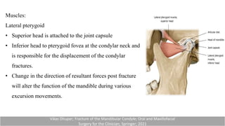
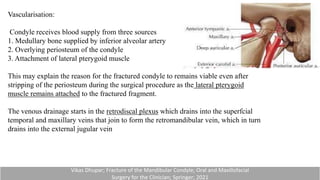
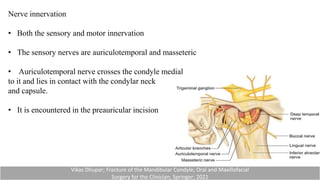
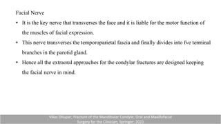
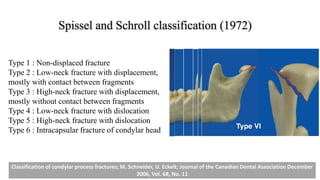
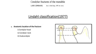
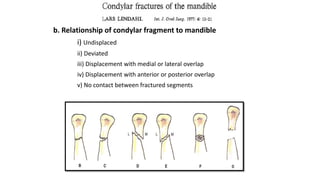
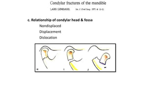
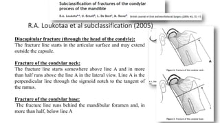
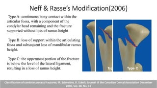
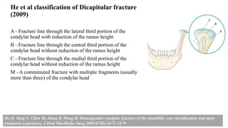
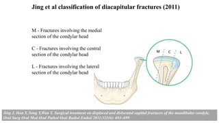
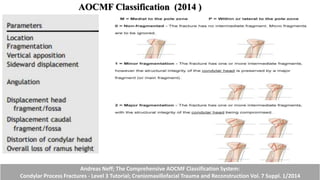
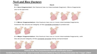
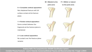
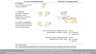
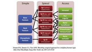
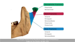
SlideShare a Scribd company logo
1 of 94
Condylar fractures
G.Kathirvel
PG - OMFS
Contents:
• Introduction
• Surgical anatomy
• Biomechanics
• Classification
• Clinical features
• Radiographic features
• Management
• Closed treatment
• Open reduction
• Surgical approaches to condylar fractures
• Complications
Introduction:
• Condylar fractures account for 17.5% to 52 %.
• It differ significantly from other fractures of the mandible owing to the functional
derangement that may affect occlusion, mastication and speech
• However, functional derangements unique to condylar fractures are very frequent sequelae
of these injuries
• This is due to the unique functional anatomy of the mandibular condyles, the specific
challenges of surgical treatment of these fractures and the various patient-specific factors
affecting the outcomes of condylar fractures.
Peterson’s Principles of Oral and Maxillofacial Surgery - Third Edition; 2011; PEOPLE’S MEDICAL PUBLISHING HOUSE—USA
Anatomy:
The mandibular condyle forms a part of the temporomandibular joint which is
unique and TMJ made of the following structures:
• Condyle of the mandible
• Squamous portion of the temporal bone
• Articular disc (contained within the TMJ)
• Ligaments
Vikas Dhupar; Fracture of the Mandibular Condyle; Oral and Maxillofacial
Surgery for the Clinician; Springer; 2021
Condyle
• The condylar head is ovoid in shape measuring approximately 15–
20 mm mediolaterally and 8–10 mm anteroposteriorly
• Articulates with the glenoid fossa present in the squamous portion
of the temporal bone to form the temporomandibular joint.
• Squamous portion of the temporal bone is as thin as 2 mm as a
result; the condylar processes maybe driven into the middle
cranial fossa following trauma.
Vikas Dhupar; Fracture of the Mandibular Condyle; Oral and Maxillofacial
Surgery for the Clinician; Springer; 2021
Articular Disc
• The squamous portion of the temporal bone and the
condyle is separated by a dense fibrous connective tissue
called the articular disc.
• The disc is firmly anchored to the condyle by the medial
and lateral collateral ligaments and it merges with the
capsule in the periphery.
• The joint space is divided into superior and inferior
compartments by the disc.
Vikas Dhupar; Fracture of the Mandibular Condyle; Oral and Maxillofacial
Surgery for the Clinician; Springer; 2021
Capsule and Ligaments
• The capsule surrounds the TMJ and is reinforced by
the medial and lateral ligaments which connect the
mandible to the temporal bone.
• The synovial membrane lines the capsule.
• This membrane produces synovial fuid which aids in
the lubrication and nourishment of the joint.
• The lateral ligament also known as temporomandibular
ligament has a horizontal and an oblique component
which stabilizes the joint.
Vikas Dhupar; Fracture of the Mandibular Condyle; Oral and Maxillofacial
Surgery for the Clinician; Springer; 2021
Capsule and Ligaments
• The strength of the lateral ligament may be partly responsible
for the fracture at the neck of condyle just below the insertion
of the ligament.
• The medial and lateral discal ligaments which are present
inside the capsule are also called collateral ligaments.
• These ligaments connect the disc to the poles of the condyle.
Accessory ligaments:
• The stylomandibular and sphenomandibular ligaments are
accessory and are not directly attached to any part of the joint.
Vikas Dhupar; Fracture of the Mandibular Condyle; Oral and Maxillofacial
Surgery for the Clinician; Springer; 2021
The stylomandibular ligament
• Runs from the styloid process to the angle of the
mandible
• It separates the parotid and submandibular salivary
glands
• Becomes taut when the mandible is protruded
The sphenomandibular ligament
• Runs from the spine of the sphenoid bone to the lingula
of mandible
• The inferior alveolar nerve descends between the
sphenomandibular ligament and the ramus of the
mandible to gain access to the mandibular foramen
• Becomes taut when the mandible is protruded
Vikas Dhupar; Fracture of the Mandibular Condyle; Oral and Maxillofacial
Surgery for the Clinician; Springer; 2021
Muscles:
Lateral pterygoid
• Superior head is attached to the joint capsule
• Inferior head to pterygoid fovea at the condylar neck and
is responsible for the displacement of the condylar
fractures.
• Change in the direction of resultant forces post fracture
will alter the function of the mandible during various
excursion movements.
Vikas Dhupar; Fracture of the Mandibular Condyle; Oral and Maxillofacial
Surgery for the Clinician; Springer; 2021
Vascularisation:
Condyle receives blood supply from three sources
1. Medullary bone supplied by inferior alveolar artery
2. Overlying periosteum of the condyle
3. Attachment of lateral pterygoid muscle
This may explain the reason for the fractured condyle to remains viable even after
stripping of the periosteum during the surgical procedure as the lateral pterygoid
muscle remains attached to the fractured fragment.
The venous drainage starts in the retrodiscal plexus which drains into the superfcial
temporal and maxillary veins that join to form the retromandibular vein, which in turn
drains into the external jugular vein
Vikas Dhupar; Fracture of the Mandibular Condyle; Oral and Maxillofacial
Surgery for the Clinician; Springer; 2021
Nerve innervation
• Both the sensory and motor innervation
• The sensory nerves are auriculotemporal and masseteric
• Auriculotemporal nerve crosses the condyle medial
to it and lies in contact with the condylar neck
and capsule.
• It is encountered in the preauricular incision
Vikas Dhupar; Fracture of the Mandibular Condyle; Oral and Maxillofacial
Surgery for the Clinician; Springer; 2021
Facial Nerve
• It is the key nerve that transverses the face and it is liable for the motor function of
the muscles of facial expression.
• This nerve transverses the temporoparietal fascia and finally divides into fve terminal
branches in the parotid gland.
• Hence all the extraoral approaches for the condylar fractures are designed keeping
the facial nerve in mind.
Vikas Dhupar; Fracture of the Mandibular Condyle; Oral and Maxillofacial
Surgery for the Clinician; Springer; 2021
Surgical anatomy:
The preauricular incision
• Al-Kayat and Bramley found the nerve was at an
average distance of 20 mm with a range of 8–35 mm
from the anterior margin of the auditory canal.
• This is the reason the preauricular incision is given in
the skin crease near the tragus or placed endurally.
Transparotid approach.
• The neck of the condyle can be exposed via the
transparotid approach.
• In this approach the condyle is reached through the
space between the temporozygomatic and buccocervical
trunks of the nerve.
• This results in a direct and safe approach to the neck of
the condyle.
Surgical anatomy:
The marginal mandibular nerve
• The marginal mandibular nerve is encountered in
the submandibular or periangular approach.
• This branch may further subdivide into two or
more branches.
• As a rule, the submandibular incision is given
2 cm below the lower border of the mandible to
prevent paresis of the lower lip.
Vikas Dhupar; Fracture of the Mandibular Condyle; Oral and Maxillofacial
Surgery for the Clinician; Springer; 2021
BIOMECHANICS
LINDAHL (1977)
HUNTING BOW CONCEPT
Classification
Wassmund classification(1934)
Type I- The angle between the head and the long axis of the ramus :10 to
45 degrees.
Type II- angle of 45 to 90 degrees, resulting in tearing of the medial
portion of the capsule.
Type III- the fragments are not in contact, and the head is displaced
mesially and forward owing to traction of the lateral pterygoid muscle.
Type IV- fractures where the condylar head articulates in an anterior
position to the articular eminence.
Type V- vertical or oblique fractures through the head of the condyle.
A.Condylar fractures
i. Without displacement of condyle
• Greenstick fracture
• Intracapsular
• Extracapsular
ii. With displacement of condyle
• Lateral
• Medial
• Forward
• Backward
iii. With overriding of fragments
iv. With dislocation in lateral or medial direction
• Intracapsular
• Complete fracture dislocation
• Complete dislodgement of the condyle
• Dislocation of the fractured part of the head of the condyle
v. With dislocation in forward direction
• Anteriorly from the articular eminence
• Posteriorly from the articular eminence
• With dislocation and displacement of the meniscus
• With comminution
• Old fracture with deformities
— Pseudoarthrosis
— Ankylosis
Thoma (1945)
B. Subcondylar fractures
i. Without displacement of fragment
ii. With displacement of fragment
By Thoma
Fracture line either extending through head or base of the condyle or neck
has been called condylar fracture , whereas in subcondylar fracture, the line
runs transversely over ascending ramus.
Mac Lennan classification (1952)
a. No Displacement - A crack fracture is seen without alteration of the normal relationship of
TM Joint
b. Deviation - simple angulation of the condylar process to the ramus
c. Displacement - Overlap occurs between the condylar process and the ramus and fractured
fragment lies lateral to the ramus.
d. Dislocation - condylar head is completely disrupted from the glenoid fossa and gets pulled
anteriorly and medially by the lateral pterygoid muscle
Rowe & Killey 's classification(1968)
a. Intracapsular Fractures or High Condylar Fractures
i. Fractures involving the articular surface
ii. Fractures above or through the anatomical neck,
which do not involve the articular surface
b. Extracapsular or Low Condylar or Sub condylar Fractures
Fracture line runs obliquly from the lowest point of curvature of the sigmoid notch
downward and backward below the neck to the upper posterior limit of the ramus.
c. Fractures associated with injury to the capsule, ligament and meniscus
d. Fractures involving adjacent bone - Roof of the glenoid fossa or tympanic plate of
the external auditory meatus.
Spissel and Schroll classification (1972)
Type 1 : Non-displaced fracture
Type 2 : Low-neck fracture with displacement,
mostly with contact between fragments
Type 3 : High-neck fracture with displacement,
mostly without contact between fragments
Type 4 : Low-neck fracture with dislocation
Type 5 : High-neck fracture with dislocation
Type 6 : Intracapsular fracture of condylar head
Classification of condylar process fractures; M. Schneider, U. Eckelt; Journal of the Canadian Dental Association December
2006, Vol. 68, No. 11
Lindahl classification(1977)
a. Anatomic location of the fracture
i) Condylar head
ii) Condylar neck
iii) Subcondylar
b. Relationship of condylar fragment to mandible
i) Undisplaced
ii) Deviated
iii) Displacement with medial or lateral overlap
iv) Displacement with anterior or posterior overlap
v) No contact between fractured segments
c. Relationship of condylar head & fossa
Nondisplaced
Displacement
Dislocation
R.A. Loukotaa et al subclassification (2005)
Diacapitular fracture (through the head of the condyle):
The fracture line starts in the articular surface and may extend
outside the capsule.
Fracture of the condylar neck:
The fracture line starts somewhere above line A and in more
than half runs above the line A in the lateral view. Line A is the
perpendicular line through the sigmoid notch to the tangent of
the ramus.
Fracture of the condylar base:
The fracture line runs behind the mandibular foramen and, in
more than half, below line A
Neff & Rasse’s Modification(2006)
Classification of condylar process fractures; M. Schneider, U. Eckelt; Journal of the Canadian Dental Association December
2006, Vol. 68, No. 11
Type A: continuous bony contact within the
articular fossa, with a component of the
condylar head remaining and the fracture
supported without loss of ramus height
Type B: loss of support within the articulating
fossa and subsequent loss of mandibular ramus
height.
Type C: the uppermost portion of the fracture
is below the level of the lateral ligament,
resulting in a loss of ramus height.
He et al classification of Dicapitular fracture
(2009)
A - Fracture line through the lateral third portion of the
condylar head with reduction of the ramus height
B - Fracture line through the central third portion of the
condylar head without reduction of the ramus height
C - Fracture line through the medial third portion of the
condylar head without reduction of the ramus height
M - A comminuted fracture with multiple fragments (usually
more than three) of the condylar head
He D, Yang C, Chen M, Jiang B, Wang B. Intracapsular condylar fracture of the mandible: our classification and open
treatment experience. J Oral Maxillofac Surg 2009;67(8):1672–1679
Jing et al classification of diacapitular fractures (2011)
M - Fractures involving the medial
section of the condylar head
C - Fractures involving the central
section of the condylar head
L - Fractures involving the lateral
section of the condylar head
Jing J, Han Y, Song Y,Wan Y. Surgical treatment on displaced and dislocated sagittal fractures of the mandibular condyle.
Oral Surg Oral Med Oral Pathol Oral Radiol Endod 2011;111(6): 693–699
AOCMF Classification (2014 )
Condylar Head fracture
Andreas Neff; The Comprehensive AOCMF Classification System:
Condylar Process Fractures - Level 3 Tutorial; Craniomaxillofacial Trauma and Reconstruction Vol. 7 Suppl. 1/2014
Neck and Base fractures
Andreas Neff; The Comprehensive AOCMF Classification System:
Condylar Process Fractures - Level 3 Tutorial; Craniomaxillofacial Trauma and Reconstruction Vol. 7 Suppl. 1/2014
Andreas Neff; The Comprehensive AOCMF Classification System:
Condylar Process Fractures - Level 3 Tutorial; Craniomaxillofacial Trauma and Reconstruction Vol. 7 Suppl. 1/2014
Andreas Neff; The Comprehensive AOCMF Classification System:
Condylar Process Fractures - Level 3 Tutorial; Craniomaxillofacial Trauma and Reconstruction Vol. 7 Suppl. 1/2014
Andreas Neff; The Comprehensive AOCMF Classification System:
Condylar Process Fractures - Level 3 Tutorial; Craniomaxillofacial Trauma and Reconstruction Vol. 7 Suppl. 1/2014
Andreas Neff; The Comprehensive AOCMF Classification System:
Condylar Process Fractures - Level 3 Tutorial; Craniomaxillofacial Trauma and Reconstruction Vol. 7 Suppl. 1/2014
Andreas Neff; The Comprehensive AOCMF Classification System:
Condylar Process Fractures - Level 3 Tutorial; Craniomaxillofacial Trauma and Reconstruction Vol. 7 Suppl. 1/2014
Distortion of condylar head articular congruency
Andreas Neff; The Comprehensive AOCMF Classification System:
Condylar Process Fractures - Level 3 Tutorial; Craniomaxillofacial Trauma and Reconstruction Vol. 7 Suppl. 1/2014
Clinical Features
Clinical Features
Condylar fractures infrequently occurred in isolation and are generally associated
with fractures of other sites of the facial bones.
Signs and symptoms:
• Pain and swelling over the preauricular region
• Ecchymosis over the mastoid region
• CSF otorrhea
• Hollowness over the condylar region
• Restricted incisal opening
• Locked mandible
Raymond.J Fonseca; Oral & maxillofacial trauma; fourth edition; 2013, Saunders, Elsevier Inc
Unilateral condylar Fractures:
• Premature occlusion on the ipsilateral side
• Open bite due to the loss of vertical height on the
contralateral side.
• Affected side deviates on opening and is common because
of the interruption of the action of the lateral pterygoid
muscle.
• Limited laterotrusive movements away from the fractured
side but may be maintained towards the fracture.
Raymond.J Fonseca; Oral & maxillofacial trauma; fourth edition; 2013, Saunders, Elsevier Inc
Bilateral condylar Fractures:
• Bilateral loss of vertical height results in anterior open bite
with posterior gagging
• Restricted mouth opening
• In bilateral condylar fractures with an associated
mandibular symphysis fracture, facial widening may result
with posterior buccal cross-bites and anterior open bite.
Raymond.J Fonseca; Oral & maxillofacial trauma; fourth edition; 2013, Saunders, Elsevier Inc
Radiographic evaluation:
Towne’s radiograph
Coronal displacement is evaluated with Towne’s radiograph
OPG
Sagittal displacement, the loss of ramus heigh is evaluated
with a panoramic radiograph
The measurement technique is as follows :
• Line drawn between gonial angles across Panorex
• Perpendicular lines to most superior aspect of condylar
heads
• The difference between the nonfractured and fractured side
equals the change in ramus height.
Raymond.J Fonseca; Oral & maxillofacial trauma; fourth edition; 2013, Saunders, Elsevier Inc
Computed tomography
Anterior posterior dimension (sagittal position)
Medial lateral dimension (coronal position)
MRI
MRI are essential to evaluate the disc injuries specially in dislocation and
intracapsular condylar fractures; however MRI are not taken as a routine practise
Raymond.J Fonseca; Oral & maxillofacial trauma; fourth edition; 2013, Saunders, Elsevier Inc
Management
CONSERVATIVE TREATMENT
• Closed treatment is treatment of condylar fractures by means other than surgical
exploration, reduction, and fixation of the fracture line (i.e., not involving an open
surgical exposure of the fracture).
• Traditionally, this has been achieved by arch bars, splints fitted over the remaining
dentition, IMF, screws, or bonded brackets.
• Once reduction of the occlusion has been achieved, a period of immobilization may
be required to encourage bony healing.
• Early mobilization is advised to minimize the risk of fibrous and bony TMJ
ankylosis.
Vikas Dhupar; Fracture of the Mandibular Condyle; Oral and Maxillofacial
Surgery for the Clinician; Springer; 2021
INDICATIONS FOR CLOSED TREATMENT
Nonsurgical treatment may be the appropriate management strategy in cases of
• Condylar neck fractures in children <12 years of age.
• High condylar neck fractures without displacement.
• Intracapsular (diacapitular) condylar fractures without loss of ramus height.
• Poor anesthetic risk.
Vikas Dhupar; Fracture of the Mandibular Condyle; Oral and Maxillofacial
Surgery for the Clinician; Springer; 2021
INDICATIONS FOR OPEN TREATMENT
APPROACHES
Emam HA, Jatana CA, Ness GM. Matching surgical approach to condylar fracture type.
Atlas Oral Maxillofac Surg Clin North Am 2017;25:55-61
Vikas Dhupar; Fracture of the Mandibular Condyle; Oral and Maxillofacial
Surgery for the Clinician; Springer; 2021
Submandibular Approach.
This is also known as the Risdon approach
Advantages:
These include the ability to distract the mandibular ramus
and direct access of the gonial angle.
Disadvantages:
• These include limited surgical site exposure (the
incision is distant from the fracture)
• Difficult to reduce medially displaced condyles
• Plate and screw fixation restricted without a
transfacial trocar.
Raymond.J Fonseca; Oral & maxillofacial trauma; fourth edition; 2013, Saunders, Elsevier Inc
Surgical Technique
Incision.
• These are 1.5 to 2 cm below the inferior border of the mandible
in or parallel to a skin crease.
• In patients with ramus height shortening, place the incision 1.5 to
2 cm below where the anticipated reduced mandible would be.
• The initial incision is placed to the depth of the platysma, with
extensive undermining in all directions.
Dissection.
• Through the platysma. Undermine and sharply dissect, being
careful to stay superficial to the superficial layer of the deep
cervical fascia.
Raymond.J Fonseca; Oral & maxillofacial trauma; fourth edition; 2013, Saunders, Elsevier Inc
• Make a small incision through the superficial layer of the deep
cervical fascia at the level of the skin incision (1.5 to 2 cm
inferior to the mandible).
• The facial artery and vein may be retracted anteriorly or
divided and ligated if necessary.
• Continue the dissection superiorly until the pterygomasseteric sling is encountered.
• Division of the pterygomasseteric sling. Sharply incise the pterygomasseteric sling with
a scalpel along the inferior border of the mandible.
• Use a periosteal elevator to expose the ramus up to the level of the TMJ capsule and
coronoid process.
Raymond.J Fonseca; Oral & maxillofacial trauma; fourth edition; 2013, Saunders, Elsevier Inc
Retromandibular Approach
Advantages.
• Short distance between the incision and the fracture site
• Best access to the fracture site
• No need for a transfacial trocar
• Facial scar is less noticeable than with a submandibular incision
• Effective in patients with edema
• Access for an osteotomy if required to reach the condyle.
Disadvantages.
The facial scar is more noticeable than with a preauricular incision.
Raymond.J Fonseca; Oral & maxillofacial trauma; fourth edition; 2013, Saunders, Elsevier Inc
Retromandibular – Trans-parotid Approach
Retromandibular- Retro-parotid- Approach
Technique
• A preauricular incision is made that extends downwards in a curvilinear
fashion in the cervicomastoid skin crease, though any variation in this
incision.
• The great auricular nerve is preserved and the flap raised in the subdermal fat
plane, superficial to the superficial musculoaponeurotic layer to allow access
to the masseter adjacent to the anteroinferior edge of the parotid gland, just
below the parotid duct.
• The area next to the anterior edge of the parotid gland is usually relatively free
of branches of the facial nerve, making this an ideal point to dissect down to
the fracture
Technique
• If a buccal branch crosses the field it is easily retracted up or down.
• The masseter is split in the direction of its fibres.
• The periosteum overlying the lateral aspect of the ramus in the
region of the condylar neck is incised and the fracture site is
exposed.
Periangular transmasseteric approach
• 4 cm long curvilinear incision, 5 mm below and along the angle of
the mandible
• After skin incision, dissection was continued in the subcutaneous
plane, upward and superficial to the platysma till the zygomatic
arch.
• The thin platysma muscle is held with tissue holding forceps and
incised obliquely to expose the masseteric fascia overlying the
masseter belly
Periangular transmasseteric approach
• The area adjacent to the anterior edge of the parotid gland is relatively
free of the branches of the facial nerve and hence ideal for further
dissection.
• The parotid tissue was retracted posteriorly with retractors positioned
horizontally, and the belly of masseter muscle was incised parallel to
the fibers of the branches of the facial nerve to expose the periosteum.
• The periosteum was incised and the direction of retractor changed
vertically to retract masseter in an upward pull manner to expose the
fractured segment
Pre-Auricular Approach
TRANSORAL APPROACH
Indications:
Fractures that involve the low condylar region.
Technique:
• An incision is made over the anterior border of the vertical
ramus, extending into the lower buccal sulcus.
• The temporalis muscle is stripped from the anterior ascending
ramus, and the masseter is stripped by subperiosteal dissection.
• A transbuccal trocar is introduced and drilling occurs
transbuccally
• the plate is introduced transorally.
TRANSORAL APPROACH
Advantage:
Avoiding facial scarring and risk of injury to the facial nerve.
Disadvantage:
• limited access, which makes fragment control difficult and the
procedure surgically more challenging.
• It may not be possible to align the posterior border perfectly
and slight errors of reduction are inevitable.
TECHNIQUES OF REDUCTION
The use of the curved elevator, Howarth’s elevator, and the
tracheostomy hook have all been advocated.
Kocher’s bone-holding forceps may be used to grasp and
reduce fragments but can crush and split the condylar head
and therefore must be used with caution.
In cases of severe and difficult reduction, the lateral
pterygoid insertion may be cut to give greater mobility and
reducibility, but preservation of the periosteal attachment to
the condylar head is mandatory to preserve a blood supply to
the proximal fragment.
Vikas Dhupar; Fracture of the Mandibular Condyle; Oral and Maxillofacial
Surgery for the Clinician; Springer; 2021
Manual digital traction: Thumb is placed on the lower
teeth and fngers support the lower border. Mandible is
pulled in inferior and anterior direction.
Bite block: A bite block is placed on the same side of the
condylar fracture in the molar region. This will result in
inferior distraction of the mandible with rotation.
Transosseous wire: A transosseous wire can be passed
along the posteroinferior border of the mandible. A traction is
applied on this wire in an inferior and anterior direction.
For easy manipulation, a plate with a single screw is fixed on
the proximal fragment. This will help in easy lateralization of
the distal fragment and prevent the fragment from slipping
back as the lower end of the plate acts as a rest on the proximal
segment.
Vikas Dhupar; Fracture of the Mandibular Condyle; Oral and Maxillofacial
Surgery for the Clinician; Springer; 2021
FIXATION
Miniplate Osteosynthesis:
Meyer’s line of osteosynthesis
In the case of low condylar fracture, two plates may be
required to achieve stability.
The posterior plate should parallel the posterior ascending
ramus, and the anterior plate parallel to sigmoid notch.
High condylar fractures may accommodate only one plate
due to bony limitations
3-D plates
Vikas Dhupar; Fracture of the Mandibular Condyle; Oral and Maxillofacial
Surgery for the Clinician; Springer; 2021
Lag Screw Osteosynthesis
Lag screw osteosynthesis was first described in condylar fractures
by Wackerbauer in 1962.
The Eckelt screw is one of the most popular lag screws
Advantage
Rapid application of rigid fixation and close approximation of the
fractured parts because of the large amount of compression
generated.
Bioresorbable Plates
Vikas Dhupar; Fracture of the Mandibular Condyle; Oral and Maxillofacial
Surgery for the Clinician; Springer; 2021
Complications
Malocclusion.
• This is often implied secondary to improper treatment, such as inadequate use of
occlusal guidance or closed reduction when open reduction is indicated.
• Patients presenting with displaced fractures, ramus shortening, and early
objective malocclusion will often have persistent centric relation discrepancies
when treated in a closed fashion.
Raymond.J Fonseca; Oral & maxillofacial trauma; fourth edition; 2013, Saunders, Elsevier Inc
Mandibular Hypomobility.
• This is related to delayed physiotherapy of the joint and has been shown to
increase the longer the patient is subjected to MMF.
• Children are more susceptible to hypomobility, as are those subjected to high-
energy injuries with capsular disruption.
Ankylosis.
• In children, ankylosis is related to severe meniscal disruption with inappropriate
physiotherapy.
• In adults, ankylosis usually results from a widened mandible, which leads to
superior lateral displacement of the condyle.
• This may be mitigated by proper reduction of the fractures restricting mandibular
widening.
Raymond.J Fonseca; Oral & maxillofacial trauma; fourth edition; 2013, Saunders, Elsevier Inc
Asymmetry.
• In children, approximately 25% of condylar fractures will produce some facial
asymmetry, whether it is hypoplasia or hyperplasia.
• In adults, deviation on opening has been noted in up to 50% of individuals following
fracture of the condyle.
Dysfunction or Degeneration.
• All injured joints are more susceptible to arthritis, and the TMJ is no different.
• Risk factors include increased age, displaced condyle, longer periods of MMF and
hypomobility secondary to capsular or meniscal injuries.
Condylar Resorption.
• Avoidable by limiting the total denudation of the blood supply and proper anatomic
reduction.
• However, difficult cases may demand replacement of the condylar head as a free bone
graft.
Raymond.J Fonseca; Oral & maxillofacial trauma; fourth edition; 2013, Saunders, Elsevier Inc
Iatrogenic Injury.
• Following surgical repair of condylar fractures, up to 15% of patients may
experience transient facial nerve weakness; however, permanent injury is
rare.
Chronic Pain.
• This occurs more commonly when condylar fractures are treated with
closed reduction.
Raymond.J Fonseca; Oral & maxillofacial trauma; fourth edition; 2013, Saunders, Elsevier Inc
Nerve injury
• During preauricular approach, a precaution should be given not to cause the injury of facial
nerve temporal branch
• Nerve injury may occur during flap elevation, fracture reduction and insertion of a metal
plate.
• If nerve injury is observed after the surgery, steroid should be immediately administered to
prevent tissue swelling, thereby shortening recovery time and preventing permanent injury.
Sialocele:
• Collection of saliva beneath the skin and subcutaneous tissue
associated with injury to glandular parenchyma or ductal system.
Clinical features:
• Salivary extravasations into the tissues causing swelling over or
adjacent to parotid gland (sialocele)
• Expanding neck mass
• Cutaneous fistula formation.
• Following transparotid approach closure includes formal closure of the
masseter and the parotid fascia - reduces the swelling and the
possibility of a sialocele
THANK YOU

More Related Content

What's hot

Zygomatic maxillary complex fracture
Zygomatic maxillary complex fractureZygomatic maxillary complex fracture
Zygomatic maxillary complex fracturejosna thankachan
 
NOE fractures
NOE fractures NOE fractures
NOE fractures anchalag8
 
Condylar Fractures
Condylar FracturesCondylar Fractures
Condylar FracturesAhmed Adawy
 
Hardware in maxillofacial trauma
Hardware in maxillofacial traumaHardware in maxillofacial trauma
Hardware in maxillofacial traumaDrChiragPatil
 
Internal derangement of tmj
Internal derangement of tmjInternal derangement of tmj
Internal derangement of tmjDrKamini Dadsena
 
Zygomatic arch fracture
Zygomatic arch fractureZygomatic arch fracture
Zygomatic arch fracturemostafa heeba
 
Mandibular Condylar fractures & its Management
Mandibular Condylar fractures & its ManagementMandibular Condylar fractures & its Management
Mandibular Condylar fractures & its ManagementMehul Hirani
 
Zygomatic Complex Fracture- ZMC
Zygomatic Complex Fracture- ZMCZygomatic Complex Fracture- ZMC
Zygomatic Complex Fracture- ZMCHimanshu Soni
 
Presurgical diagnosis and treatment planing of orthognathic surgery, Dr. Lide...
Presurgical diagnosis and treatment planing of orthognathic surgery, Dr. Lide...Presurgical diagnosis and treatment planing of orthognathic surgery, Dr. Lide...
Presurgical diagnosis and treatment planing of orthognathic surgery, Dr. Lide...LIDETU AFEWORK
 
Aocmf classification . 2014, level 3 , condylar fracture ,
Aocmf classification . 2014, level 3 , condylar fracture ,Aocmf classification . 2014, level 3 , condylar fracture ,
Aocmf classification . 2014, level 3 , condylar fracture ,DrJayTrivedi
 
Temporomandibular joint disorders IV
Temporomandibular joint disorders IVTemporomandibular joint disorders IV
Temporomandibular joint disorders IVIAU Dent
 
7. fractures of middle third of facial skeleton
7. fractures of middle third of facial skeleton7. fractures of middle third of facial skeleton
7. fractures of middle third of facial skeletonDr. Samarth Johari
 
Mandibular fractures
Mandibular fracturesMandibular fractures
Mandibular fractureschaitanyeah
 
Management of condylar fractures
Management of condylar fracturesManagement of condylar fractures
Management of condylar fracturesdralimohammedhasan
 
Condylar fractures
Condylar fracturesCondylar fractures
Condylar fracturesZeeshan Arif
 

What's hot (20)

Zygomatic maxillary complex fracture
Zygomatic maxillary complex fractureZygomatic maxillary complex fracture
Zygomatic maxillary complex fracture
 
NOE fractures
NOE fractures NOE fractures
NOE fractures
 
Condylar Fractures
Condylar FracturesCondylar Fractures
Condylar Fractures
 
FRONTAL BONE FRACTURE AND ITS MANAGEMENT.pptx
FRONTAL BONE FRACTURE AND ITS MANAGEMENT.pptxFRONTAL BONE FRACTURE AND ITS MANAGEMENT.pptx
FRONTAL BONE FRACTURE AND ITS MANAGEMENT.pptx
 
Hardware in maxillofacial trauma
Hardware in maxillofacial traumaHardware in maxillofacial trauma
Hardware in maxillofacial trauma
 
Internal derangement of tmj
Internal derangement of tmjInternal derangement of tmj
Internal derangement of tmj
 
Zygomatic arch fracture
Zygomatic arch fractureZygomatic arch fracture
Zygomatic arch fracture
 
Mandibular Condylar fractures & its Management
Mandibular Condylar fractures & its ManagementMandibular Condylar fractures & its Management
Mandibular Condylar fractures & its Management
 
Zygomatic Complex Fracture- ZMC
Zygomatic Complex Fracture- ZMCZygomatic Complex Fracture- ZMC
Zygomatic Complex Fracture- ZMC
 
Presurgical diagnosis and treatment planing of orthognathic surgery, Dr. Lide...
Presurgical diagnosis and treatment planing of orthognathic surgery, Dr. Lide...Presurgical diagnosis and treatment planing of orthognathic surgery, Dr. Lide...
Presurgical diagnosis and treatment planing of orthognathic surgery, Dr. Lide...
 
Aocmf classification . 2014, level 3 , condylar fracture ,
Aocmf classification . 2014, level 3 , condylar fracture ,Aocmf classification . 2014, level 3 , condylar fracture ,
Aocmf classification . 2014, level 3 , condylar fracture ,
 
Mandible # brief
Mandible # brief Mandible # brief
Mandible # brief
 
Temporomandibular joint disorders IV
Temporomandibular joint disorders IVTemporomandibular joint disorders IV
Temporomandibular joint disorders IV
 
Le Fort Fractures
Le Fort FracturesLe Fort Fractures
Le Fort Fractures
 
7. fractures of middle third of facial skeleton
7. fractures of middle third of facial skeleton7. fractures of middle third of facial skeleton
7. fractures of middle third of facial skeleton
 
04 frontal sinus FRACTURE
04 frontal sinus FRACTURE04 frontal sinus FRACTURE
04 frontal sinus FRACTURE
 
Mandibular fractures
Mandibular fracturesMandibular fractures
Mandibular fractures
 
Management of condylar fractures
Management of condylar fracturesManagement of condylar fractures
Management of condylar fractures
 
Condylar fractures
Condylar fracturesCondylar fractures
Condylar fractures
 
3 approaches to the tmj
3 approaches to the tmj3 approaches to the tmj
3 approaches to the tmj
 

Similar to Condylar fracture.pptx

5 NOE FRACTURE seminar 5.pptx
5 NOE FRACTURE seminar 5.pptx5 NOE FRACTURE seminar 5.pptx
5 NOE FRACTURE seminar 5.pptxsneha
 
Surgical anatomy of Noe complex in context of trauma
Surgical anatomy of Noe complex in context of traumaSurgical anatomy of Noe complex in context of trauma
Surgical anatomy of Noe complex in context of traumaDr. Hani Yousuf
 
Anatomical Landmarks Mandibular prosthodontics
Anatomical Landmarks Mandibular prosthodonticsAnatomical Landmarks Mandibular prosthodontics
Anatomical Landmarks Mandibular prosthodonticsSUBHRAKANTI PANDIT
 
Tmj and prosthodontic implications
Tmj and prosthodontic implicationsTmj and prosthodontic implications
Tmj and prosthodontic implicationsPramod Chahar
 
Subluxation and dislocation of temporo mandibular joint
Subluxation and dislocation of temporo mandibular jointSubluxation and dislocation of temporo mandibular joint
Subluxation and dislocation of temporo mandibular jointDr. Akshay Shah
 
Functional anatomy of mandible
Functional anatomy of mandibleFunctional anatomy of mandible
Functional anatomy of mandibleFarakath Khan
 
Functional anatomy of mandible
Functional anatomy of mandibleFunctional anatomy of mandible
Functional anatomy of mandibleFarakath Khan
 
Management of Mandibular fractures.pptx
Management of Mandibular fractures.pptxManagement of Mandibular fractures.pptx
Management of Mandibular fractures.pptxAminat Farayola
 
Condylar fractures /certified fixed orthodontic courses by Indian dental aca...
Condylar fractures  /certified fixed orthodontic courses by Indian dental aca...Condylar fractures  /certified fixed orthodontic courses by Indian dental aca...
Condylar fractures /certified fixed orthodontic courses by Indian dental aca...Indian dental academy
 
TMJ ankylosis and reconstruction.pptx
TMJ ankylosis and reconstruction.pptxTMJ ankylosis and reconstruction.pptx
TMJ ankylosis and reconstruction.pptxKathirvelGopalakrish
 
Lefort 2 fracture
Lefort 2 fractureLefort 2 fracture
Lefort 2 fracturejoyaljoice
 

Similar to Condylar fracture.pptx (20)

Condylar fractures
Condylar fracturesCondylar fractures
Condylar fractures
 
5 NOE FRACTURE seminar 5.pptx
5 NOE FRACTURE seminar 5.pptx5 NOE FRACTURE seminar 5.pptx
5 NOE FRACTURE seminar 5.pptx
 
Condylar #
Condylar #Condylar #
Condylar #
 
Surgical anatomy of Noe complex in context of trauma
Surgical anatomy of Noe complex in context of traumaSurgical anatomy of Noe complex in context of trauma
Surgical anatomy of Noe complex in context of trauma
 
Anatomical Landmarks Mandibular prosthodontics
Anatomical Landmarks Mandibular prosthodonticsAnatomical Landmarks Mandibular prosthodontics
Anatomical Landmarks Mandibular prosthodontics
 
tmj ana,phy,dys.pptx
tmj ana,phy,dys.pptxtmj ana,phy,dys.pptx
tmj ana,phy,dys.pptx
 
Anatomy of mandible
Anatomy of mandibleAnatomy of mandible
Anatomy of mandible
 
Tmj ankylosis
Tmj ankylosisTmj ankylosis
Tmj ankylosis
 
TMJ.pptx
TMJ.pptxTMJ.pptx
TMJ.pptx
 
Ramus osteotomy
Ramus osteotomy Ramus osteotomy
Ramus osteotomy
 
TMD-FL.pdf
TMD-FL.pdfTMD-FL.pdf
TMD-FL.pdf
 
Tmj and prosthodontic implications
Tmj and prosthodontic implicationsTmj and prosthodontic implications
Tmj and prosthodontic implications
 
TMJ Ankylosis.pptx
TMJ Ankylosis.pptxTMJ Ankylosis.pptx
TMJ Ankylosis.pptx
 
Subluxation and dislocation of temporo mandibular joint
Subluxation and dislocation of temporo mandibular jointSubluxation and dislocation of temporo mandibular joint
Subluxation and dislocation of temporo mandibular joint
 
Functional anatomy of mandible
Functional anatomy of mandibleFunctional anatomy of mandible
Functional anatomy of mandible
 
Functional anatomy of mandible
Functional anatomy of mandibleFunctional anatomy of mandible
Functional anatomy of mandible
 
Management of Mandibular fractures.pptx
Management of Mandibular fractures.pptxManagement of Mandibular fractures.pptx
Management of Mandibular fractures.pptx
 
Condylar fractures /certified fixed orthodontic courses by Indian dental aca...
Condylar fractures  /certified fixed orthodontic courses by Indian dental aca...Condylar fractures  /certified fixed orthodontic courses by Indian dental aca...
Condylar fractures /certified fixed orthodontic courses by Indian dental aca...
 
TMJ ankylosis and reconstruction.pptx
TMJ ankylosis and reconstruction.pptxTMJ ankylosis and reconstruction.pptx
TMJ ankylosis and reconstruction.pptx
 
Lefort 2 fracture
Lefort 2 fractureLefort 2 fracture
Lefort 2 fracture
 

More from KathirvelGopalakrish (14)

Mandibular osteotomies.pptx
Mandibular osteotomies.pptxMandibular osteotomies.pptx
Mandibular osteotomies.pptx
 
Midface fracture.pptx
Midface fracture.pptxMidface fracture.pptx
Midface fracture.pptx
 
Primary assessment in maxillofacial trauma.pptx
Primary assessment in maxillofacial trauma.pptxPrimary assessment in maxillofacial trauma.pptx
Primary assessment in maxillofacial trauma.pptx
 
Rhinoplasty.pptx
Rhinoplasty.pptxRhinoplasty.pptx
Rhinoplasty.pptx
 
Preop assessment and operation theatre protocols.pptx
Preop assessment and operation theatre protocols.pptxPreop assessment and operation theatre protocols.pptx
Preop assessment and operation theatre protocols.pptx
 
Osteomyelitis of jaw
Osteomyelitis of jawOsteomyelitis of jaw
Osteomyelitis of jaw
 
Maxillary canine and molar impaction
Maxillary canine and molar impactionMaxillary canine and molar impaction
Maxillary canine and molar impaction
 
Maxillary nerve block
Maxillary nerve blockMaxillary nerve block
Maxillary nerve block
 
Anatomy of orbit
Anatomy of orbitAnatomy of orbit
Anatomy of orbit
 
General anesthesia
General anesthesiaGeneral anesthesia
General anesthesia
 
Unconsciousness and syncope - Management
Unconsciousness and syncope - ManagementUnconsciousness and syncope - Management
Unconsciousness and syncope - Management
 
Anterior triangles of neck
Anterior triangles of neckAnterior triangles of neck
Anterior triangles of neck
 
Facial nerve
Facial nerveFacial nerve
Facial nerve
 
Vascular supply of face and neck
Vascular supply of face and neckVascular supply of face and neck
Vascular supply of face and neck
 

Recently uploaded

Call Girls Kochi Just Call 9907093804 Top Class Call Girl Service Available
Call Girls Kochi Just Call 9907093804 Top Class Call Girl Service AvailableCall Girls Kochi Just Call 9907093804 Top Class Call Girl Service Available
Call Girls Kochi Just Call 9907093804 Top Class Call Girl Service AvailableDipal Arora
 
Call Girls Faridabad Just Call 9907093804 Top Class Call Girl Service Available
Call Girls Faridabad Just Call 9907093804 Top Class Call Girl Service AvailableCall Girls Faridabad Just Call 9907093804 Top Class Call Girl Service Available
Call Girls Faridabad Just Call 9907093804 Top Class Call Girl Service AvailableDipal Arora
 
Call Girls Bhubaneswar Just Call 9907093804 Top Class Call Girl Service Avail...
Call Girls Bhubaneswar Just Call 9907093804 Top Class Call Girl Service Avail...Call Girls Bhubaneswar Just Call 9907093804 Top Class Call Girl Service Avail...
Call Girls Bhubaneswar Just Call 9907093804 Top Class Call Girl Service Avail...Dipal Arora
 
Russian Call Girls in Jaipur Riya WhatsApp ❤8445551418 VIP Call Girls Jaipur
Russian Call Girls in Jaipur Riya WhatsApp ❤8445551418 VIP Call Girls JaipurRussian Call Girls in Jaipur Riya WhatsApp ❤8445551418 VIP Call Girls Jaipur
Russian Call Girls in Jaipur Riya WhatsApp ❤8445551418 VIP Call Girls Jaipurparulsinha
 
Call Girls Ooty Just Call 9907093804 Top Class Call Girl Service Available
Call Girls Ooty Just Call 9907093804 Top Class Call Girl Service AvailableCall Girls Ooty Just Call 9907093804 Top Class Call Girl Service Available
Call Girls Ooty Just Call 9907093804 Top Class Call Girl Service AvailableDipal Arora
 
Russian Escorts Girls Nehru Place ZINATHI 🔝9711199012 ☪ 24/7 Call Girls Delhi
Russian Escorts Girls  Nehru Place ZINATHI 🔝9711199012 ☪ 24/7 Call Girls DelhiRussian Escorts Girls  Nehru Place ZINATHI 🔝9711199012 ☪ 24/7 Call Girls Delhi
Russian Escorts Girls Nehru Place ZINATHI 🔝9711199012 ☪ 24/7 Call Girls DelhiAlinaDevecerski
 
Night 7k to 12k Navi Mumbai Call Girl Photo 👉 BOOK NOW 9833363713 👈 ♀️ night ...
Night 7k to 12k Navi Mumbai Call Girl Photo 👉 BOOK NOW 9833363713 👈 ♀️ night ...Night 7k to 12k Navi Mumbai Call Girl Photo 👉 BOOK NOW 9833363713 👈 ♀️ night ...
Night 7k to 12k Navi Mumbai Call Girl Photo 👉 BOOK NOW 9833363713 👈 ♀️ night ...aartirawatdelhi
 
VIP Russian Call Girls in Varanasi Samaira 8250192130 Independent Escort Serv...
VIP Russian Call Girls in Varanasi Samaira 8250192130 Independent Escort Serv...VIP Russian Call Girls in Varanasi Samaira 8250192130 Independent Escort Serv...
VIP Russian Call Girls in Varanasi Samaira 8250192130 Independent Escort Serv...Neha Kaur
 
Vip Call Girls Anna Salai Chennai 👉 8250192130 ❣️💯 Top Class Girls Available
Vip Call Girls Anna Salai Chennai 👉 8250192130 ❣️💯 Top Class Girls AvailableVip Call Girls Anna Salai Chennai 👉 8250192130 ❣️💯 Top Class Girls Available
Vip Call Girls Anna Salai Chennai 👉 8250192130 ❣️💯 Top Class Girls AvailableNehru place Escorts
 
(👑VVIP ISHAAN ) Russian Call Girls Service Navi Mumbai🖕9920874524🖕Independent...
(👑VVIP ISHAAN ) Russian Call Girls Service Navi Mumbai🖕9920874524🖕Independent...(👑VVIP ISHAAN ) Russian Call Girls Service Navi Mumbai🖕9920874524🖕Independent...
(👑VVIP ISHAAN ) Russian Call Girls Service Navi Mumbai🖕9920874524🖕Independent...Taniya Sharma
 
Call Girls Jabalpur Just Call 9907093804 Top Class Call Girl Service Available
Call Girls Jabalpur Just Call 9907093804 Top Class Call Girl Service AvailableCall Girls Jabalpur Just Call 9907093804 Top Class Call Girl Service Available
Call Girls Jabalpur Just Call 9907093804 Top Class Call Girl Service AvailableDipal Arora
 
Call Girls Coimbatore Just Call 9907093804 Top Class Call Girl Service Available
Call Girls Coimbatore Just Call 9907093804 Top Class Call Girl Service AvailableCall Girls Coimbatore Just Call 9907093804 Top Class Call Girl Service Available
Call Girls Coimbatore Just Call 9907093804 Top Class Call Girl Service AvailableDipal Arora
 
Call Girls Service Surat Samaira ❤️🍑 8250192130 👄 Independent Escort Service ...
Call Girls Service Surat Samaira ❤️🍑 8250192130 👄 Independent Escort Service ...Call Girls Service Surat Samaira ❤️🍑 8250192130 👄 Independent Escort Service ...
Call Girls Service Surat Samaira ❤️🍑 8250192130 👄 Independent Escort Service ...CALL GIRLS
 
Call Girls Bareilly Just Call 9907093804 Top Class Call Girl Service Available
Call Girls Bareilly Just Call 9907093804 Top Class Call Girl Service AvailableCall Girls Bareilly Just Call 9907093804 Top Class Call Girl Service Available
Call Girls Bareilly Just Call 9907093804 Top Class Call Girl Service AvailableDipal Arora
 
Call Girls Siliguri Just Call 9907093804 Top Class Call Girl Service Available
Call Girls Siliguri Just Call 9907093804 Top Class Call Girl Service AvailableCall Girls Siliguri Just Call 9907093804 Top Class Call Girl Service Available
Call Girls Siliguri Just Call 9907093804 Top Class Call Girl Service AvailableDipal Arora
 
VIP Service Call Girls Sindhi Colony 📳 7877925207 For 18+ VIP Call Girl At Th...
VIP Service Call Girls Sindhi Colony 📳 7877925207 For 18+ VIP Call Girl At Th...VIP Service Call Girls Sindhi Colony 📳 7877925207 For 18+ VIP Call Girl At Th...
VIP Service Call Girls Sindhi Colony 📳 7877925207 For 18+ VIP Call Girl At Th...jageshsingh5554
 
(Rocky) Jaipur Call Girl - 09521753030 Escorts Service 50% Off with Cash ON D...
(Rocky) Jaipur Call Girl - 09521753030 Escorts Service 50% Off with Cash ON D...(Rocky) Jaipur Call Girl - 09521753030 Escorts Service 50% Off with Cash ON D...
(Rocky) Jaipur Call Girl - 09521753030 Escorts Service 50% Off with Cash ON D...indiancallgirl4rent
 
Bangalore Call Girl Whatsapp Number 100% Complete Your Sexual Needs
Bangalore Call Girl Whatsapp Number 100% Complete Your Sexual NeedsBangalore Call Girl Whatsapp Number 100% Complete Your Sexual Needs
Bangalore Call Girl Whatsapp Number 100% Complete Your Sexual NeedsGfnyt
 
Call Girls Varanasi Just Call 9907093804 Top Class Call Girl Service Available
Call Girls Varanasi Just Call 9907093804 Top Class Call Girl Service AvailableCall Girls Varanasi Just Call 9907093804 Top Class Call Girl Service Available
Call Girls Varanasi Just Call 9907093804 Top Class Call Girl Service AvailableDipal Arora
 
♛VVIP Hyderabad Call Girls Chintalkunta🖕7001035870🖕Riya Kappor Top Call Girl ...
♛VVIP Hyderabad Call Girls Chintalkunta🖕7001035870🖕Riya Kappor Top Call Girl ...♛VVIP Hyderabad Call Girls Chintalkunta🖕7001035870🖕Riya Kappor Top Call Girl ...
♛VVIP Hyderabad Call Girls Chintalkunta🖕7001035870🖕Riya Kappor Top Call Girl ...astropune
 

Recently uploaded (20)

Call Girls Kochi Just Call 9907093804 Top Class Call Girl Service Available
Call Girls Kochi Just Call 9907093804 Top Class Call Girl Service AvailableCall Girls Kochi Just Call 9907093804 Top Class Call Girl Service Available
Call Girls Kochi Just Call 9907093804 Top Class Call Girl Service Available
 
Call Girls Faridabad Just Call 9907093804 Top Class Call Girl Service Available
Call Girls Faridabad Just Call 9907093804 Top Class Call Girl Service AvailableCall Girls Faridabad Just Call 9907093804 Top Class Call Girl Service Available
Call Girls Faridabad Just Call 9907093804 Top Class Call Girl Service Available
 
Call Girls Bhubaneswar Just Call 9907093804 Top Class Call Girl Service Avail...
Call Girls Bhubaneswar Just Call 9907093804 Top Class Call Girl Service Avail...Call Girls Bhubaneswar Just Call 9907093804 Top Class Call Girl Service Avail...
Call Girls Bhubaneswar Just Call 9907093804 Top Class Call Girl Service Avail...
 
Russian Call Girls in Jaipur Riya WhatsApp ❤8445551418 VIP Call Girls Jaipur
Russian Call Girls in Jaipur Riya WhatsApp ❤8445551418 VIP Call Girls JaipurRussian Call Girls in Jaipur Riya WhatsApp ❤8445551418 VIP Call Girls Jaipur
Russian Call Girls in Jaipur Riya WhatsApp ❤8445551418 VIP Call Girls Jaipur
 
Call Girls Ooty Just Call 9907093804 Top Class Call Girl Service Available
Call Girls Ooty Just Call 9907093804 Top Class Call Girl Service AvailableCall Girls Ooty Just Call 9907093804 Top Class Call Girl Service Available
Call Girls Ooty Just Call 9907093804 Top Class Call Girl Service Available
 
Russian Escorts Girls Nehru Place ZINATHI 🔝9711199012 ☪ 24/7 Call Girls Delhi
Russian Escorts Girls  Nehru Place ZINATHI 🔝9711199012 ☪ 24/7 Call Girls DelhiRussian Escorts Girls  Nehru Place ZINATHI 🔝9711199012 ☪ 24/7 Call Girls Delhi
Russian Escorts Girls Nehru Place ZINATHI 🔝9711199012 ☪ 24/7 Call Girls Delhi
 
Night 7k to 12k Navi Mumbai Call Girl Photo 👉 BOOK NOW 9833363713 👈 ♀️ night ...
Night 7k to 12k Navi Mumbai Call Girl Photo 👉 BOOK NOW 9833363713 👈 ♀️ night ...Night 7k to 12k Navi Mumbai Call Girl Photo 👉 BOOK NOW 9833363713 👈 ♀️ night ...
Night 7k to 12k Navi Mumbai Call Girl Photo 👉 BOOK NOW 9833363713 👈 ♀️ night ...
 
VIP Russian Call Girls in Varanasi Samaira 8250192130 Independent Escort Serv...
VIP Russian Call Girls in Varanasi Samaira 8250192130 Independent Escort Serv...VIP Russian Call Girls in Varanasi Samaira 8250192130 Independent Escort Serv...
VIP Russian Call Girls in Varanasi Samaira 8250192130 Independent Escort Serv...
 
Vip Call Girls Anna Salai Chennai 👉 8250192130 ❣️💯 Top Class Girls Available
Vip Call Girls Anna Salai Chennai 👉 8250192130 ❣️💯 Top Class Girls AvailableVip Call Girls Anna Salai Chennai 👉 8250192130 ❣️💯 Top Class Girls Available
Vip Call Girls Anna Salai Chennai 👉 8250192130 ❣️💯 Top Class Girls Available
 
(👑VVIP ISHAAN ) Russian Call Girls Service Navi Mumbai🖕9920874524🖕Independent...
(👑VVIP ISHAAN ) Russian Call Girls Service Navi Mumbai🖕9920874524🖕Independent...(👑VVIP ISHAAN ) Russian Call Girls Service Navi Mumbai🖕9920874524🖕Independent...
(👑VVIP ISHAAN ) Russian Call Girls Service Navi Mumbai🖕9920874524🖕Independent...
 
Call Girls Jabalpur Just Call 9907093804 Top Class Call Girl Service Available
Call Girls Jabalpur Just Call 9907093804 Top Class Call Girl Service AvailableCall Girls Jabalpur Just Call 9907093804 Top Class Call Girl Service Available
Call Girls Jabalpur Just Call 9907093804 Top Class Call Girl Service Available
 
Call Girls Coimbatore Just Call 9907093804 Top Class Call Girl Service Available
Call Girls Coimbatore Just Call 9907093804 Top Class Call Girl Service AvailableCall Girls Coimbatore Just Call 9907093804 Top Class Call Girl Service Available
Call Girls Coimbatore Just Call 9907093804 Top Class Call Girl Service Available
 
Call Girls Service Surat Samaira ❤️🍑 8250192130 👄 Independent Escort Service ...
Call Girls Service Surat Samaira ❤️🍑 8250192130 👄 Independent Escort Service ...Call Girls Service Surat Samaira ❤️🍑 8250192130 👄 Independent Escort Service ...
Call Girls Service Surat Samaira ❤️🍑 8250192130 👄 Independent Escort Service ...
 
Call Girls Bareilly Just Call 9907093804 Top Class Call Girl Service Available
Call Girls Bareilly Just Call 9907093804 Top Class Call Girl Service AvailableCall Girls Bareilly Just Call 9907093804 Top Class Call Girl Service Available
Call Girls Bareilly Just Call 9907093804 Top Class Call Girl Service Available
 
Call Girls Siliguri Just Call 9907093804 Top Class Call Girl Service Available
Call Girls Siliguri Just Call 9907093804 Top Class Call Girl Service AvailableCall Girls Siliguri Just Call 9907093804 Top Class Call Girl Service Available
Call Girls Siliguri Just Call 9907093804 Top Class Call Girl Service Available
 
VIP Service Call Girls Sindhi Colony 📳 7877925207 For 18+ VIP Call Girl At Th...
VIP Service Call Girls Sindhi Colony 📳 7877925207 For 18+ VIP Call Girl At Th...VIP Service Call Girls Sindhi Colony 📳 7877925207 For 18+ VIP Call Girl At Th...
VIP Service Call Girls Sindhi Colony 📳 7877925207 For 18+ VIP Call Girl At Th...
 
(Rocky) Jaipur Call Girl - 09521753030 Escorts Service 50% Off with Cash ON D...
(Rocky) Jaipur Call Girl - 09521753030 Escorts Service 50% Off with Cash ON D...(Rocky) Jaipur Call Girl - 09521753030 Escorts Service 50% Off with Cash ON D...
(Rocky) Jaipur Call Girl - 09521753030 Escorts Service 50% Off with Cash ON D...
 
Bangalore Call Girl Whatsapp Number 100% Complete Your Sexual Needs
Bangalore Call Girl Whatsapp Number 100% Complete Your Sexual NeedsBangalore Call Girl Whatsapp Number 100% Complete Your Sexual Needs
Bangalore Call Girl Whatsapp Number 100% Complete Your Sexual Needs
 
Call Girls Varanasi Just Call 9907093804 Top Class Call Girl Service Available
Call Girls Varanasi Just Call 9907093804 Top Class Call Girl Service AvailableCall Girls Varanasi Just Call 9907093804 Top Class Call Girl Service Available
Call Girls Varanasi Just Call 9907093804 Top Class Call Girl Service Available
 
♛VVIP Hyderabad Call Girls Chintalkunta🖕7001035870🖕Riya Kappor Top Call Girl ...
♛VVIP Hyderabad Call Girls Chintalkunta🖕7001035870🖕Riya Kappor Top Call Girl ...♛VVIP Hyderabad Call Girls Chintalkunta🖕7001035870🖕Riya Kappor Top Call Girl ...
♛VVIP Hyderabad Call Girls Chintalkunta🖕7001035870🖕Riya Kappor Top Call Girl ...
 

Condylar fracture.pptx

  • 2. Contents: • Introduction • Surgical anatomy • Biomechanics • Classification • Clinical features • Radiographic features • Management • Closed treatment • Open reduction • Surgical approaches to condylar fractures • Complications
  • 3. Introduction: • Condylar fractures account for 17.5% to 52 %. • It differ significantly from other fractures of the mandible owing to the functional derangement that may affect occlusion, mastication and speech • However, functional derangements unique to condylar fractures are very frequent sequelae of these injuries • This is due to the unique functional anatomy of the mandibular condyles, the specific challenges of surgical treatment of these fractures and the various patient-specific factors affecting the outcomes of condylar fractures. Peterson’s Principles of Oral and Maxillofacial Surgery - Third Edition; 2011; PEOPLE’S MEDICAL PUBLISHING HOUSE—USA
  • 4. Anatomy: The mandibular condyle forms a part of the temporomandibular joint which is unique and TMJ made of the following structures: • Condyle of the mandible • Squamous portion of the temporal bone • Articular disc (contained within the TMJ) • Ligaments Vikas Dhupar; Fracture of the Mandibular Condyle; Oral and Maxillofacial Surgery for the Clinician; Springer; 2021
  • 5. Condyle • The condylar head is ovoid in shape measuring approximately 15– 20 mm mediolaterally and 8–10 mm anteroposteriorly • Articulates with the glenoid fossa present in the squamous portion of the temporal bone to form the temporomandibular joint. • Squamous portion of the temporal bone is as thin as 2 mm as a result; the condylar processes maybe driven into the middle cranial fossa following trauma. Vikas Dhupar; Fracture of the Mandibular Condyle; Oral and Maxillofacial Surgery for the Clinician; Springer; 2021
  • 6. Articular Disc • The squamous portion of the temporal bone and the condyle is separated by a dense fibrous connective tissue called the articular disc. • The disc is firmly anchored to the condyle by the medial and lateral collateral ligaments and it merges with the capsule in the periphery. • The joint space is divided into superior and inferior compartments by the disc. Vikas Dhupar; Fracture of the Mandibular Condyle; Oral and Maxillofacial Surgery for the Clinician; Springer; 2021
  • 7. Capsule and Ligaments • The capsule surrounds the TMJ and is reinforced by the medial and lateral ligaments which connect the mandible to the temporal bone. • The synovial membrane lines the capsule. • This membrane produces synovial fuid which aids in the lubrication and nourishment of the joint. • The lateral ligament also known as temporomandibular ligament has a horizontal and an oblique component which stabilizes the joint. Vikas Dhupar; Fracture of the Mandibular Condyle; Oral and Maxillofacial Surgery for the Clinician; Springer; 2021
  • 8. Capsule and Ligaments • The strength of the lateral ligament may be partly responsible for the fracture at the neck of condyle just below the insertion of the ligament. • The medial and lateral discal ligaments which are present inside the capsule are also called collateral ligaments. • These ligaments connect the disc to the poles of the condyle. Accessory ligaments: • The stylomandibular and sphenomandibular ligaments are accessory and are not directly attached to any part of the joint. Vikas Dhupar; Fracture of the Mandibular Condyle; Oral and Maxillofacial Surgery for the Clinician; Springer; 2021
  • 9. The stylomandibular ligament • Runs from the styloid process to the angle of the mandible • It separates the parotid and submandibular salivary glands • Becomes taut when the mandible is protruded The sphenomandibular ligament • Runs from the spine of the sphenoid bone to the lingula of mandible • The inferior alveolar nerve descends between the sphenomandibular ligament and the ramus of the mandible to gain access to the mandibular foramen • Becomes taut when the mandible is protruded Vikas Dhupar; Fracture of the Mandibular Condyle; Oral and Maxillofacial Surgery for the Clinician; Springer; 2021
  • 10. Muscles: Lateral pterygoid • Superior head is attached to the joint capsule • Inferior head to pterygoid fovea at the condylar neck and is responsible for the displacement of the condylar fractures. • Change in the direction of resultant forces post fracture will alter the function of the mandible during various excursion movements. Vikas Dhupar; Fracture of the Mandibular Condyle; Oral and Maxillofacial Surgery for the Clinician; Springer; 2021
  • 11. Vascularisation: Condyle receives blood supply from three sources 1. Medullary bone supplied by inferior alveolar artery 2. Overlying periosteum of the condyle 3. Attachment of lateral pterygoid muscle This may explain the reason for the fractured condyle to remains viable even after stripping of the periosteum during the surgical procedure as the lateral pterygoid muscle remains attached to the fractured fragment. The venous drainage starts in the retrodiscal plexus which drains into the superfcial temporal and maxillary veins that join to form the retromandibular vein, which in turn drains into the external jugular vein Vikas Dhupar; Fracture of the Mandibular Condyle; Oral and Maxillofacial Surgery for the Clinician; Springer; 2021
  • 12. Nerve innervation • Both the sensory and motor innervation • The sensory nerves are auriculotemporal and masseteric • Auriculotemporal nerve crosses the condyle medial to it and lies in contact with the condylar neck and capsule. • It is encountered in the preauricular incision Vikas Dhupar; Fracture of the Mandibular Condyle; Oral and Maxillofacial Surgery for the Clinician; Springer; 2021
  • 13. Facial Nerve • It is the key nerve that transverses the face and it is liable for the motor function of the muscles of facial expression. • This nerve transverses the temporoparietal fascia and finally divides into fve terminal branches in the parotid gland. • Hence all the extraoral approaches for the condylar fractures are designed keeping the facial nerve in mind. Vikas Dhupar; Fracture of the Mandibular Condyle; Oral and Maxillofacial Surgery for the Clinician; Springer; 2021
  • 14. Surgical anatomy: The preauricular incision • Al-Kayat and Bramley found the nerve was at an average distance of 20 mm with a range of 8–35 mm from the anterior margin of the auditory canal. • This is the reason the preauricular incision is given in the skin crease near the tragus or placed endurally. Transparotid approach. • The neck of the condyle can be exposed via the transparotid approach. • In this approach the condyle is reached through the space between the temporozygomatic and buccocervical trunks of the nerve. • This results in a direct and safe approach to the neck of the condyle.
  • 15. Surgical anatomy: The marginal mandibular nerve • The marginal mandibular nerve is encountered in the submandibular or periangular approach. • This branch may further subdivide into two or more branches. • As a rule, the submandibular incision is given 2 cm below the lower border of the mandible to prevent paresis of the lower lip. Vikas Dhupar; Fracture of the Mandibular Condyle; Oral and Maxillofacial Surgery for the Clinician; Springer; 2021
  • 16.
  • 21. Wassmund classification(1934) Type I- The angle between the head and the long axis of the ramus :10 to 45 degrees. Type II- angle of 45 to 90 degrees, resulting in tearing of the medial portion of the capsule. Type III- the fragments are not in contact, and the head is displaced mesially and forward owing to traction of the lateral pterygoid muscle. Type IV- fractures where the condylar head articulates in an anterior position to the articular eminence. Type V- vertical or oblique fractures through the head of the condyle.
  • 22. A.Condylar fractures i. Without displacement of condyle • Greenstick fracture • Intracapsular • Extracapsular ii. With displacement of condyle • Lateral • Medial • Forward • Backward iii. With overriding of fragments iv. With dislocation in lateral or medial direction • Intracapsular • Complete fracture dislocation • Complete dislodgement of the condyle • Dislocation of the fractured part of the head of the condyle v. With dislocation in forward direction • Anteriorly from the articular eminence • Posteriorly from the articular eminence • With dislocation and displacement of the meniscus • With comminution • Old fracture with deformities — Pseudoarthrosis — Ankylosis Thoma (1945)
  • 23. B. Subcondylar fractures i. Without displacement of fragment ii. With displacement of fragment By Thoma Fracture line either extending through head or base of the condyle or neck has been called condylar fracture , whereas in subcondylar fracture, the line runs transversely over ascending ramus.
  • 24. Mac Lennan classification (1952) a. No Displacement - A crack fracture is seen without alteration of the normal relationship of TM Joint b. Deviation - simple angulation of the condylar process to the ramus c. Displacement - Overlap occurs between the condylar process and the ramus and fractured fragment lies lateral to the ramus. d. Dislocation - condylar head is completely disrupted from the glenoid fossa and gets pulled anteriorly and medially by the lateral pterygoid muscle
  • 25. Rowe & Killey 's classification(1968) a. Intracapsular Fractures or High Condylar Fractures i. Fractures involving the articular surface ii. Fractures above or through the anatomical neck, which do not involve the articular surface b. Extracapsular or Low Condylar or Sub condylar Fractures Fracture line runs obliquly from the lowest point of curvature of the sigmoid notch downward and backward below the neck to the upper posterior limit of the ramus. c. Fractures associated with injury to the capsule, ligament and meniscus d. Fractures involving adjacent bone - Roof of the glenoid fossa or tympanic plate of the external auditory meatus.
  • 26. Spissel and Schroll classification (1972) Type 1 : Non-displaced fracture Type 2 : Low-neck fracture with displacement, mostly with contact between fragments Type 3 : High-neck fracture with displacement, mostly without contact between fragments Type 4 : Low-neck fracture with dislocation Type 5 : High-neck fracture with dislocation Type 6 : Intracapsular fracture of condylar head Classification of condylar process fractures; M. Schneider, U. Eckelt; Journal of the Canadian Dental Association December 2006, Vol. 68, No. 11
  • 27. Lindahl classification(1977) a. Anatomic location of the fracture i) Condylar head ii) Condylar neck iii) Subcondylar
  • 28. b. Relationship of condylar fragment to mandible i) Undisplaced ii) Deviated iii) Displacement with medial or lateral overlap iv) Displacement with anterior or posterior overlap v) No contact between fractured segments
  • 29. c. Relationship of condylar head & fossa Nondisplaced Displacement Dislocation
  • 30. R.A. Loukotaa et al subclassification (2005) Diacapitular fracture (through the head of the condyle): The fracture line starts in the articular surface and may extend outside the capsule. Fracture of the condylar neck: The fracture line starts somewhere above line A and in more than half runs above the line A in the lateral view. Line A is the perpendicular line through the sigmoid notch to the tangent of the ramus. Fracture of the condylar base: The fracture line runs behind the mandibular foramen and, in more than half, below line A
  • 31. Neff & Rasse’s Modification(2006) Classification of condylar process fractures; M. Schneider, U. Eckelt; Journal of the Canadian Dental Association December 2006, Vol. 68, No. 11 Type A: continuous bony contact within the articular fossa, with a component of the condylar head remaining and the fracture supported without loss of ramus height Type B: loss of support within the articulating fossa and subsequent loss of mandibular ramus height. Type C: the uppermost portion of the fracture is below the level of the lateral ligament, resulting in a loss of ramus height.
  • 32. He et al classification of Dicapitular fracture (2009) A - Fracture line through the lateral third portion of the condylar head with reduction of the ramus height B - Fracture line through the central third portion of the condylar head without reduction of the ramus height C - Fracture line through the medial third portion of the condylar head without reduction of the ramus height M - A comminuted fracture with multiple fragments (usually more than three) of the condylar head He D, Yang C, Chen M, Jiang B, Wang B. Intracapsular condylar fracture of the mandible: our classification and open treatment experience. J Oral Maxillofac Surg 2009;67(8):1672–1679
  • 33. Jing et al classification of diacapitular fractures (2011) M - Fractures involving the medial section of the condylar head C - Fractures involving the central section of the condylar head L - Fractures involving the lateral section of the condylar head Jing J, Han Y, Song Y,Wan Y. Surgical treatment on displaced and dislocated sagittal fractures of the mandibular condyle. Oral Surg Oral Med Oral Pathol Oral Radiol Endod 2011;111(6): 693–699
  • 34. AOCMF Classification (2014 ) Condylar Head fracture Andreas Neff; The Comprehensive AOCMF Classification System: Condylar Process Fractures - Level 3 Tutorial; Craniomaxillofacial Trauma and Reconstruction Vol. 7 Suppl. 1/2014
  • 35. Neck and Base fractures Andreas Neff; The Comprehensive AOCMF Classification System: Condylar Process Fractures - Level 3 Tutorial; Craniomaxillofacial Trauma and Reconstruction Vol. 7 Suppl. 1/2014
  • 36. Andreas Neff; The Comprehensive AOCMF Classification System: Condylar Process Fractures - Level 3 Tutorial; Craniomaxillofacial Trauma and Reconstruction Vol. 7 Suppl. 1/2014
  • 37. Andreas Neff; The Comprehensive AOCMF Classification System: Condylar Process Fractures - Level 3 Tutorial; Craniomaxillofacial Trauma and Reconstruction Vol. 7 Suppl. 1/2014
  • 38. Andreas Neff; The Comprehensive AOCMF Classification System: Condylar Process Fractures - Level 3 Tutorial; Craniomaxillofacial Trauma and Reconstruction Vol. 7 Suppl. 1/2014
  • 39. Andreas Neff; The Comprehensive AOCMF Classification System: Condylar Process Fractures - Level 3 Tutorial; Craniomaxillofacial Trauma and Reconstruction Vol. 7 Suppl. 1/2014
  • 40. Andreas Neff; The Comprehensive AOCMF Classification System: Condylar Process Fractures - Level 3 Tutorial; Craniomaxillofacial Trauma and Reconstruction Vol. 7 Suppl. 1/2014
  • 41. Distortion of condylar head articular congruency Andreas Neff; The Comprehensive AOCMF Classification System: Condylar Process Fractures - Level 3 Tutorial; Craniomaxillofacial Trauma and Reconstruction Vol. 7 Suppl. 1/2014
  • 43. Clinical Features Condylar fractures infrequently occurred in isolation and are generally associated with fractures of other sites of the facial bones. Signs and symptoms: • Pain and swelling over the preauricular region • Ecchymosis over the mastoid region • CSF otorrhea • Hollowness over the condylar region • Restricted incisal opening • Locked mandible Raymond.J Fonseca; Oral & maxillofacial trauma; fourth edition; 2013, Saunders, Elsevier Inc
  • 44. Unilateral condylar Fractures: • Premature occlusion on the ipsilateral side • Open bite due to the loss of vertical height on the contralateral side. • Affected side deviates on opening and is common because of the interruption of the action of the lateral pterygoid muscle. • Limited laterotrusive movements away from the fractured side but may be maintained towards the fracture. Raymond.J Fonseca; Oral & maxillofacial trauma; fourth edition; 2013, Saunders, Elsevier Inc
  • 45. Bilateral condylar Fractures: • Bilateral loss of vertical height results in anterior open bite with posterior gagging • Restricted mouth opening • In bilateral condylar fractures with an associated mandibular symphysis fracture, facial widening may result with posterior buccal cross-bites and anterior open bite. Raymond.J Fonseca; Oral & maxillofacial trauma; fourth edition; 2013, Saunders, Elsevier Inc
  • 46. Radiographic evaluation: Towne’s radiograph Coronal displacement is evaluated with Towne’s radiograph OPG Sagittal displacement, the loss of ramus heigh is evaluated with a panoramic radiograph The measurement technique is as follows : • Line drawn between gonial angles across Panorex • Perpendicular lines to most superior aspect of condylar heads • The difference between the nonfractured and fractured side equals the change in ramus height. Raymond.J Fonseca; Oral & maxillofacial trauma; fourth edition; 2013, Saunders, Elsevier Inc
  • 47. Computed tomography Anterior posterior dimension (sagittal position) Medial lateral dimension (coronal position) MRI MRI are essential to evaluate the disc injuries specially in dislocation and intracapsular condylar fractures; however MRI are not taken as a routine practise Raymond.J Fonseca; Oral & maxillofacial trauma; fourth edition; 2013, Saunders, Elsevier Inc
  • 49.
  • 50. CONSERVATIVE TREATMENT • Closed treatment is treatment of condylar fractures by means other than surgical exploration, reduction, and fixation of the fracture line (i.e., not involving an open surgical exposure of the fracture). • Traditionally, this has been achieved by arch bars, splints fitted over the remaining dentition, IMF, screws, or bonded brackets. • Once reduction of the occlusion has been achieved, a period of immobilization may be required to encourage bony healing. • Early mobilization is advised to minimize the risk of fibrous and bony TMJ ankylosis. Vikas Dhupar; Fracture of the Mandibular Condyle; Oral and Maxillofacial Surgery for the Clinician; Springer; 2021
  • 51. INDICATIONS FOR CLOSED TREATMENT Nonsurgical treatment may be the appropriate management strategy in cases of • Condylar neck fractures in children <12 years of age. • High condylar neck fractures without displacement. • Intracapsular (diacapitular) condylar fractures without loss of ramus height. • Poor anesthetic risk. Vikas Dhupar; Fracture of the Mandibular Condyle; Oral and Maxillofacial Surgery for the Clinician; Springer; 2021
  • 52. INDICATIONS FOR OPEN TREATMENT
  • 53.
  • 54.
  • 55.
  • 57.
  • 58. Emam HA, Jatana CA, Ness GM. Matching surgical approach to condylar fracture type. Atlas Oral Maxillofac Surg Clin North Am 2017;25:55-61
  • 59. Vikas Dhupar; Fracture of the Mandibular Condyle; Oral and Maxillofacial Surgery for the Clinician; Springer; 2021
  • 60. Submandibular Approach. This is also known as the Risdon approach Advantages: These include the ability to distract the mandibular ramus and direct access of the gonial angle. Disadvantages: • These include limited surgical site exposure (the incision is distant from the fracture) • Difficult to reduce medially displaced condyles • Plate and screw fixation restricted without a transfacial trocar. Raymond.J Fonseca; Oral & maxillofacial trauma; fourth edition; 2013, Saunders, Elsevier Inc
  • 61. Surgical Technique Incision. • These are 1.5 to 2 cm below the inferior border of the mandible in or parallel to a skin crease. • In patients with ramus height shortening, place the incision 1.5 to 2 cm below where the anticipated reduced mandible would be. • The initial incision is placed to the depth of the platysma, with extensive undermining in all directions. Dissection. • Through the platysma. Undermine and sharply dissect, being careful to stay superficial to the superficial layer of the deep cervical fascia. Raymond.J Fonseca; Oral & maxillofacial trauma; fourth edition; 2013, Saunders, Elsevier Inc
  • 62. • Make a small incision through the superficial layer of the deep cervical fascia at the level of the skin incision (1.5 to 2 cm inferior to the mandible). • The facial artery and vein may be retracted anteriorly or divided and ligated if necessary. • Continue the dissection superiorly until the pterygomasseteric sling is encountered. • Division of the pterygomasseteric sling. Sharply incise the pterygomasseteric sling with a scalpel along the inferior border of the mandible. • Use a periosteal elevator to expose the ramus up to the level of the TMJ capsule and coronoid process. Raymond.J Fonseca; Oral & maxillofacial trauma; fourth edition; 2013, Saunders, Elsevier Inc
  • 63.
  • 64. Retromandibular Approach Advantages. • Short distance between the incision and the fracture site • Best access to the fracture site • No need for a transfacial trocar • Facial scar is less noticeable than with a submandibular incision • Effective in patients with edema • Access for an osteotomy if required to reach the condyle. Disadvantages. The facial scar is more noticeable than with a preauricular incision. Raymond.J Fonseca; Oral & maxillofacial trauma; fourth edition; 2013, Saunders, Elsevier Inc
  • 67. Technique • A preauricular incision is made that extends downwards in a curvilinear fashion in the cervicomastoid skin crease, though any variation in this incision. • The great auricular nerve is preserved and the flap raised in the subdermal fat plane, superficial to the superficial musculoaponeurotic layer to allow access to the masseter adjacent to the anteroinferior edge of the parotid gland, just below the parotid duct. • The area next to the anterior edge of the parotid gland is usually relatively free of branches of the facial nerve, making this an ideal point to dissect down to the fracture
  • 68. Technique • If a buccal branch crosses the field it is easily retracted up or down. • The masseter is split in the direction of its fibres. • The periosteum overlying the lateral aspect of the ramus in the region of the condylar neck is incised and the fracture site is exposed.
  • 69. Periangular transmasseteric approach • 4 cm long curvilinear incision, 5 mm below and along the angle of the mandible • After skin incision, dissection was continued in the subcutaneous plane, upward and superficial to the platysma till the zygomatic arch. • The thin platysma muscle is held with tissue holding forceps and incised obliquely to expose the masseteric fascia overlying the masseter belly
  • 70. Periangular transmasseteric approach • The area adjacent to the anterior edge of the parotid gland is relatively free of the branches of the facial nerve and hence ideal for further dissection. • The parotid tissue was retracted posteriorly with retractors positioned horizontally, and the belly of masseter muscle was incised parallel to the fibers of the branches of the facial nerve to expose the periosteum. • The periosteum was incised and the direction of retractor changed vertically to retract masseter in an upward pull manner to expose the fractured segment
  • 71.
  • 73.
  • 74.
  • 75.
  • 76. TRANSORAL APPROACH Indications: Fractures that involve the low condylar region. Technique: • An incision is made over the anterior border of the vertical ramus, extending into the lower buccal sulcus. • The temporalis muscle is stripped from the anterior ascending ramus, and the masseter is stripped by subperiosteal dissection. • A transbuccal trocar is introduced and drilling occurs transbuccally • the plate is introduced transorally.
  • 77. TRANSORAL APPROACH Advantage: Avoiding facial scarring and risk of injury to the facial nerve. Disadvantage: • limited access, which makes fragment control difficult and the procedure surgically more challenging. • It may not be possible to align the posterior border perfectly and slight errors of reduction are inevitable.
  • 78. TECHNIQUES OF REDUCTION The use of the curved elevator, Howarth’s elevator, and the tracheostomy hook have all been advocated. Kocher’s bone-holding forceps may be used to grasp and reduce fragments but can crush and split the condylar head and therefore must be used with caution. In cases of severe and difficult reduction, the lateral pterygoid insertion may be cut to give greater mobility and reducibility, but preservation of the periosteal attachment to the condylar head is mandatory to preserve a blood supply to the proximal fragment. Vikas Dhupar; Fracture of the Mandibular Condyle; Oral and Maxillofacial Surgery for the Clinician; Springer; 2021
  • 79. Manual digital traction: Thumb is placed on the lower teeth and fngers support the lower border. Mandible is pulled in inferior and anterior direction. Bite block: A bite block is placed on the same side of the condylar fracture in the molar region. This will result in inferior distraction of the mandible with rotation. Transosseous wire: A transosseous wire can be passed along the posteroinferior border of the mandible. A traction is applied on this wire in an inferior and anterior direction. For easy manipulation, a plate with a single screw is fixed on the proximal fragment. This will help in easy lateralization of the distal fragment and prevent the fragment from slipping back as the lower end of the plate acts as a rest on the proximal segment. Vikas Dhupar; Fracture of the Mandibular Condyle; Oral and Maxillofacial Surgery for the Clinician; Springer; 2021
  • 80.
  • 81. FIXATION Miniplate Osteosynthesis: Meyer’s line of osteosynthesis In the case of low condylar fracture, two plates may be required to achieve stability. The posterior plate should parallel the posterior ascending ramus, and the anterior plate parallel to sigmoid notch. High condylar fractures may accommodate only one plate due to bony limitations 3-D plates Vikas Dhupar; Fracture of the Mandibular Condyle; Oral and Maxillofacial Surgery for the Clinician; Springer; 2021
  • 82. Lag Screw Osteosynthesis Lag screw osteosynthesis was first described in condylar fractures by Wackerbauer in 1962. The Eckelt screw is one of the most popular lag screws Advantage Rapid application of rigid fixation and close approximation of the fractured parts because of the large amount of compression generated. Bioresorbable Plates Vikas Dhupar; Fracture of the Mandibular Condyle; Oral and Maxillofacial Surgery for the Clinician; Springer; 2021
  • 83.
  • 85. Malocclusion. • This is often implied secondary to improper treatment, such as inadequate use of occlusal guidance or closed reduction when open reduction is indicated. • Patients presenting with displaced fractures, ramus shortening, and early objective malocclusion will often have persistent centric relation discrepancies when treated in a closed fashion. Raymond.J Fonseca; Oral & maxillofacial trauma; fourth edition; 2013, Saunders, Elsevier Inc
  • 86. Mandibular Hypomobility. • This is related to delayed physiotherapy of the joint and has been shown to increase the longer the patient is subjected to MMF. • Children are more susceptible to hypomobility, as are those subjected to high- energy injuries with capsular disruption. Ankylosis. • In children, ankylosis is related to severe meniscal disruption with inappropriate physiotherapy. • In adults, ankylosis usually results from a widened mandible, which leads to superior lateral displacement of the condyle. • This may be mitigated by proper reduction of the fractures restricting mandibular widening. Raymond.J Fonseca; Oral & maxillofacial trauma; fourth edition; 2013, Saunders, Elsevier Inc
  • 87. Asymmetry. • In children, approximately 25% of condylar fractures will produce some facial asymmetry, whether it is hypoplasia or hyperplasia. • In adults, deviation on opening has been noted in up to 50% of individuals following fracture of the condyle. Dysfunction or Degeneration. • All injured joints are more susceptible to arthritis, and the TMJ is no different. • Risk factors include increased age, displaced condyle, longer periods of MMF and hypomobility secondary to capsular or meniscal injuries. Condylar Resorption. • Avoidable by limiting the total denudation of the blood supply and proper anatomic reduction. • However, difficult cases may demand replacement of the condylar head as a free bone graft. Raymond.J Fonseca; Oral & maxillofacial trauma; fourth edition; 2013, Saunders, Elsevier Inc
  • 88. Iatrogenic Injury. • Following surgical repair of condylar fractures, up to 15% of patients may experience transient facial nerve weakness; however, permanent injury is rare. Chronic Pain. • This occurs more commonly when condylar fractures are treated with closed reduction. Raymond.J Fonseca; Oral & maxillofacial trauma; fourth edition; 2013, Saunders, Elsevier Inc
  • 89. Nerve injury • During preauricular approach, a precaution should be given not to cause the injury of facial nerve temporal branch • Nerve injury may occur during flap elevation, fracture reduction and insertion of a metal plate. • If nerve injury is observed after the surgery, steroid should be immediately administered to prevent tissue swelling, thereby shortening recovery time and preventing permanent injury.
  • 90. Sialocele: • Collection of saliva beneath the skin and subcutaneous tissue associated with injury to glandular parenchyma or ductal system. Clinical features: • Salivary extravasations into the tissues causing swelling over or adjacent to parotid gland (sialocele) • Expanding neck mass • Cutaneous fistula formation. • Following transparotid approach closure includes formal closure of the masseter and the parotid fascia - reduces the swelling and the possibility of a sialocele
  • 91.
  • 92.
  • 93.

Editor's Notes

  1. Fonseca
  2. Object, individua
  3. Arbeitsgemeinschaft Osteosynthesefragen
  4. fonseca
  5. Peter ward
  6. peter ward
  7. Fonseca
  8. Fonseca
  9. Surgical Technique Incision.  This is made in the skin fold along the entire length of the ear. It extends superiorly to the top of the helix and may include the anterior extension. Incise to the depth of the superficial layer of the temporalis fascia. Dissection to the Joint Capsule.  Dissect along the anterior portion of the external auditory cartilage to avoid damage to the superficial temporal vessels and auriculotemporal nerve above the zygomatic arch and the parotid below the zygomatic arch . Above the zygomatic arch, bluntly dissect approximately 1.5 to 2 cm anteriorly at the level of the superficial layer of the temporalis fascia
  10. Below the zygomatic arch, bluntly dissect parallel to and along the external auditory cartilage. Incise the superficial layer of the temporalis fascia just anterior to the tragus at the zygoma, continuing in the anterior superior direction. Carry the dissection along the posterior side of the superficial layer of the temporalis fascia to the zygoma; then incise and reflect the periosteum laterally. Carry the incision of the posteriormost portion of the dissected superficial layer of the temporalis fascia inferiorly, parallel to the original skin incision. Reflect and further dissect the subperiosteal flap anteriorly until the entire TMJ capsule is revealed.
  11. peter ward
  12. peter ward
  13. Fonseca