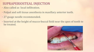
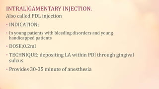
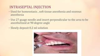
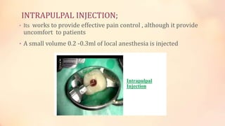
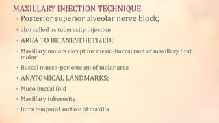
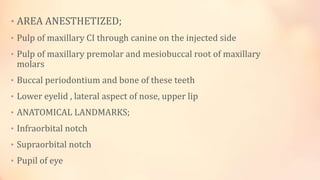
LOCAL ANESTHESIA
TECHNIQUE
SARDAR USMAN.
ROLL NO 39.
FINAL YEAR BDS.
KMU-IDS,KOHAT.
SUPRAPERIOSTEAL INJECTION
• Also called as local infiltration.
• Pulpal and soft tissue anesthesia in maxillary anterior teeth.
• 27 guage needle recommended.
• Inserted at the height of mucco-buccal field near the apex of tooth to
be treated.
INTRALIGAMENTARY INJECTION.
Also called PDL injection
• INDICATION;
• In young patients with bleeding disorders and young
handicapped patients
• DOSE;0.2ml
• TECHNIQUE; depositing LA within PDl through gingival
sulcus
• Provides 30-35 minute of anesthesia
INTRAOSSEOUS INJECTION;
• Aim of Intro-sseous anesthesia is to inject local anesthetic
solution into cancellous bone adjacent to the apex of tooth by
piercing buccal gingiva and bone in relation to the tooth to be
anesthetized.
• It can be used as a supplemental technique with mandibular
nerve blocks to enhance deep pulpal anesthesia or as primary
technique so that patient do not experience numb lips and
tongue.
INTRASEPTAL INJECTION
• Used for homeostasis , soft tissue anesthesia and osseous
anesthesia
• Use 27 guage needle and insert perpendicular to the area to be
anesthetized at 90 degree angle
• Slowly deposit 0.2 ml solution
INTRAPULPAL INJECTION;
• Its works to provide effective pain control , although it provide
uncomfort to patients
• A small volume 0.2 -0.3ml of local anesthesia is injected
MAXILLARY INJECTION TECHNIQUE
• Posterior superior alveolar nerve block;
• also called as tuberosity injection
• AREA TO BE ANESTHETIZED;
• Maxillary molars except for mesio-buccal root of maxillary first
molar
• Buccal mucco-periosteum of molar area
• ANATOMICAL LANDMARKS;
• Muco buccal fold
• Maxillary tuberosity
• Infra temporal surface of maxilla
• Zygomatic process of maxilla
• Anterior border and coronoid process of mandible
• POSITION OF patient;
• The patient is placed of maxillary occlusal plane at 45 degree
with floor.
• POSITION OF OPERATOR;
• For left handed dentist ,9 o clock position
• In right sided dentist 7 o clock position
TECHNIQUE;
• Retract the cheek and prepare the injection
• The needle is introduced into the height of the muco buccal fold
above the 2nd molar
• Advance the needle slowly upward backward and inward
• The depth of them needle insertion is 16 mm for adults and 14 mm
for children following careful aspiration
• If blood comes out then retract and try again but if you get blood
also next time then abort the technique
• If no blood comes out then deposit 1.5 ml of the anesthetic solution
and wait for 5 minutes
MIDDLE SUPERIOR ALVEOLAR NERVE BLOCK
• AREA ANESTHETIZED;
• Pulp of maxillary ist and 2nd premolar and mesio buccal root of ist
molar
• Buccal periodontal tissue and bone of these teeth.
• Anatomical Landmarks;
• Mucco buccal fold above maxillary 2nd premolar
• POSITION OF PATIENT;
• 45 degree occlusal plane w.r.t floor
• POSITION OF OPERATOR;
• Right side MSAN 9 o clock
• Left Side MSAN 7 o clock
PATIENT POSITION;
Patient is placed with maxillary occlusal plane 45 degree with floor
TECHNIQUE;
Stretch the lip for visibility
Insert the needle at the height of muccobuccal fold above
maxillary 2nd premolar
deposit 0.9-1.2ml of solution for 30 seconds
ANTERIOR SUPERIOR ALVEOLAR NERVE BLOCK
• Also called infra orbital nerve block
• AREA ANESTHETIZED;
• ASA
• MSA
• Infra orbital
• Inferior palpebral
• Lateral nasal
• Superior labial
• AREA ANESTHETIZED;
• Pulp of maxillary CI through canine on the injected side
• Pulp of maxillary premolar and mesiobuccal root of maxillary
molars
• Buccal periodontium and bone of these teeth
• Lower eyelid , lateral aspect of nose, upper lip
• ANATOMICAL LANDMARKS;
• Infraorbital notch
• Supraorbital notch
• Pupil of eye
• Muccobuccal fold
• Anterior teeth.
• POSSITION OF PATIENT;
• Patient occlusal plane is 45 degree with floor
• POSITION OF OPERATOR;
• For right side ASA 9 o clock position
• For left side ASA 7 o clock position
TECHNIQUES;
• BISUSPID APPROACH.
• Needle is inserted parallel with supraorbital notch , pupil of
eye, infra orbital notch , 2nd bicuspid tooth
• CENTRAL INCISOR APPROACH.
• The needle bisects the crown of central incisors from mesio incisal angle
to disto-gingival angle
• The needle should not penetrate more than ¾ inch
GREATER PALATINE NERVE BLOCK
• NERVE ANESTHETIZED;
• Greater palatine nerve.
• AREA ANESTHETIZED;
• Posterior area of hard palate and its overlying tissue
• Anteriorly as far as ist premolar and medially to the midline
• ANATOMICAL LAND MARKS;
• Greater palatine foramen and junction of maxillary alveolar process and
palatine bone.
POSITION OF PATIENT;
Patient should be in supine position
• Ask the patient to do following
• 1)open wide mouth 2)extend the neck 3)turn the head to left or right
• OPERATOR POSITION:
• For Right side 7 o clock position.
• For left side 11 o clock position.
• TECHNIQUE;
• Locate greater palatine foramen.
• Place the cotton swab at the junction of maxillary alveolar
process and hard palate , then palpate posteriorly by pressing
firmly into tissue with swab
• Depression is created by foramen where swab falls .
• Foramen is located mostly distal to maxillary second molar
• prepare tissue for injection just 1-2 mm anterior to greater palatine
foramen
• Apply topical anesthetic and weight for 2 minutes
• Direct the syringe in mouth from opposite side with needle approaching
injection side at right angle.
• Depth of penetration is 5mm.
NASOPALATINE NERVE BLOCK.
• Also called incisive and sphenopalatine nerve block
• NERVE ANESTHTIZED.
• Naso palatine nerve
• AREA ANESTHETIZED.
• Mucosa of anterior part of palate opposite to anterior teeth.
• POSITION OF PATIENT;
• Patient should be supine in position.
• Occlusal plane should be parallel to operator shoulder.
• POSITION OF OPERATOR;
• 9 o clock.
TECHNIQUE
• The point of needle insertion is incisive foramen i.e. the crest of incisive
papilla .
• The direction of needle insertion into the crest of incisive papilla
between the upper centrals making 45° to the palatal mucosa Ask the
patient to mouth wide open The labi o lingual crest injection is made to
anaesthetize the incisive papilla first.
• The needle is oriented parallel with the labial O alveolar plate with the
needle directed towards crest of incisive papilla.
• 4mm needle is penetrated.
• 0.45 ml of anesthetic solution is injected.
MANDIBULAR NERVE INJECTIONS.
• INFERIOR ALVELOAR NERVE BLOCK.
• NERVE ANESTHETIZE;
• Inferior alveolar nerve
• Incisive nerve
• Mental nerve
• Lingual nerve.
• AREAS ANESTHETIZED;
• Mandibular teeth.
• Body body of mandible.
• Inferior portion of ramus
• Buccal mucoperiosteum
• Anterior 2/3 of tongue
• Floor of tongue
• Lingual soft tissue and periosteum
• ANATOMICAL LANDMARKS;
• Coronoid notch
• Pterygomandibular raphe
• Occlusal plane.
• POSITION OF PATIENT;
• Position should be in supine position
• Occlusal plane should be near to operator shoulder .
POSITION OF PATIENT.
• For right IANB is 7 o clock
For left IANB is 9 o clock
TECHNIQUE;
MENTAL NERVE BLOCK
• NERVE ANESTHETIZED;
• Mental nerve.
• AREA ANESTHETIZED;
• Lower lip.
• Muccobuccal fold anterior to mental foramen.
• ANATOMICAL LANDMARKS;
• Mandibular bicuspids.
• INDICATION;
• Where manipulation of buccal soft tissue anterior to mental
foramen
CONTRAINDICATION;
• Acute inflammation.
• TECHNIQUES;
• Needle is inserted at apex of 2nd premolar or between 1st or 2nd
premolars,
LONG BUCCAL NERVE;
• NERVE ANESTHETIZED;
• Long buccal nerve
• AREA ANESTHETIZED.
• Buccal mucous membrane and muccoperiosteum of
mandibulars molars.
• ANATOMICAL LANDMARKS;
Mandibular molars and muccobuccal folds ;
INDICATIONS;
Surgery of mandibular molars and adjunct to INAB.
TECHNIQUES;
Inserted into buccal mucosa distal to 3rd molar.
GOW GATES MANDIBULAR NERVE BLOCK
• NERVES ANESTHETIZED;
• IAN
• Mental
• Incisive
• Mylohyoid nerve
• Auriculotemporal nerve
• AREA ANESTHETIZED.
• Mandibular teeth to midline
• Buccal mucosa ipsilaterally
• Body of mandible and skin over zygoma and cheek.
LANDMARKS
• EXTRAORALS;
• Corner of mouth
• Tragus
• Inter tragus notch
• INTRAORAL;
• Mesio palatal cusp of 2M.
• Soft tissue distal to 2nd maxillary molar.
• AREA OF INSERTION ; mucous membrane on mesial side of mandibular ramus ,on line
from the intratragic notch just distal to maxillary 2nd molar.
• TARGET AREA ; lateral side of neck just below insertion of lateral pterygoid.
• Needle endpoint is lateral aspect of anterior portion of condyle , just inferior to lateral
pterygoid muscle.
• Needle is inserted to a depth of 25 to 30 mm
VAZIRANI AKINOSI NERVE BLOCK;
• INDICATION;
• Unilateral severe trsimus.
• NERVE ANESTHETIZED;
• Inferior alveolar nerve
• Mental nerve
• Incisive nerve
• Mylohyoid nerve.
AREA OF INSERTION;
• Soft tissue in lingual border of mandibular ramus directly
adjacent to maxillary tuberosity at the height of muccogingival
junction adjacent to height of 3rd molar.
• TARGET AREA;
• Soft tissue at the medial border of ramus where it run towards
mandibular foramen
• The depth of insertion depends on anterio posterior size of
ramus, however in adult in adult according to thumb rule the
hub of needle should be opposite to mesial aspect of maxillary
2nd molar.
LOCAL ANESTHESIA TECHNIQUE.pptx

LOCAL ANESTHESIA TECHNIQUE.pptx

  • 1.
    LOCAL ANESTHESIA TECHNIQUE SARDAR USMAN. ROLLNO 39. FINAL YEAR BDS. KMU-IDS,KOHAT.
  • 2.
    SUPRAPERIOSTEAL INJECTION • Alsocalled as local infiltration. • Pulpal and soft tissue anesthesia in maxillary anterior teeth. • 27 guage needle recommended. • Inserted at the height of mucco-buccal field near the apex of tooth to be treated.
  • 3.
    INTRALIGAMENTARY INJECTION. Also calledPDL injection • INDICATION; • In young patients with bleeding disorders and young handicapped patients • DOSE;0.2ml • TECHNIQUE; depositing LA within PDl through gingival sulcus • Provides 30-35 minute of anesthesia
  • 5.
    INTRAOSSEOUS INJECTION; • Aimof Intro-sseous anesthesia is to inject local anesthetic solution into cancellous bone adjacent to the apex of tooth by piercing buccal gingiva and bone in relation to the tooth to be anesthetized. • It can be used as a supplemental technique with mandibular nerve blocks to enhance deep pulpal anesthesia or as primary technique so that patient do not experience numb lips and tongue.
  • 6.
    INTRASEPTAL INJECTION • Usedfor homeostasis , soft tissue anesthesia and osseous anesthesia • Use 27 guage needle and insert perpendicular to the area to be anesthetized at 90 degree angle • Slowly deposit 0.2 ml solution
  • 7.
    INTRAPULPAL INJECTION; • Itsworks to provide effective pain control , although it provide uncomfort to patients • A small volume 0.2 -0.3ml of local anesthesia is injected
  • 8.
    MAXILLARY INJECTION TECHNIQUE •Posterior superior alveolar nerve block; • also called as tuberosity injection • AREA TO BE ANESTHETIZED; • Maxillary molars except for mesio-buccal root of maxillary first molar • Buccal mucco-periosteum of molar area • ANATOMICAL LANDMARKS; • Muco buccal fold • Maxillary tuberosity • Infra temporal surface of maxilla
  • 9.
    • Zygomatic processof maxilla • Anterior border and coronoid process of mandible • POSITION OF patient; • The patient is placed of maxillary occlusal plane at 45 degree with floor. • POSITION OF OPERATOR; • For left handed dentist ,9 o clock position • In right sided dentist 7 o clock position
  • 10.
    TECHNIQUE; • Retract thecheek and prepare the injection • The needle is introduced into the height of the muco buccal fold above the 2nd molar • Advance the needle slowly upward backward and inward • The depth of them needle insertion is 16 mm for adults and 14 mm for children following careful aspiration • If blood comes out then retract and try again but if you get blood also next time then abort the technique • If no blood comes out then deposit 1.5 ml of the anesthetic solution and wait for 5 minutes
  • 12.
    MIDDLE SUPERIOR ALVEOLARNERVE BLOCK • AREA ANESTHETIZED; • Pulp of maxillary ist and 2nd premolar and mesio buccal root of ist molar • Buccal periodontal tissue and bone of these teeth. • Anatomical Landmarks; • Mucco buccal fold above maxillary 2nd premolar • POSITION OF PATIENT; • 45 degree occlusal plane w.r.t floor • POSITION OF OPERATOR; • Right side MSAN 9 o clock • Left Side MSAN 7 o clock
  • 13.
    PATIENT POSITION; Patient isplaced with maxillary occlusal plane 45 degree with floor TECHNIQUE; Stretch the lip for visibility Insert the needle at the height of muccobuccal fold above maxillary 2nd premolar deposit 0.9-1.2ml of solution for 30 seconds
  • 15.
    ANTERIOR SUPERIOR ALVEOLARNERVE BLOCK • Also called infra orbital nerve block • AREA ANESTHETIZED; • ASA • MSA • Infra orbital • Inferior palpebral • Lateral nasal • Superior labial
  • 16.
    • AREA ANESTHETIZED; •Pulp of maxillary CI through canine on the injected side • Pulp of maxillary premolar and mesiobuccal root of maxillary molars • Buccal periodontium and bone of these teeth • Lower eyelid , lateral aspect of nose, upper lip • ANATOMICAL LANDMARKS; • Infraorbital notch • Supraorbital notch • Pupil of eye
  • 17.
    • Muccobuccal fold •Anterior teeth. • POSSITION OF PATIENT; • Patient occlusal plane is 45 degree with floor • POSITION OF OPERATOR; • For right side ASA 9 o clock position • For left side ASA 7 o clock position
  • 18.
    TECHNIQUES; • BISUSPID APPROACH. •Needle is inserted parallel with supraorbital notch , pupil of eye, infra orbital notch , 2nd bicuspid tooth • CENTRAL INCISOR APPROACH. • The needle bisects the crown of central incisors from mesio incisal angle to disto-gingival angle • The needle should not penetrate more than ¾ inch
  • 20.
    GREATER PALATINE NERVEBLOCK • NERVE ANESTHETIZED; • Greater palatine nerve. • AREA ANESTHETIZED; • Posterior area of hard palate and its overlying tissue • Anteriorly as far as ist premolar and medially to the midline • ANATOMICAL LAND MARKS; • Greater palatine foramen and junction of maxillary alveolar process and palatine bone. POSITION OF PATIENT; Patient should be in supine position
  • 21.
    • Ask thepatient to do following • 1)open wide mouth 2)extend the neck 3)turn the head to left or right • OPERATOR POSITION: • For Right side 7 o clock position. • For left side 11 o clock position. • TECHNIQUE; • Locate greater palatine foramen. • Place the cotton swab at the junction of maxillary alveolar process and hard palate , then palpate posteriorly by pressing firmly into tissue with swab • Depression is created by foramen where swab falls . • Foramen is located mostly distal to maxillary second molar
  • 22.
    • prepare tissuefor injection just 1-2 mm anterior to greater palatine foramen • Apply topical anesthetic and weight for 2 minutes • Direct the syringe in mouth from opposite side with needle approaching injection side at right angle. • Depth of penetration is 5mm.
  • 23.
    NASOPALATINE NERVE BLOCK. •Also called incisive and sphenopalatine nerve block • NERVE ANESTHTIZED. • Naso palatine nerve • AREA ANESTHETIZED. • Mucosa of anterior part of palate opposite to anterior teeth. • POSITION OF PATIENT; • Patient should be supine in position. • Occlusal plane should be parallel to operator shoulder. • POSITION OF OPERATOR; • 9 o clock.
  • 24.
    TECHNIQUE • The pointof needle insertion is incisive foramen i.e. the crest of incisive papilla . • The direction of needle insertion into the crest of incisive papilla between the upper centrals making 45° to the palatal mucosa Ask the patient to mouth wide open The labi o lingual crest injection is made to anaesthetize the incisive papilla first. • The needle is oriented parallel with the labial O alveolar plate with the needle directed towards crest of incisive papilla. • 4mm needle is penetrated. • 0.45 ml of anesthetic solution is injected.
  • 26.
    MANDIBULAR NERVE INJECTIONS. •INFERIOR ALVELOAR NERVE BLOCK. • NERVE ANESTHETIZE; • Inferior alveolar nerve • Incisive nerve • Mental nerve • Lingual nerve. • AREAS ANESTHETIZED; • Mandibular teeth. • Body body of mandible. • Inferior portion of ramus
  • 27.
    • Buccal mucoperiosteum •Anterior 2/3 of tongue • Floor of tongue • Lingual soft tissue and periosteum • ANATOMICAL LANDMARKS; • Coronoid notch • Pterygomandibular raphe • Occlusal plane. • POSITION OF PATIENT; • Position should be in supine position • Occlusal plane should be near to operator shoulder .
  • 28.
    POSITION OF PATIENT. •For right IANB is 7 o clock For left IANB is 9 o clock
  • 29.
  • 30.
    MENTAL NERVE BLOCK •NERVE ANESTHETIZED; • Mental nerve. • AREA ANESTHETIZED; • Lower lip. • Muccobuccal fold anterior to mental foramen. • ANATOMICAL LANDMARKS; • Mandibular bicuspids. • INDICATION; • Where manipulation of buccal soft tissue anterior to mental foramen
  • 31.
    CONTRAINDICATION; • Acute inflammation. •TECHNIQUES; • Needle is inserted at apex of 2nd premolar or between 1st or 2nd premolars,
  • 32.
    LONG BUCCAL NERVE; •NERVE ANESTHETIZED; • Long buccal nerve • AREA ANESTHETIZED. • Buccal mucous membrane and muccoperiosteum of mandibulars molars. • ANATOMICAL LANDMARKS; Mandibular molars and muccobuccal folds ; INDICATIONS; Surgery of mandibular molars and adjunct to INAB.
  • 33.
    TECHNIQUES; Inserted into buccalmucosa distal to 3rd molar.
  • 34.
    GOW GATES MANDIBULARNERVE BLOCK • NERVES ANESTHETIZED; • IAN • Mental • Incisive • Mylohyoid nerve • Auriculotemporal nerve • AREA ANESTHETIZED. • Mandibular teeth to midline • Buccal mucosa ipsilaterally • Body of mandible and skin over zygoma and cheek.
  • 35.
    LANDMARKS • EXTRAORALS; • Cornerof mouth • Tragus • Inter tragus notch • INTRAORAL; • Mesio palatal cusp of 2M. • Soft tissue distal to 2nd maxillary molar. • AREA OF INSERTION ; mucous membrane on mesial side of mandibular ramus ,on line from the intratragic notch just distal to maxillary 2nd molar. • TARGET AREA ; lateral side of neck just below insertion of lateral pterygoid. • Needle endpoint is lateral aspect of anterior portion of condyle , just inferior to lateral pterygoid muscle.
  • 36.
    • Needle isinserted to a depth of 25 to 30 mm
  • 37.
    VAZIRANI AKINOSI NERVEBLOCK; • INDICATION; • Unilateral severe trsimus. • NERVE ANESTHETIZED; • Inferior alveolar nerve • Mental nerve • Incisive nerve • Mylohyoid nerve.
  • 38.
    AREA OF INSERTION; •Soft tissue in lingual border of mandibular ramus directly adjacent to maxillary tuberosity at the height of muccogingival junction adjacent to height of 3rd molar. • TARGET AREA; • Soft tissue at the medial border of ramus where it run towards mandibular foramen • The depth of insertion depends on anterio posterior size of ramus, however in adult in adult according to thumb rule the hub of needle should be opposite to mesial aspect of maxillary 2nd molar.