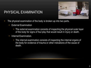
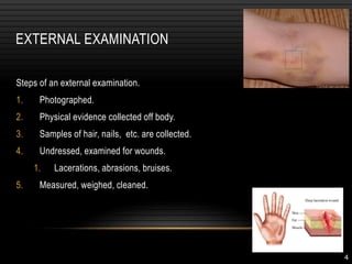
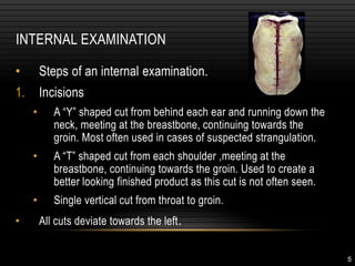
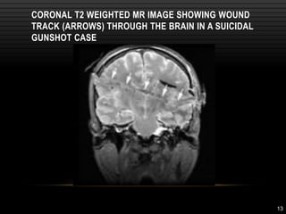
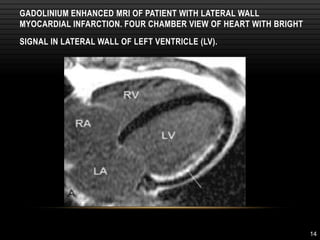
MINIMALLY INVASIVE AUTOPSY
2
WHAT IS AN AUTOPSY?
• An autopsy is a post mortem
examination preformed on a
corpse to determine the cause
and manner of death.
• The prefix 'auto-' means 'self',
and so autopsy means 'to see
for oneself‘.
3
PHYSICAL EXAMINATION
• The physical examination of the body is broken up into two parts.
• External Examination
• The external examination consists of inspecting the physical outer layer
of the body for signs of foul play that would result in injury or death.
• Internal Examination.
• The internal examination consists of inspecting the internal organs of
the body for evidence of trauma or other indications of the cause of
death.
4
EXTERNAL EXAMINATION
Steps of an external examination.
1. Photographed.
2. Physical evidence collected off body.
3. Samples of hair, nails, etc. are collected.
4. Undressed, examined for wounds.
1. Lacerations, abrasions, bruises.
5. Measured, weighed, cleaned.
5
INTERNAL EXAMINATION
• Steps of an internal examination.
1. Incisions
• A “Y” shaped cut from behind each ear and running down the
neck, meeting at the breastbone, continuing towards the
groin. Most often used in cases of suspected strangulation.
• A “T” shaped cut from each shoulder ,meeting at the
breastbone, continuing towards the groin. Used to create a
better looking finished product as this cut is not often seen.
• Single vertical cut from throat to groin.
• All cuts deviate towards the left.
6
INTERNAL EXAMINATION (CONT’D)
2. Cuts
1. The chest cavity is cut open using shears.
2. The ribs are sawed away, letting them be lifted off the body,
exposing the heart and lungs.
3. Removal
1. En masse technique of Letulle – All organs removed at once.
2. En bloc method of Ghon – organs divided into four groups
and removed in sections.
4. All removed organs are now weighed and examined for
unusual markings or signs.
7
5. Brain examination
1. An incision is made from a point behind one ear, over the top
of the head, to a point behind the opposite ear.
2. The scalp is pulled away from the skull, creating tow flaps.
The front flap goes over the face, the rear flap over the neck.
3. The skull is then cut with an electric saw to create a cover
that can be pulled off to expose the brain.
4. The brain si then cut from the spinal cord and lifted out of the
skull for further examination.
INTERNAL EXAMINATION (CONT’D)
8
9
MINIMALLY INVASIVE AUTOPSY
TECHNIQUES AVAILABLE:
• External examination
• Clinical history and medical records
• Imaging techniques – xray, MRI, CT, Ultrasonography
• Aspiration of blood, urine and other fluids.
• FNAC or needle biopsy and histology
• Laparoscopy and thoracoscopy
10
POST- MORTEM XRAY
• Identification purposes.
• Diagnosis of traumatic bone lesions
• Identification of bullets and other foreign bodies
• Identification of gas in body cavities, vessels and other sites.
11
POST-MORTEM MRI
• Depends on the presence of protons within water content of body, radiofrequency pulse
sequences and gradient magnetic fields applied within the main static magnetic field
• Motion sensitive, thus best resolution in immobile subjects
• Offers non-invasive high resolution imaging of soft tissues
• Provides a comprehensive digital record of each case for future reference.
• Sensitivity is higher for cns abnormalities
12
POST-MORTEM MRI
• In fetal, perinatal and pediatric autopsies
- To identify bone age
- Detect skeletal abnormalities
- Discover or confirm skeletal fractures in birth trauma or child abuse
- - cns abnormaliies
13
CORONAL T2 WEIGHTED MR IMAGE SHOWING WOUND
TRACK (ARROWS) THROUGH THE BRAIN IN A SUICIDAL
GUNSHOT CASE
14
GADOLINIUM ENHANCED MRI OF PATIENT WITH LATERAL WALL
MYOCARDIAL INFARCTION. FOUR CHAMBER VIEW OF HEART WITH BRIGHT
SIGNAL IN LATERAL WALL OF LEFT VENTRICLE (LV).
15
MERITS AND DEMERITS
Merits
• observer independent
• reproducible
• non invasive
• illustrative visualization
• forensic telemedicine consultation
16
Demerits:
MRI was unable to
• image coronary artery lesions
• differentiate thrombus from postmortem clot
• differentiate pulmonary oedema from pneumonic exudates
• detect perinatal cardiac malformations
• lack of validation
• interpretative problems in advanced decomposition
• considerably high costs
• time consuming
17
POST-MORTEM CT
• Radiologically producing thin transverse sectional images through a body or anatomical
region.
• X ray beam passed through body and detected using a circular array of photomultiplier
tubes
• Scan results analyzed for absorption patterns (100HU- -1000HU)
18
POST MORTEM CT
MERITS
• More accurate than MRI in identifying cause of death
• More widely available, less expensive and quicker to do.
• Can also be combined with angiography for detection of vascular pathologies
• Useful in identifying haemorrhages – haemopericardium, aneurysms and pulmonary
tumours over 10mm in diameter
19
DEMERITS OF CT
• interpretative problems in advanced decomposition
• considerably high costs
• time consuming
20
SIEMENS DEFINITION CT SCANNER
Scan length: 2 m
10 GB under a minute
21
VIRTUAL AUTOPSY - WHAT IS IT?
Specifically, this is a minimally invasive computerized 2D or
3D reconstruction of a body created postmortem by a
combination of modern imaging technologies
22
• Before autopsy, two dimensional images taken via computed
tomography are reconstructed into three dimensional images
with the help of advanced software.
• Can be combined with angiography for visualizaion of vascular
lesions.
• Gives better orientation of structures.
23
BENEFITS OF THE VIRTUAL AUTOPSY
• Clear graphics
• Permanency
• Paperless
• Minimally invasive
• Click back and forth between
images
• Can be merged with
photogrammetric data
• Trace bullet/knife path
• Toolmark analysis
• Detect air/gas pockets
• Detect water/blood inhalation
24
DRAWBACKS OF THE VIRTUAL AUTOPSY
• Cost of equipment
• Cost of training
• Perceived infallibility
• Requires manipulation
• Infancy stage
• Inability to diagnose natural deaths
• Inability to diagnose toxicological
deaths
• Not a replacement for external
examination
25
LETHAL GUNSHOT WOUND TO THE CHEST
CT IMAGE SHOWS THE ENTRY WOUND AS AN IRREGULAR LINEAR
SOFT-TISSUE DEFECT IN THE RIGHT ANTERIOR CHEST WITH
ASSOCIATED SUBCUTANEOUS GAS
25
26
SURFACE-RENDERED IMAGE OF CT DATA
SHOWS THE ENTRY WOUND LATERAL TO THE
RIGHT NIPPLE
26
27
FEATURES OF A GUNSHOT WOUND TO THE HEAD AT VIRTUAL
AUTOPSY
CT IMAGES OF SHOW MULTIPLE CALVARIAL FRACTURES
POSTERIOR SETTLING OF THE BRAIN AND PNEUMOCEPHALUS.
METALLIC FRAGMENTS AND FOCI OF GAS ARE PRESENT IN THE LEFT
CEREBELLUM
27
28
THREE-DIMENSIONAL VOLUME-RENDERED IMAGE OF THE
SKULL SHOWS THE COMMINUTED FRACTURE OF THE
POSTERIOR SKULL
28
29
LETHAL GUNSHOT WOUND TO THE CHEST
29
30
RECONSTRUCTION IMAGE SHOWS MIDLINE
FRONTAL ENTRANCE AND RIGHT OCCIPITAL EXIT
30
31
THREE-DIMENSIONAL RECONSTRUCTION IMAGE OF THORAX
VIEWED FROM POSTERIOR SHOWS BONE FRACTURE DEFECTS
IN RIGHT SIXTH RIB AND MEDIAL MARGIN OF RIGHT SCAPULA
31
32
33
CHALLENGE OF VIRTUAL AUTOPSIES
• Gigabytes of data – do not fit in memory
• Full body and high resolution details
34
CASE 1
• Man acts aggressively when police
arrives
• Police officer fires gun, man is dead on
arrival
• Police officer accused for not warning
• Warning shot ricocheted as indicated by
bullet fragments
35
CASE 2
• Routine procedure to control
respiration for surgery
• A needle was inserted to
remove air from the lung cavity
(sack)
• Accidentally the heart stopped
• Physical autopsy fails to
explain the cause of death
• Virtual autopsy reveals air in
unexpected parts of the body
36
FORENSIC AUTOPSY WORKFLOW
Physical Autopsy
Crime Scene Investigation
Police
Forensics
CT Scan
DVR Visualization
Visual Exploration
Radiology
37
CONCLUSIONS
• The Virtual Autopsy procedure
• Enables digital exploration of human cadavers
• Is already an important forensic tool
• May in some cases replace traditional autopsies
• Honors religious boundaries and demands from relatives
• Full body volume rendering on commodity PCs
• High quality and interactivity at high resolution.
• Preserving full quality of volume data
38
THANK YOU

MINIMALLY_INVASIVE_AUTOPSY (post mortem).ppt

  • 1.
  • 2.
    2 WHAT IS ANAUTOPSY? • An autopsy is a post mortem examination preformed on a corpse to determine the cause and manner of death. • The prefix 'auto-' means 'self', and so autopsy means 'to see for oneself‘.
  • 3.
    3 PHYSICAL EXAMINATION • Thephysical examination of the body is broken up into two parts. • External Examination • The external examination consists of inspecting the physical outer layer of the body for signs of foul play that would result in injury or death. • Internal Examination. • The internal examination consists of inspecting the internal organs of the body for evidence of trauma or other indications of the cause of death.
  • 4.
    4 EXTERNAL EXAMINATION Steps ofan external examination. 1. Photographed. 2. Physical evidence collected off body. 3. Samples of hair, nails, etc. are collected. 4. Undressed, examined for wounds. 1. Lacerations, abrasions, bruises. 5. Measured, weighed, cleaned.
  • 5.
    5 INTERNAL EXAMINATION • Stepsof an internal examination. 1. Incisions • A “Y” shaped cut from behind each ear and running down the neck, meeting at the breastbone, continuing towards the groin. Most often used in cases of suspected strangulation. • A “T” shaped cut from each shoulder ,meeting at the breastbone, continuing towards the groin. Used to create a better looking finished product as this cut is not often seen. • Single vertical cut from throat to groin. • All cuts deviate towards the left.
  • 6.
    6 INTERNAL EXAMINATION (CONT’D) 2.Cuts 1. The chest cavity is cut open using shears. 2. The ribs are sawed away, letting them be lifted off the body, exposing the heart and lungs. 3. Removal 1. En masse technique of Letulle – All organs removed at once. 2. En bloc method of Ghon – organs divided into four groups and removed in sections. 4. All removed organs are now weighed and examined for unusual markings or signs.
  • 7.
    7 5. Brain examination 1.An incision is made from a point behind one ear, over the top of the head, to a point behind the opposite ear. 2. The scalp is pulled away from the skull, creating tow flaps. The front flap goes over the face, the rear flap over the neck. 3. The skull is then cut with an electric saw to create a cover that can be pulled off to expose the brain. 4. The brain si then cut from the spinal cord and lifted out of the skull for further examination. INTERNAL EXAMINATION (CONT’D)
  • 8.
  • 9.
    9 MINIMALLY INVASIVE AUTOPSY TECHNIQUESAVAILABLE: • External examination • Clinical history and medical records • Imaging techniques – xray, MRI, CT, Ultrasonography • Aspiration of blood, urine and other fluids. • FNAC or needle biopsy and histology • Laparoscopy and thoracoscopy
  • 10.
    10 POST- MORTEM XRAY •Identification purposes. • Diagnosis of traumatic bone lesions • Identification of bullets and other foreign bodies • Identification of gas in body cavities, vessels and other sites.
  • 11.
    11 POST-MORTEM MRI • Dependson the presence of protons within water content of body, radiofrequency pulse sequences and gradient magnetic fields applied within the main static magnetic field • Motion sensitive, thus best resolution in immobile subjects • Offers non-invasive high resolution imaging of soft tissues • Provides a comprehensive digital record of each case for future reference. • Sensitivity is higher for cns abnormalities
  • 12.
    12 POST-MORTEM MRI • Infetal, perinatal and pediatric autopsies - To identify bone age - Detect skeletal abnormalities - Discover or confirm skeletal fractures in birth trauma or child abuse - - cns abnormaliies
  • 13.
    13 CORONAL T2 WEIGHTEDMR IMAGE SHOWING WOUND TRACK (ARROWS) THROUGH THE BRAIN IN A SUICIDAL GUNSHOT CASE
  • 14.
    14 GADOLINIUM ENHANCED MRIOF PATIENT WITH LATERAL WALL MYOCARDIAL INFARCTION. FOUR CHAMBER VIEW OF HEART WITH BRIGHT SIGNAL IN LATERAL WALL OF LEFT VENTRICLE (LV).
  • 15.
    15 MERITS AND DEMERITS Merits •observer independent • reproducible • non invasive • illustrative visualization • forensic telemedicine consultation
  • 16.
    16 Demerits: MRI was unableto • image coronary artery lesions • differentiate thrombus from postmortem clot • differentiate pulmonary oedema from pneumonic exudates • detect perinatal cardiac malformations • lack of validation • interpretative problems in advanced decomposition • considerably high costs • time consuming
  • 17.
    17 POST-MORTEM CT • Radiologicallyproducing thin transverse sectional images through a body or anatomical region. • X ray beam passed through body and detected using a circular array of photomultiplier tubes • Scan results analyzed for absorption patterns (100HU- -1000HU)
  • 18.
    18 POST MORTEM CT MERITS •More accurate than MRI in identifying cause of death • More widely available, less expensive and quicker to do. • Can also be combined with angiography for detection of vascular pathologies • Useful in identifying haemorrhages – haemopericardium, aneurysms and pulmonary tumours over 10mm in diameter
  • 19.
    19 DEMERITS OF CT •interpretative problems in advanced decomposition • considerably high costs • time consuming
  • 20.
    20 SIEMENS DEFINITION CTSCANNER Scan length: 2 m 10 GB under a minute
  • 21.
    21 VIRTUAL AUTOPSY -WHAT IS IT? Specifically, this is a minimally invasive computerized 2D or 3D reconstruction of a body created postmortem by a combination of modern imaging technologies
  • 22.
    22 • Before autopsy,two dimensional images taken via computed tomography are reconstructed into three dimensional images with the help of advanced software. • Can be combined with angiography for visualizaion of vascular lesions. • Gives better orientation of structures.
  • 23.
    23 BENEFITS OF THEVIRTUAL AUTOPSY • Clear graphics • Permanency • Paperless • Minimally invasive • Click back and forth between images • Can be merged with photogrammetric data • Trace bullet/knife path • Toolmark analysis • Detect air/gas pockets • Detect water/blood inhalation
  • 24.
    24 DRAWBACKS OF THEVIRTUAL AUTOPSY • Cost of equipment • Cost of training • Perceived infallibility • Requires manipulation • Infancy stage • Inability to diagnose natural deaths • Inability to diagnose toxicological deaths • Not a replacement for external examination
  • 25.
    25 LETHAL GUNSHOT WOUNDTO THE CHEST CT IMAGE SHOWS THE ENTRY WOUND AS AN IRREGULAR LINEAR SOFT-TISSUE DEFECT IN THE RIGHT ANTERIOR CHEST WITH ASSOCIATED SUBCUTANEOUS GAS 25
  • 26.
    26 SURFACE-RENDERED IMAGE OFCT DATA SHOWS THE ENTRY WOUND LATERAL TO THE RIGHT NIPPLE 26
  • 27.
    27 FEATURES OF AGUNSHOT WOUND TO THE HEAD AT VIRTUAL AUTOPSY CT IMAGES OF SHOW MULTIPLE CALVARIAL FRACTURES POSTERIOR SETTLING OF THE BRAIN AND PNEUMOCEPHALUS. METALLIC FRAGMENTS AND FOCI OF GAS ARE PRESENT IN THE LEFT CEREBELLUM 27
  • 28.
    28 THREE-DIMENSIONAL VOLUME-RENDERED IMAGEOF THE SKULL SHOWS THE COMMINUTED FRACTURE OF THE POSTERIOR SKULL 28
  • 29.
    29 LETHAL GUNSHOT WOUNDTO THE CHEST 29
  • 30.
    30 RECONSTRUCTION IMAGE SHOWSMIDLINE FRONTAL ENTRANCE AND RIGHT OCCIPITAL EXIT 30
  • 31.
    31 THREE-DIMENSIONAL RECONSTRUCTION IMAGEOF THORAX VIEWED FROM POSTERIOR SHOWS BONE FRACTURE DEFECTS IN RIGHT SIXTH RIB AND MEDIAL MARGIN OF RIGHT SCAPULA 31
  • 32.
  • 33.
    33 CHALLENGE OF VIRTUALAUTOPSIES • Gigabytes of data – do not fit in memory • Full body and high resolution details
  • 34.
    34 CASE 1 • Manacts aggressively when police arrives • Police officer fires gun, man is dead on arrival • Police officer accused for not warning • Warning shot ricocheted as indicated by bullet fragments
  • 35.
    35 CASE 2 • Routineprocedure to control respiration for surgery • A needle was inserted to remove air from the lung cavity (sack) • Accidentally the heart stopped • Physical autopsy fails to explain the cause of death • Virtual autopsy reveals air in unexpected parts of the body
  • 36.
    36 FORENSIC AUTOPSY WORKFLOW PhysicalAutopsy Crime Scene Investigation Police Forensics CT Scan DVR Visualization Visual Exploration Radiology
  • 37.
    37 CONCLUSIONS • The VirtualAutopsy procedure • Enables digital exploration of human cadavers • Is already an important forensic tool • May in some cases replace traditional autopsies • Honors religious boundaries and demands from relatives • Full body volume rendering on commodity PCs • High quality and interactivity at high resolution. • Preserving full quality of volume data
  • 38.