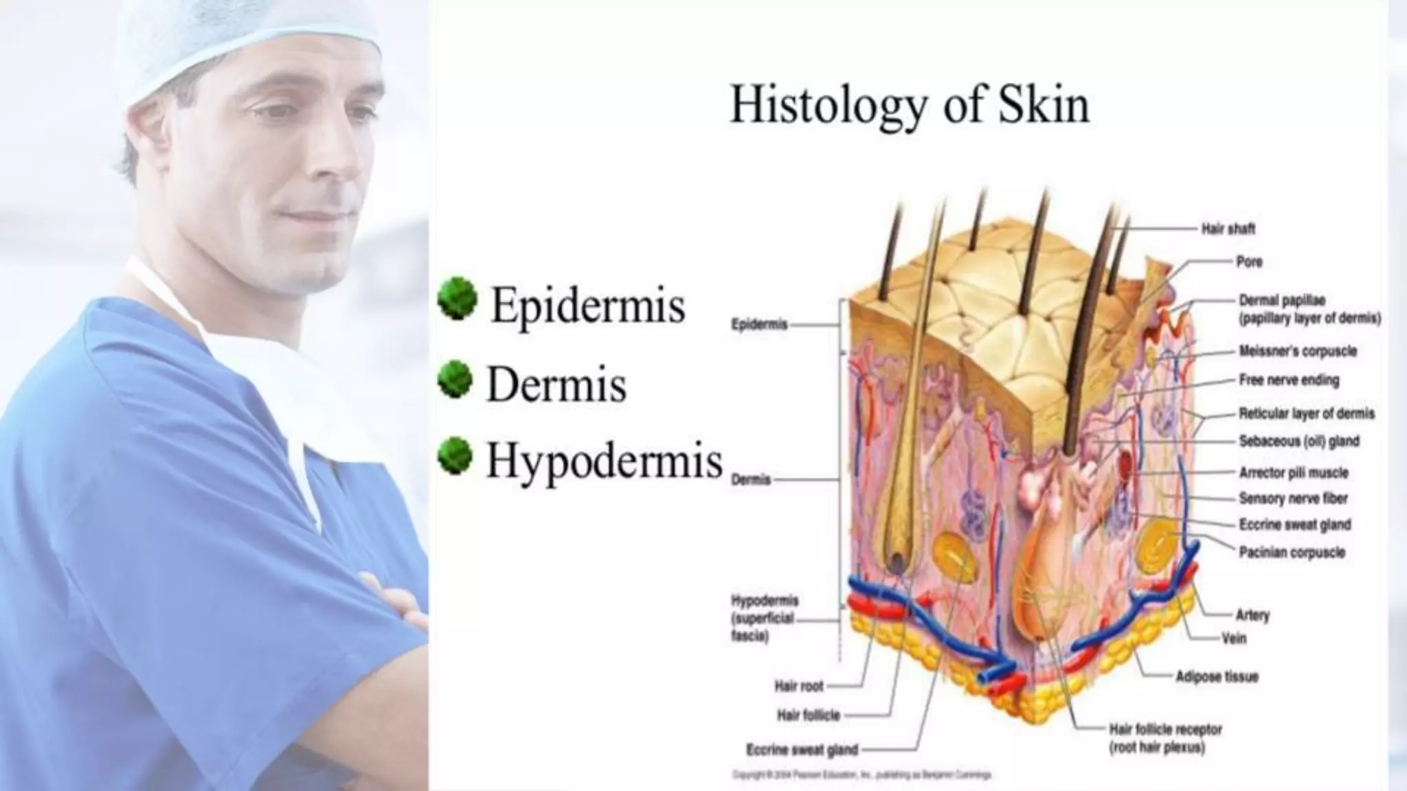
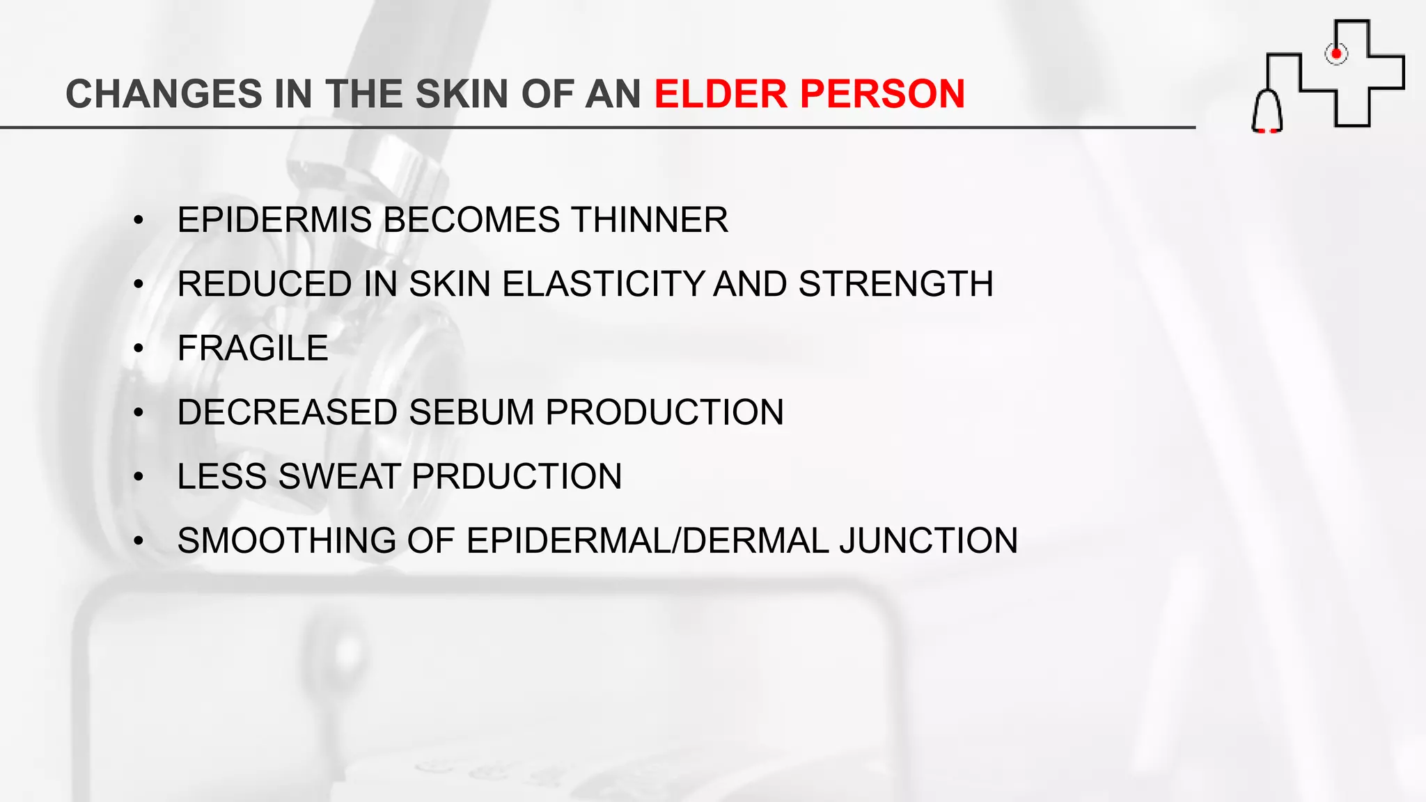
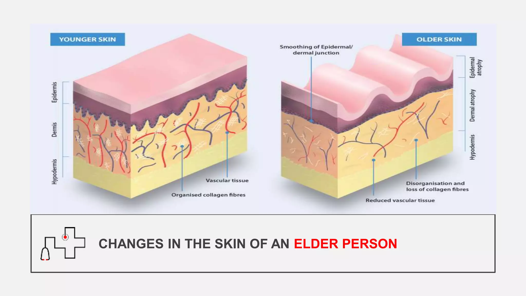
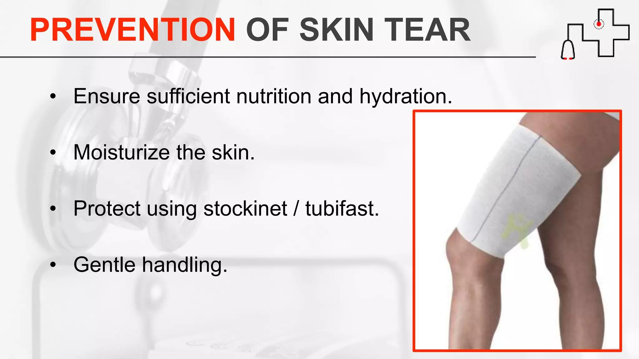
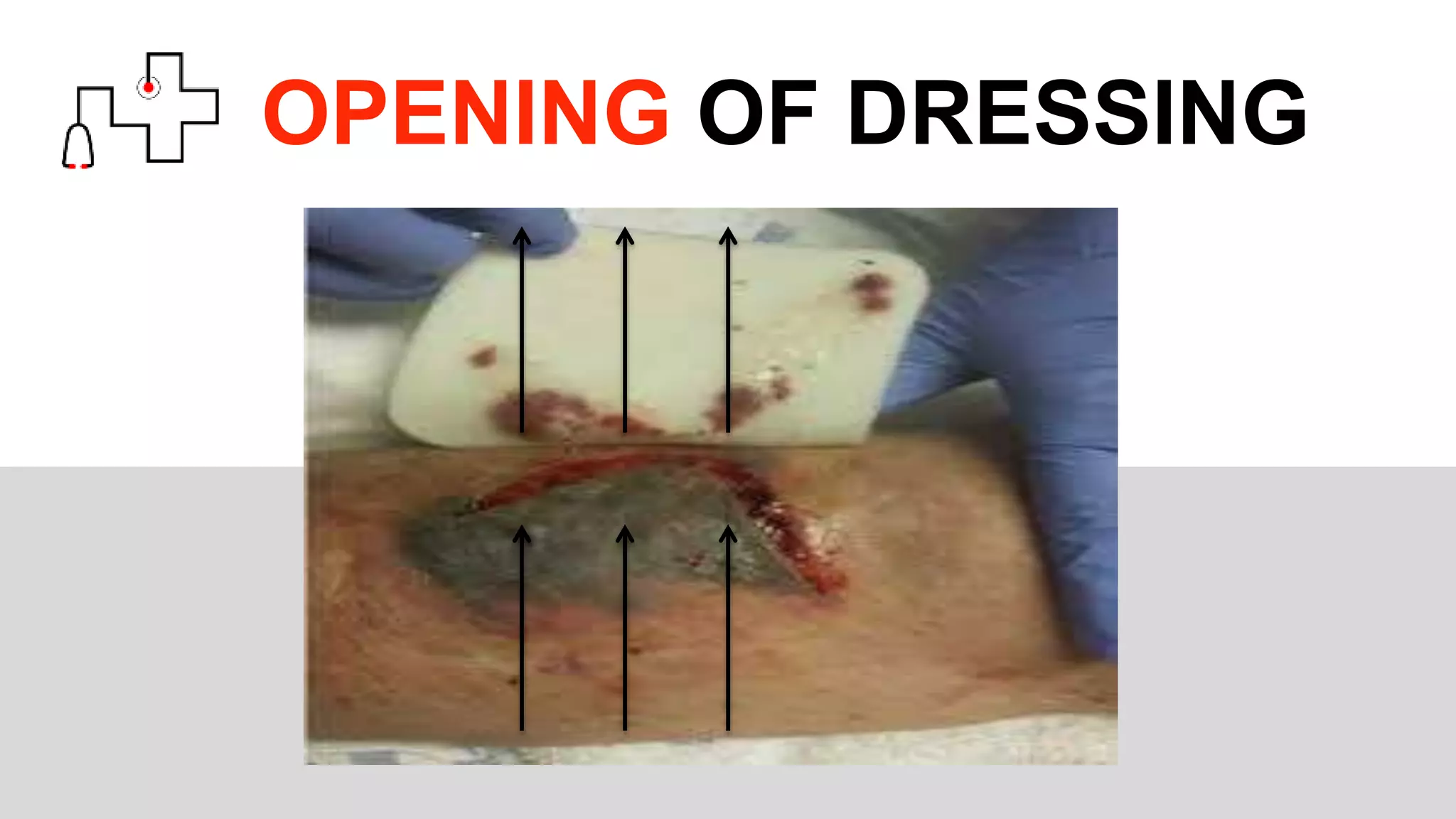
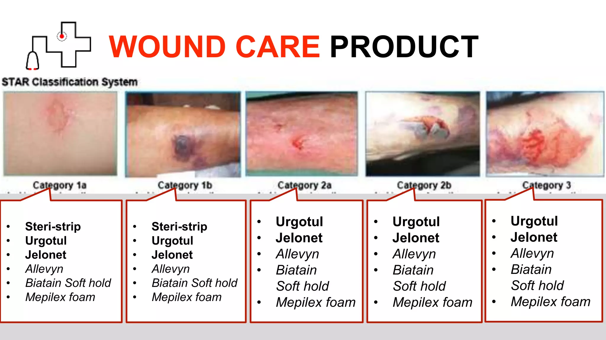
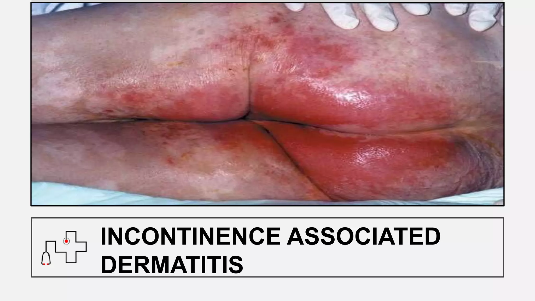
Managing Basic Wound Care in
Nursing Home
By EN John and NA Felix (SCH)
OBJECTIVE
To understand the following:
• STAR skin tear classification
• IAD ( Incontinence associated
dermatitis
• Crusting technique
• Pressure Injury
To understand the wound products available
and their applications.
CHANGES IN THE SKIN OF AN ELDER PERSON
• EPIDERMIS BECOMES THINNER
• REDUCED IN SKIN ELASTICITY AND STRENGTH
• FRAGILE
• DECREASED SEBUM PRODUCTION
• LESS SWEAT PRDUCTION
• SMOOTHING OF EPIDERMAL/DERMAL JUNCTION
CHANGES IN THE SKIN OF AN ELDER PERSON
SKIN TEAR
“A traumatic wound occurring principally on the extremities of
older adults, as a result of friction alone or shearing and friction
forces which separate the epidermis from the dermis (partial
thickness wound) or which separate both the epidermis and
the dermis from underlying structures (full thickness wound)”
http://www.woundsaustralia.com.au
FULL THICKNESS VS PARTIAL THICKNESS
SKIN TEAR
FULL THICKNESS SKIN TEAR
https://etmcourse.com/injury-description-in-the-ed/
These tears may occur due to shearing
and friction forces or a blunt trauma both
epidermis and the dermis to separate fro
m the underlying structures
PARTIAL THICKNESS SKIN TEAR
https://www.google.com.sg/search?rlz=1C1CHBD_enSG731SG731&biw=1163&bih=559&tbm=isch&sa=1&ei=mo67Wp6ECsz6vASBz7-I
DA&q=skin+tear+1a&oq=skin+tear+1a&gs_l=psy-ab.3..0.462660.465443.0.465578.12.11.0.1.1.0.157.945.7j3.10.0....0...1c.1.64.psy-ab..
1.11.942...0i67k1.0.i2Q-vu0_Kbo#imgrc=7iOuVEWGYoxmfM:
These tears may occur due to shearing
and friction forces or a blunt trauma,
causing the epidermis to separate from
the dermis.
S.T.A.R. SKIN TEAR CLASSIFICTAION
https://www.researchgate.net/figure/The-STAR-Skin-Tear-Classification-23_fig2_263895366
SKIN TEAR
ASSESSMENT
SIZE
FRAGILITY
CLASSIFICATION
CATEGORY 1a & 1b MANAGEMENT
• Stop Bleeding
• Clean the wound and surrounding with NaCl
• Realign the skin flap
– Allow skin to be natural primary dressing
– Do not stretch skin flap
• Decide on wound product
– Steri-strip - To secure skin flap
– Urgotul / Jelonet - To ensure moist environment for healing
CATEGORY 1a & 1b MANAGEMENT
• Gauze as secondary dressing and secure.
– Avoid adhesive on skin if fragile.
• Leave dressing on for 3 days for Cat 1a.
• Leave dressing on for 2 days for Cat 1b.
• Remove steri-strip in the direction of the flap.
• Observe for signs of infection.
– Pain
– Purulent discharges
– Odour
CATEGORY 2a & 2b MANAGEMENT
• Stop bleeding
• Clean the wound and surrounding with NaCl
• Moisten flap with normal saline if needed.
• Realign skin flap.
• Apply steri-strip only if skin flap can reach edge.
• Apply urgotul / Jelonet.
CATEGORY 2a & 2b MANAGEMENT
• Gauze as secondary dressing and secure.
– Avoid adhesive on skin if fragile.
• Leave dressing on for 3 days for Cat 2a.
• Leave dressing on for 2 days for Cat 2b.
• Change PRN if dressing is soaked.
• Observe for signs of infection:
• Pain
• Purulent discharges
• Odour
CATEGORY 3 MANAGEMENT
• Stop bleeding
• Clean the wound and surrounding with NaCl.
• Urgotul / Jelonet as primary dressing.
PREVENTION OF SKIN TEAR
• Ensure sufficient nutrition and hydration.
• Moisturize the skin.
• Protect using stockinet / tubifast.
• Gentle handling.
OPENING OF DRESSING
WOUND CARE PRODUCT
• Steri-strip
• Urgotul
• Jelonet
• Allevyn
• Biatain Soft hold
• Mepilex foam
• Steri-strip
• Urgotul
• Jelonet
• Allevyn
• Biatain Soft hold
• Mepilex foam
• Urgotul
• Jelonet
• Allevyn
• Biatain
Soft hold
• Mepilex foam
• Urgotul
• Jelonet
• Allevyn
• Biatain
Soft hold
• Mepilex foam
• Urgotul
• Jelonet
• Allevyn
• Biatain
Soft hold
• Mepilex foam
INCONTINENCE ASSOCIATED
DERMATITIS
CONTRIBUTING FACTORS
• Urea is converted into ammonia which increases the pH of the skin.
• Prolonged exposure to feces and urine.
• Cleaning using abrasive wash cloth.
• Poor cleaning technique.
NURSING INTERVENTIONS
• Use soft wipes or cotton wool to clean with water.
• Use soap with pH level close to skin if possible.
• Identify the cause of incontinent.
• E.g. lactose intolerance, antibiotics, Etc.
• Indwelling catheter
• Use barrier cream with zinc.
• e.g. Cavilon cream, secura cream, Aze cream, etc…
• Use crusting technique.
WHAT ARE Pressure Injuries?
Localized area/s of tissue necrosis that develops when soft tissue is
compressed between a bony prominent area and external surface for
a prolonged period of time.
RISK FACTORS PRESSURE INJURY
• IMMOBILITY
• IMPAIRED SENSORY PERCEPTION / COGNITION
• DECREASED TISSUE PERFUSION
• POOR NUTRINIONAL STATUS
• FRICTION AND SHEARING
• INCREASED SKIN MOISTURE
• GENERAL HEALTH STATUS / UNDERLYING DESEASES
• EQUIPMENTS
WOUND ASSESSMENT
WOUND ASSESSMENT
LOCATION UNDERMINING/TUNNELING
STAGE MEASUREMENT
TISSUE TYPE COLOUR
DRAINAGE/ EXUDATE ODOUR
PER-WOUND ANATOMICAL STRUCTURE
PAIN
MEASUREING WOUND
UNDERMINING & TUNNELING
SINUS TRACT / TUNNELING
WOUND DEPTH
LENGTH AND WIDTH
FISTULA
is an abnormal passage
between a hollow organ and
the skin surface, or between
two hollow organs.
NECROTIC WOUND
AVASCULAR HARDENED TISSUE
(ESCHAR)
SLOUGHY (YELLOW)
FIBROUS DEVITALIZED TISSUE
GRANULATING WOUND
HEALTHY GRANULATING TISSUE
EPITHILIALAZING
NEW SKIN
HYPERGRANULATION
FRIABLE
BLEEDING GRANULATION TISSUE
INFECTED
• BLEEDS EASILY
• PAIN
• ERYTHEMA
• PURULENT DISCHARGES
• ODOR
DESCRIBING EXUDATES
• Serous Fluid – Clear to pale yellow
• Hemoserous – Blood mixed with serous Fluid
• Sanguineous – Red and Bloody
• Purulent – Thick, can be yellowish to greenish
• Haemopurulent - Blood mixed with purulent
discharges
FACTORS AFFECTING WOUND HEALING
• Age
o  Skin Turgor
o Collagen
o Circulation
• Medical condition
o Chronic illness
o Cognitive impairment
o Non-compliance
o Edema
o Osteomyelitis
• Smoking/ alcohol
• Nutritional compromise
• Hydration
• Immobility
• Pressure
• Sensory compromise
• Technique of wound care
• Wound products
PURPOSE WOUND DRESSING
• Support autolytic debridement.
• Absorb and contain exudate.
• Create moist environment to promote healing.
• Control and prevent bleeding.
• Control odour.
• Protect the wound.
BASIC PRINCIPLES OF WOUND MANAGEMENT
Tissue
– Well vascularized
– Free of devitalized tissue
Infection / Inflammation
– Free of signs of infection
Moisture Balance
– Not too wet (Maceration) or too dry
Edge of wound
– Signs of undermining
UNDERSTANDING WOUND DRESSING
• Cleansing solution
• Primary wound dressing
Product that is placed directly on the wound.
 E.g. Alginate, urgotul, iodosorb, etc…
• Secondary Wound dressing
Product that is placed on Primary dressing covering the
wound.
 E.g. Gauze, Gamgee, etc…
CLEANSING SOLUTIONS
Cleansing solutions Shelf life once ope
ned
Advantages Disadvantages
Sodium Cholride 0.9
%
• 24 hours • Isotonic to body tissue.
• Economical
• No antiseptic properties
Chlorhexidine Glucon
ate
• 24 hours • Bactericidal & Bacteriostati
c against gram +/- bacteria
• Low toxicity to granulating
tissue
• May cause skin sensitivity
• Inactivated by soap or
povidine iodine
Octenisept
(octenidine
dihydrochloride)
• Not more then 3
years but not mo
re then expiry da
te.
• Broad spectrum of antisept
ic efficacy. Effective agains
t gram +/- bacteria, fungi, v
iruses & protozoa.
• Fast action (1 min) and lon
g erm effect (24hr remane
nce)
• Risk of cell toxicity @ 4%
concentration
PRIMARY WOUND DRESSING
• Silver (Ag) dressings
– Topical Antimicrobial agent.
– Reduces the need for antibiotics.
– Silver is released into wound bed when in contact with exudate
• Alginate (From calcium salts of seaweed)
– Loosens slough
– Controls exudate (Highly absorbant)
– Becomes gel when in contact with exudate. Keeps wound moist.
– Hemostatic properties
PRIMARY WOUND DRESSING
• Hydrofibre (Aquacel)
– Controls exudate (Highly absorbant)
– Becomes gel when in contact with exudate.
Keeps wound moist.
• Hydrogels (Duoderm/ intrasite)
– Facilitate autolytic debridement.
– Hydrates dry wound bed.
– Soften and loosens slough and necrotic wound debris /
eschar.
PRIMARY WOUND DRESSING
• Hydrogels (Duoderm/ intrasite)
– Facilitate autolytic debridement.
– Hydrates dry wound bed.
– Soften and loosens slough and necrotic wound debris / eschar.
• Hypertonic Dressing (Mesalt)
– 20% sodium Chloride
– Osmotic pressure to debride and absorb exudate
– Loosens slough
– NOT Antimicrobial
PRIMARY WOUND DRESSING
• Tulle Gras Dressing
– Jelonet: Gauze cloth impregnated with paraffin for non-traumatic
removal
– Bactigras: Gauze cloth dressing impregnated with soft paraffin and
0.5% chlorhexidine acetate.
• UrgoTul Dressing
– Polyester mesh impregnated with hydrocolloid and petroleum jelly p
articles.
– Stimulates fibroblast proliferation
• Foam (usually made from Semi-permeable polyurethane)
– Absorbs low to moderate amount of exudates.
– Conforms to body’s contour
– Cushions
– Creates moist environment
– Permeable to gases and water vapor
– Acts as bacterial barrier
PRIMARY WOUND DRESSING
• Hydrocolloid Dressing
– The layer when in contact with exudate / fluid will swell and form a gel.
– Acts as bacterial barrier.
– Softens Eschar and cause demarcation
– Manage minimal exudating wounds.
– Not for infected wound.
SECONDARY DRESSING
• Gauze
• Gamgee
• Foam dressing
SKIN BARRIER PRODUCTS
CREAM / SPRAY
• Protect skin from exudate, Urine, Faeces.
• Maintains moisture
CLASSIFICATIONS
OF PRESSURE INJURY
STAGE l
USUALLY OVER BONY PROMINENCE
INTACT SKIN
ON LOCALAIZED AREA
NON-BLANCHABLE REDNESS
WARM TO TOUCH
SKIN INCREASED TEMPERATURE
DUSKY BLUE GRAY
MAY PROGRESS TO
PAIN OR DISCOMFORT
MAY VERBALIZE
STAGE ll
ULCER DISCHARGES
SKIN BREAKS
ABRASION, BLISTER, OR SHALLOW
CRATER
WARM TO TOUCH
SKIN INCREASED TEMPERATURE
MAY DEVELOP INFECTION
PINK WOUND BED W/O SLOUGH
SHALLOW OPEN ULCER WITH RED
STAGE lll
TISSUE LOSS
FULL THICKNESS
SUBCUTANEOUS FAT
NOT EXPOSED
BONE, TENDON, OR MUSCLES
UNDERMINING OR TUNNELING
MAY INCLUDE
BUT DOES NOT OBSCURETHE
DEPTH OF TISSUE LOSS
SLOUGH MAY BE PRESENT
MAY BE VISIBLE
STAGE lV
TISSUE LOSS
FULL THICKNESS
SUBCUTANEOUS FAT
EXPOSED
BONE, TENDON, OR MUSCLES
UNDERMINING OR TUNNELING
OFTEN INVOLVES
ON SOME PART OF THE WOUND
SLOUGH OR ESCHAR MAY BE
PRESENT
MAY BE VISIBLE
UNSTAGEABLE
TISSUE LOSS
FULL THICKNESS
BASE OF ULCER IS COVERED BY
OF ULCER
UNABLE TO DETERMINE EXTENT
SERVES AS “BODY’S NATURAL COVER”
STABLE ESCHAR ON HEELS
SLOUGH AND/OR ESCHAR
DEEP TISSUE INJURY (DEPTH UNKNOWN)
LOCALIZED AREA OF DISCOLORED
INTACT SKIN
PURPLE OR MAROON
BLOOD FILLED BLISTER DUE TO
UNDERLYING DOFT TISSUE FROM
PRESSURE AND/OR SHEER
•Avoid positioning on area of
erythema whenever possible.
•Keep skin clean and dry.
•Moisturizer to hydrate dry skin
•Do not massage or vigorously
rub skin that is at risk of PI.
•Continence management plan
•Cleanse skin promptly after episo
des of incontinence.
•Barrier product. (e.g. cream or s
pray)
• Off Load
PREVENTIVE SKIN CARE
SITTING RESIDENT
• Limit the time seated in a chair to 3x/day n
of more then 60mins each time.
• Do not use ring or donut shaped devices

Managing Basic Wound Care in Nursing Home

  • 1.
    Managing Basic WoundCare in Nursing Home By EN John and NA Felix (SCH)
  • 2.
    OBJECTIVE To understand thefollowing: • STAR skin tear classification • IAD ( Incontinence associated dermatitis • Crusting technique • Pressure Injury To understand the wound products available and their applications.
  • 4.
    CHANGES IN THESKIN OF AN ELDER PERSON
  • 5.
    CHANGES IN THESKIN OF AN ELDER PERSON • EPIDERMIS BECOMES THINNER • REDUCED IN SKIN ELASTICITY AND STRENGTH • FRAGILE • DECREASED SEBUM PRODUCTION • LESS SWEAT PRDUCTION • SMOOTHING OF EPIDERMAL/DERMAL JUNCTION
  • 6.
    CHANGES IN THESKIN OF AN ELDER PERSON
  • 7.
    SKIN TEAR “A traumaticwound occurring principally on the extremities of older adults, as a result of friction alone or shearing and friction forces which separate the epidermis from the dermis (partial thickness wound) or which separate both the epidermis and the dermis from underlying structures (full thickness wound)” http://www.woundsaustralia.com.au
  • 8.
    FULL THICKNESS VSPARTIAL THICKNESS SKIN TEAR
  • 9.
    FULL THICKNESS SKINTEAR https://etmcourse.com/injury-description-in-the-ed/ These tears may occur due to shearing and friction forces or a blunt trauma both epidermis and the dermis to separate fro m the underlying structures
  • 10.
    PARTIAL THICKNESS SKINTEAR https://www.google.com.sg/search?rlz=1C1CHBD_enSG731SG731&biw=1163&bih=559&tbm=isch&sa=1&ei=mo67Wp6ECsz6vASBz7-I DA&q=skin+tear+1a&oq=skin+tear+1a&gs_l=psy-ab.3..0.462660.465443.0.465578.12.11.0.1.1.0.157.945.7j3.10.0....0...1c.1.64.psy-ab.. 1.11.942...0i67k1.0.i2Q-vu0_Kbo#imgrc=7iOuVEWGYoxmfM: These tears may occur due to shearing and friction forces or a blunt trauma, causing the epidermis to separate from the dermis.
  • 11.
    S.T.A.R. SKIN TEARCLASSIFICTAION https://www.researchgate.net/figure/The-STAR-Skin-Tear-Classification-23_fig2_263895366
  • 12.
  • 13.
    CATEGORY 1a &1b MANAGEMENT • Stop Bleeding • Clean the wound and surrounding with NaCl • Realign the skin flap – Allow skin to be natural primary dressing – Do not stretch skin flap • Decide on wound product – Steri-strip - To secure skin flap – Urgotul / Jelonet - To ensure moist environment for healing
  • 14.
    CATEGORY 1a &1b MANAGEMENT • Gauze as secondary dressing and secure. – Avoid adhesive on skin if fragile. • Leave dressing on for 3 days for Cat 1a. • Leave dressing on for 2 days for Cat 1b. • Remove steri-strip in the direction of the flap. • Observe for signs of infection. – Pain – Purulent discharges – Odour
  • 15.
    CATEGORY 2a &2b MANAGEMENT • Stop bleeding • Clean the wound and surrounding with NaCl • Moisten flap with normal saline if needed. • Realign skin flap. • Apply steri-strip only if skin flap can reach edge. • Apply urgotul / Jelonet.
  • 16.
    CATEGORY 2a &2b MANAGEMENT • Gauze as secondary dressing and secure. – Avoid adhesive on skin if fragile. • Leave dressing on for 3 days for Cat 2a. • Leave dressing on for 2 days for Cat 2b. • Change PRN if dressing is soaked. • Observe for signs of infection: • Pain • Purulent discharges • Odour
  • 17.
    CATEGORY 3 MANAGEMENT •Stop bleeding • Clean the wound and surrounding with NaCl. • Urgotul / Jelonet as primary dressing.
  • 18.
    PREVENTION OF SKINTEAR • Ensure sufficient nutrition and hydration. • Moisturize the skin. • Protect using stockinet / tubifast. • Gentle handling.
  • 19.
  • 20.
    WOUND CARE PRODUCT •Steri-strip • Urgotul • Jelonet • Allevyn • Biatain Soft hold • Mepilex foam • Steri-strip • Urgotul • Jelonet • Allevyn • Biatain Soft hold • Mepilex foam • Urgotul • Jelonet • Allevyn • Biatain Soft hold • Mepilex foam • Urgotul • Jelonet • Allevyn • Biatain Soft hold • Mepilex foam • Urgotul • Jelonet • Allevyn • Biatain Soft hold • Mepilex foam
  • 21.
  • 22.
    CONTRIBUTING FACTORS • Ureais converted into ammonia which increases the pH of the skin. • Prolonged exposure to feces and urine. • Cleaning using abrasive wash cloth. • Poor cleaning technique.
  • 23.
    NURSING INTERVENTIONS • Usesoft wipes or cotton wool to clean with water. • Use soap with pH level close to skin if possible. • Identify the cause of incontinent. • E.g. lactose intolerance, antibiotics, Etc. • Indwelling catheter • Use barrier cream with zinc. • e.g. Cavilon cream, secura cream, Aze cream, etc… • Use crusting technique.
  • 24.
    WHAT ARE PressureInjuries? Localized area/s of tissue necrosis that develops when soft tissue is compressed between a bony prominent area and external surface for a prolonged period of time.
  • 25.
    RISK FACTORS PRESSUREINJURY • IMMOBILITY • IMPAIRED SENSORY PERCEPTION / COGNITION • DECREASED TISSUE PERFUSION • POOR NUTRINIONAL STATUS • FRICTION AND SHEARING • INCREASED SKIN MOISTURE • GENERAL HEALTH STATUS / UNDERLYING DESEASES • EQUIPMENTS
  • 26.
  • 27.
    WOUND ASSESSMENT LOCATION UNDERMINING/TUNNELING STAGEMEASUREMENT TISSUE TYPE COLOUR DRAINAGE/ EXUDATE ODOUR PER-WOUND ANATOMICAL STRUCTURE PAIN
  • 28.
  • 29.
    SINUS TRACT /TUNNELING
  • 30.
  • 31.
  • 32.
    FISTULA is an abnormalpassage between a hollow organ and the skin surface, or between two hollow organs.
  • 33.
  • 34.
  • 35.
  • 36.
  • 37.
  • 38.
  • 39.
    INFECTED • BLEEDS EASILY •PAIN • ERYTHEMA • PURULENT DISCHARGES • ODOR
  • 40.
    DESCRIBING EXUDATES • SerousFluid – Clear to pale yellow • Hemoserous – Blood mixed with serous Fluid • Sanguineous – Red and Bloody • Purulent – Thick, can be yellowish to greenish • Haemopurulent - Blood mixed with purulent discharges
  • 41.
    FACTORS AFFECTING WOUNDHEALING • Age o  Skin Turgor o Collagen o Circulation • Medical condition o Chronic illness o Cognitive impairment o Non-compliance o Edema o Osteomyelitis • Smoking/ alcohol • Nutritional compromise • Hydration • Immobility • Pressure • Sensory compromise • Technique of wound care • Wound products
  • 42.
    PURPOSE WOUND DRESSING •Support autolytic debridement. • Absorb and contain exudate. • Create moist environment to promote healing. • Control and prevent bleeding. • Control odour. • Protect the wound.
  • 43.
    BASIC PRINCIPLES OFWOUND MANAGEMENT Tissue – Well vascularized – Free of devitalized tissue Infection / Inflammation – Free of signs of infection Moisture Balance – Not too wet (Maceration) or too dry Edge of wound – Signs of undermining
  • 44.
    UNDERSTANDING WOUND DRESSING •Cleansing solution • Primary wound dressing Product that is placed directly on the wound.  E.g. Alginate, urgotul, iodosorb, etc… • Secondary Wound dressing Product that is placed on Primary dressing covering the wound.  E.g. Gauze, Gamgee, etc…
  • 45.
    CLEANSING SOLUTIONS Cleansing solutionsShelf life once ope ned Advantages Disadvantages Sodium Cholride 0.9 % • 24 hours • Isotonic to body tissue. • Economical • No antiseptic properties Chlorhexidine Glucon ate • 24 hours • Bactericidal & Bacteriostati c against gram +/- bacteria • Low toxicity to granulating tissue • May cause skin sensitivity • Inactivated by soap or povidine iodine Octenisept (octenidine dihydrochloride) • Not more then 3 years but not mo re then expiry da te. • Broad spectrum of antisept ic efficacy. Effective agains t gram +/- bacteria, fungi, v iruses & protozoa. • Fast action (1 min) and lon g erm effect (24hr remane nce) • Risk of cell toxicity @ 4% concentration
  • 46.
    PRIMARY WOUND DRESSING •Silver (Ag) dressings – Topical Antimicrobial agent. – Reduces the need for antibiotics. – Silver is released into wound bed when in contact with exudate • Alginate (From calcium salts of seaweed) – Loosens slough – Controls exudate (Highly absorbant) – Becomes gel when in contact with exudate. Keeps wound moist. – Hemostatic properties
  • 47.
    PRIMARY WOUND DRESSING •Hydrofibre (Aquacel) – Controls exudate (Highly absorbant) – Becomes gel when in contact with exudate. Keeps wound moist. • Hydrogels (Duoderm/ intrasite) – Facilitate autolytic debridement. – Hydrates dry wound bed. – Soften and loosens slough and necrotic wound debris / eschar.
  • 48.
    PRIMARY WOUND DRESSING •Hydrogels (Duoderm/ intrasite) – Facilitate autolytic debridement. – Hydrates dry wound bed. – Soften and loosens slough and necrotic wound debris / eschar. • Hypertonic Dressing (Mesalt) – 20% sodium Chloride – Osmotic pressure to debride and absorb exudate – Loosens slough – NOT Antimicrobial
  • 49.
    PRIMARY WOUND DRESSING •Tulle Gras Dressing – Jelonet: Gauze cloth impregnated with paraffin for non-traumatic removal – Bactigras: Gauze cloth dressing impregnated with soft paraffin and 0.5% chlorhexidine acetate. • UrgoTul Dressing – Polyester mesh impregnated with hydrocolloid and petroleum jelly p articles. – Stimulates fibroblast proliferation
  • 50.
    • Foam (usuallymade from Semi-permeable polyurethane) – Absorbs low to moderate amount of exudates. – Conforms to body’s contour – Cushions – Creates moist environment – Permeable to gases and water vapor – Acts as bacterial barrier PRIMARY WOUND DRESSING
  • 51.
    PRIMARY WOUND DRESSING •Hydrocolloid Dressing – The layer when in contact with exudate / fluid will swell and form a gel. – Acts as bacterial barrier. – Softens Eschar and cause demarcation – Manage minimal exudating wounds. – Not for infected wound.
  • 52.
    SECONDARY DRESSING • Gauze •Gamgee • Foam dressing
  • 53.
    SKIN BARRIER PRODUCTS CREAM/ SPRAY • Protect skin from exudate, Urine, Faeces. • Maintains moisture
  • 54.
  • 55.
    STAGE l USUALLY OVERBONY PROMINENCE INTACT SKIN ON LOCALAIZED AREA NON-BLANCHABLE REDNESS WARM TO TOUCH SKIN INCREASED TEMPERATURE DUSKY BLUE GRAY MAY PROGRESS TO PAIN OR DISCOMFORT MAY VERBALIZE
  • 56.
    STAGE ll ULCER DISCHARGES SKINBREAKS ABRASION, BLISTER, OR SHALLOW CRATER WARM TO TOUCH SKIN INCREASED TEMPERATURE MAY DEVELOP INFECTION PINK WOUND BED W/O SLOUGH SHALLOW OPEN ULCER WITH RED
  • 57.
    STAGE lll TISSUE LOSS FULLTHICKNESS SUBCUTANEOUS FAT NOT EXPOSED BONE, TENDON, OR MUSCLES UNDERMINING OR TUNNELING MAY INCLUDE BUT DOES NOT OBSCURETHE DEPTH OF TISSUE LOSS SLOUGH MAY BE PRESENT MAY BE VISIBLE
  • 58.
    STAGE lV TISSUE LOSS FULLTHICKNESS SUBCUTANEOUS FAT EXPOSED BONE, TENDON, OR MUSCLES UNDERMINING OR TUNNELING OFTEN INVOLVES ON SOME PART OF THE WOUND SLOUGH OR ESCHAR MAY BE PRESENT MAY BE VISIBLE
  • 59.
    UNSTAGEABLE TISSUE LOSS FULL THICKNESS BASEOF ULCER IS COVERED BY OF ULCER UNABLE TO DETERMINE EXTENT SERVES AS “BODY’S NATURAL COVER” STABLE ESCHAR ON HEELS SLOUGH AND/OR ESCHAR
  • 60.
    DEEP TISSUE INJURY(DEPTH UNKNOWN) LOCALIZED AREA OF DISCOLORED INTACT SKIN PURPLE OR MAROON BLOOD FILLED BLISTER DUE TO UNDERLYING DOFT TISSUE FROM PRESSURE AND/OR SHEER
  • 61.
    •Avoid positioning onarea of erythema whenever possible. •Keep skin clean and dry. •Moisturizer to hydrate dry skin •Do not massage or vigorously rub skin that is at risk of PI. •Continence management plan •Cleanse skin promptly after episo des of incontinence. •Barrier product. (e.g. cream or s pray) • Off Load PREVENTIVE SKIN CARE
  • 62.
    SITTING RESIDENT • Limitthe time seated in a chair to 3x/day n of more then 60mins each time. • Do not use ring or donut shaped devices

Editor's Notes

  • #4 Skin The skin is the largest organ in the body and is made up of three main layers; the epidermis, dermis and hypodermis. The skin has a number of very important functions: protection, sensation, thermo-regulation, secretion of sebum, sweat and cerumen and synthesis of Vitamin D. The skin is the body’s main protective barrier against invasive micro-organisms, toxins and UV light. It also protects the internal tissues and organs and helps maintain homeostasis17,18. The average thickness of the skin is 1-2mm and this varies according to the anatomical site. Epidermis The epidermis is very thin: approximately 0.1 mm. It receives oxygen and nutrients via the dermis as the epidermis does not have its own blood supply. The epidermis is firmly attached to the dermis at the dermo-epidermal junction. As skin ages the epidermis gradually thins, particularly after the age of 70with a flattened interface between the epidermis and the dermis. This reduces its resistance to shearing forces. Thinning makes the skin more susceptible to the mechanical forces such as friction and shear. Dermis The dermis is composed of connective tissue and other components such as blood vessels, lymphatics, macrophages, endothelial cells and fibroblasts. A reduction in collagen and elastin makes it more susceptible to friction and shearing forces. During the ageing process there is approximately 20% loss in the thickness of the dermal layer. The thinning of the dermis also causes a reduction in the blood supply to the area as well as a reduction in the number of nerve endings and collagen. This in turn leads to a decrease in sensation, temperature control, rigidity and moisture control. Hypodermis The subcutaneous layer or hypodermis lies below the dermis. This layer is made of adipose tissue and connective tissue. As skin loses its elasticity and strength, its protective function is reduced. Alterations in the vascularity and thickness of the hypodermis with advanced age contributes to the skin’s susceptibility to trauma. In addition, the vascular capillaries become more fragile, which can lead to vascular lesions such as ecchymosis (bruising) and senile purpura.
  • #6 With aging, the outer skin layer (epidermis) thins, even though the number of cell layers remains unchanged. The number of pigment-containing cells (melanocytes) decreases. The remaining melanocytes increase in size. Aging skin looks thinner, paler, and clear (translucent). Large pigmented spots, including age spots, liver spots, or lentigos, may appear in sun-exposed areas. Changes in the connective tissue reduce the skin's strength and elasticity. This is known as elastosis. It is more noticeable in sun-exposed areas (solar elastosis). Elastosis produces the leathery, weather-beaten appearance common to farmers, sailors, and others who spend a large amount of time outdoors. The blood vessels of the dermis become more fragile. This leads to bruising, bleeding under the skin (often called senile purpura), cherry angiomas, and similar conditions. Sebaceous glands produce less oil as you age. Men experience a minimal decrease, most often after the age of 80. Women gradually produce less oil beginning after menopause. This can make it harder to keep the skin moist, resulting in dryness and itchiness. EFFECT OF CHANGES As you age, you are at increased risk for skin injury. Your skin is thinner, more fragile, and you lose the protective fat layer. You also may be less able to sense touch, pressure, vibration, heat, and cold. Rubbing or pulling on the skin can cause skin tears. Fragile blood vessels can break easily. Bruises, flat collections of blood (purpura), and raised collections of blood (hematomas) may form after even a minor injury. Pressure ulcers can be caused by skin changes, loss of the fat layer, reduced activity, poor nutrition, and illnesses. Sores are most easily seen on the outside surface of the forearms, but they can occur anywhere on the body. Aging skin repairs itself more slowly than younger skin. Wound healing may be up to 4 times slower. This contributes to pressure ulcers and infections. Diabetes, blood vessel changes, lowered immunity, and other factors also affect healing. The subcutaneous fat layer thins so it has less insulation and padding. This increases your risk of skin injury and reduces your ability to maintain body temperature. Because you have less natural insulation, you can get hypothermia in cold weather. Some medicines are absorbed by the fat layer. Losing this layer changes the way that these medicines work. The sweat glands produce less sweat. This makes it harder to keep cool. Your risk for overheating or developing heat stroke increases. Growths such as skin tags, warts, rough patches (keratoses), and other blemishes are more common in older people
  • #10 Both Epidermis and the Dermis separate from the underlying structures.
  • #11 Epidermis separated from Dermis
  • #13 Size of skin tear. Fragility of surrounding skin. Classification of skin tear category.
  • #24 Crusting tOstomy power followed by spray 3X.
  • #26 Prolonged pressure on tissue Immobility, compromised mobility Loss of protective reflexes, sensory deficit/loss Poor skin perfusion, edema Malnutrition, hypoproteinemia, anemia, vitamin deficiency Friction, shearing forces, trauma Incontinence of urine or feces Altered skin moisture: excessively dry, excessively moist Advanced age, debilitation Equipment: casts, traction, restraints
  • #34 Black necrotic wounds (non-ischemic) Level of exudate: low.  Necrotic tissue or eschar consists of an accumulation of dead cells, tissue and cellular debris. Removal of wound eschar through appropriate debridement techniques (surgical or medical) is a prerequisite for the wound healing processes to start. Necrotic. This is a fancy term for dead tissue. Necrotic tissue occurs when certain skin cells in or on one part of the wound die off , either due to an infection, disease or age. Skin cells typically live for only two to three weeks, so if skin has been stuck under a bandage or dressing for this length or longer, there will be some dead tissue on the wound site.
  • #35  Sloughy.  is a type of necrotic tissue. As the name suggests, sloughy tissue is separating itself from the body/wound site, and is often stringy. Because most, if not all, of the sloughy tissue is already dead, it is often white, yellow or grey in color.
  • #36  new connective tissue that is created when the surface area is healing from an injury or wound.  granulating tissue will be light red or pink in hue, and will be moist granulating flesh is healthy, and means that your body is working to provide a strong, protective new layer of flesh.
  • #38 Cauterize Silver nitrate Low-dose cortisone cream or tape to promote collagen breakdown Topical corticosteroids are not approved or indicated for open wounds or hypergranulation tissue. This method of treatmentis rarely successful. Surgical lasers.
  • #39 Friable, bleeding granulation tissue despite gentle handling of and the non adhesive nature of wound management materials used. Unexpected pain and/or tenderness either at the time of dressing change or reported by the patient as associated specifically with the wound even when the wound dressing is in place.
  • #47 BOX 1: Antimicrobial agents (modified from14–16) Antimicrobial – any agent that kills or prevents the multiplication of microorganisms, eg bacteria or fungi. Antimicrobials may be antibiotics, antiseptics or disinfectants Antibiotics – agents that act selectively against bacteria and may be administered systemically or sometimes topically (although topical antibiotics are not recommended for wounds). They usually have one specific target of disruptive activity in bacterial cells and act against a narrower range of bacteria than antiseptics. Development of resistance to antibiotics is an increasing problem Antiseptics – chemical agents that can be applied topically to skin or wounds. They are relatively nonselective agents that inhibit multiplication of, or kill, microorganisms. They may also have toxic effects on tissue cells, which has led to controversy and reduced their widespread use. Development of resistance to antiseptics is unknown in wound care. Antiseptics are often referred to as 'topical antimicrobials' even though the term also applies to topical antibiotics Disinfectants – relatively non-selective agents often with multiple sites of action that kill a wide range of microorganisms including bacteria and fungi. Disinfectants are generally not suitable for use on body tissues because they are toxic to human cells http://www.woundsinternational.com/media/issues/567/files/content_10381.pdf
  • #48 BOX 1: Antimicrobial agents (modified from14–16) Antimicrobial – any agent that kills or prevents the multiplication of microorganisms, eg bacteria or fungi. Antimicrobials may be antibiotics, antiseptics or disinfectants Antibiotics – agents that act selectively against bacteria and may be administered systemically or sometimes topically (although topical antibiotics are not recommended for wounds). They usually have one specific target of disruptive activity in bacterial cells and act against a narrower range of bacteria than antiseptics. Development of resistance to antibiotics is an increasing problem Antiseptics – chemical agents that can be applied topically to skin or wounds. They are relatively nonselective agents that inhibit multiplication of, or kill, microorganisms. They may also have toxic effects on tissue cells, which has led to controversy and reduced their widespread use. Development of resistance to antiseptics is unknown in wound care. Antiseptics are often referred to as 'topical antimicrobials' even though the term also applies to topical antibiotics Disinfectants – relatively non-selective agents often with multiple sites of action that kill a wide range of microorganisms including bacteria and fungi. Disinfectants are generally not suitable for use on body tissues because they are toxic to human cells http://www.woundsinternational.com/media/issues/567/files/content_10381.pdf
  • #56 Generally, a stage I pressure ulcer is an area of nonblanchable erythema, tissue swelling, and congestion, and the patient complains of discomfort. The skin temperature is elevated because of the increased vasodilation. The redness progresses to a dusky, cyanotic blue-gray appearance, which is the result of skin capillary occlusion and subcutaneous weakening.
  • #57 Wound discharge is normal in the healing process ( if clear fluid) INFECTION ( pus / yellowish discharge )\ A stage II pressure ulcer exhibits a break in the skin through the epidermis or the dermis. An abrasion, blister, or shallow crater may be seen. Necrosis occurs along with venous sludging and thrombosis and edema with cellular extravasation and infiltration
  • #58 A stage III pressure ulcer extends into the subcutaneous tissues. Clinically, a deep crater with or without undermining of adjacent tissues is noted.
  • #59 A stage IV pressure ulcer extends into the underlying structures, including the muscle and, possibly, the bone. The skin lesion may appear insignificant when in reality, beneath the small surface ulcer is a large undermined area of necrotic tissue. The appearance of purulent drainage or foul odor suggests an infection. With an extensive pressure ulcer, deep pockets of infection are often present. Drying and crusting of exudate may be present. Infection of a pressure ulcer may advance to osteomyelitis, pyarthrosis (pus formation within a joint cavity), sepsis, and septic shock.
  • #60 Full thickness skin and tissue loss in which the extent of tissue damage within the ulcer cannot be confirmed because it is obscured by slough or eschar. If slough or eschar is removed, a Stage 3 or Stage 4 pressure injury will be revealed. Stable eschar (i.e. dry, adherent, intact without erythema or fluctuance) on an ischemic limb or the heel(s) should not be softened or removed.1
  • #61 Intact or non-intact skin with localized area of persistent non-blanchable deep red, maroon, purple discoloration or epidermal separation revealing a dark wound bed or blood filled blister. Pain and temperature change often precede skin color changes. Discoloration may appear differently in darkly pigmented skin. This injury results from intense and/or prolonged pressure and shear forces at the bone-muscle interface. The wound may evolve rapidly to reveal the actual extent of tissue injury, or may resolve without tissue loss. If necrotic tissue, subcutaneous tissue, granulation tissue, fascia, muscle or other underlying structures are visible, this indicates a full thickness pressure injury (Unstageable, Stage 3 or Stage 4). Do not use DTPI to describe vascular, traumatic, neuropathic, or dermatologic conditions.1  DTPI should be offloaded as soon as it is discovered. With reperfusion, some injured and ischemic tissue may recover. For additional descriptions and time sequencing of DTPI that evolve into full-thickness pressure injuries