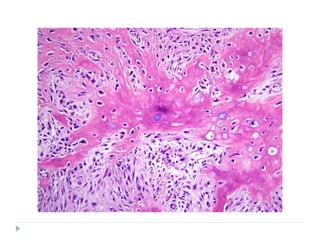
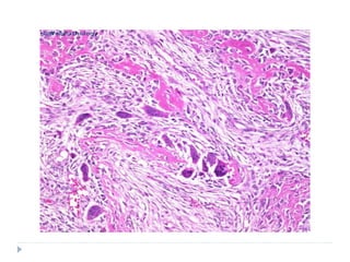
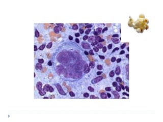
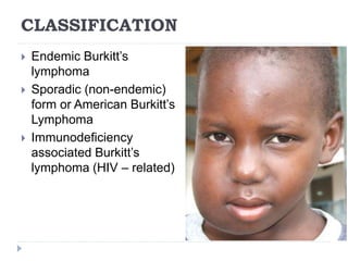
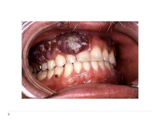
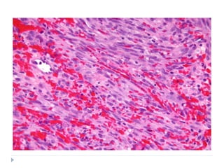
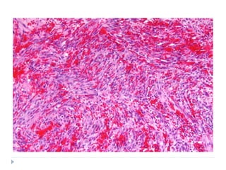
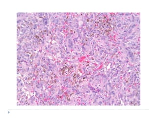
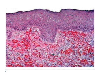
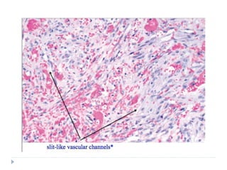
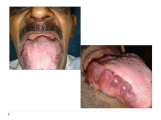
MALIGNANT CONNECTIVE
TISSUE TUMOURS
DR R AMRUTHA

 FIBROSARCOMA
 OSTEOSARCOMA
 HODGKIN’S LYMPHOMA
 BURKITT’S LYMPHOMA
 KAPOSI’S SARCOMA
 PLASMACYTOMA
 MULTIPLE MYELOMA
MALIGNANT TUMOURS
FIBROSARCOMA
 Malignant tumour of fibroblasts
 Can occur as
 soft tissue mass or as
 a primary bone tumour
 or secondary bone tumour
 Etiology
 No definite cause is known
 May arise from pre-existing lesions like
 Neurofibromas in neurofibromatosis
 Fibrous dysplasia
 Chronic osteomyelitis
 Bone infarcts
 Paget’s disease of bone
 Previously irradiated areas of bone
CLASSIFICATION
 SOFT TISSUE FIBROSARCOMA: occurring in the
soft tissues
 FIBROSARCOMA OF BONE
 PRIMARY
 PERIPHERAL: Arising from the periosteum
 CENTRAL : Arising from the medullary cavity
 SECONDARY: Arises from the pre-existing lesions or in
previously irradiated areas
CLINICAL FEATURES
 Represent 10% of musculo-skeletal sarcomas and
5% of all primary tumours of bone
 Age: In adults 35-55 years
 Infantile form: occurs in children < 20 years
 Gender: fibrosarcoma of bone has male predilection
 Location: most common in the extremities
 10% occur in head and neck
CLINICAL PRESENTATION
 SOFT TISSUE FIBROSARCOMA
 Large painless mass deep to the fascia
 Has ill-defined margins
 FIBROSARCOMA OF BONE
 Present with pain and swelling
 May grow to a large size and can cause pathological
fractures
 PRIOR H/O IRRADIATION AND OTHER DISEASE HAVE
TO BE ENQUIRED: COULD BE SECONDARY
FIBROSARCOMA
 Infantile form has excellent prognosis, unlike the
fibrosarcoma of adults
HISTOPATHOLOGY
 They vary in histologic grade
 WELL DIFFERENTIATED
 Multiple plump fibroblasts with pale eosinophilic cytoplasm and
deeply stained spindled nuclei
 Malignant cells are dispersed in rich collagen background
 Histologically normal mitotic figures are seen in few numbers
 Cells and nuclei are not pleomorphic
 INTERMEDIATE GRADE
 Cellular
 Have HERRING BONE PATTERN: parallel sheets of cell
arranged in intertwining whorls
 Slight degree of cellular pleomorphism
 Moderate amount of mature collagen
HISTOPATHOLOGY
 HIGH GRADE
 Very cellular with marked cellular atypia
 Marked mitotic activity
 Sparse matrix
 Multinucleated giant cells may be seen
 NO MALIGNANT OSTEOID FORMATION SHOULD BE
SEEN
 Extremely anaplastic and pleomorphic cells with bizzare
nuclei
TREATMENT AND PROGNOSIS
 Radical surgery including removal of invaded muscle
and bone
 Chemotherapy and radiotherapy can also be used in
conjunction with surgery
 Survival rate:
 5-year: 65%
 10-year: < 30%
 Secondary fibrosarcoma: < 10% at 10 years
OSTEOSARCOMA
 Also known as osteogenic sarcoma
 Malignancy of mesenchymal cells that have the
ability to produce osteoid or immature bone
 Etiopathogenesis
 Retinoblastoma (RB) gene mutation
 TRAUMA
 Bone dysplasias: fibrous dysplasia, Paget’s disease of
bone
 Li Fraumeni syndrome
 Rothmund Thompson syndrome
 Radiation therapy for some benign lesions like giant cell
granuloma
CLINICAL FEATURES
 Age: bimodal age distribution is seen
 First peak: 10 – 25 years (dramatic increase in
adolescence corresponds with the growth spurt)
 Second peak: after 50 years
 Gender: Slight male predilection
 M:F = 1.25 : 1
 Location: majority occurs in intramedullary location
 location also corresponds with the areas of bone growth
 Initial peak is more common in long bones
 In older patients axial skeleton and flat bones are most
frequently involved
 Paget’s disease and previous irradiation are associated
with increased prevalence
CLINICAL PRESENTATION
 Swelling and pain; particularly with activity of the involved
bone
ORAL MANIFESTATIONS
 Osteosarcomas of jaw are uncommon (6-8%)
 Most common in 3rd and 4th decades (10 -15 years older
than the osteosarcomas of long bones)
 Mandible and maxilla are involved with equal frequency
 Mandibular tumours: posterior body and ramus of mandible
 Maxillary lesions: alveolar ridge, sinus floor and palate
 Swelling and pain are the most common symptoms
 Loosening of teeth and paraesthesia are also seen
 Nasal obstruction is seen with maxillary tumours
RADIOGRAPHIC FEATURES
 Three classic features are seen
 SUNRAY OR SUNBURST APPEARANCE: Small streaks
of bone radiate outward
 Due to osteophytic bone production on the surface of the lesion
 Appreciated best on occlusal radiographs
 Tumour may grow within the PDL space causing
resorption of the adjacent bone: resulting in uniform
widening of the PDL space
 This is an early radiographic change
 CODMAN’S TRIANGLE: In long bones affected with
osteosarcoma the periosteum is elevated over the
expanding tumour mass in a tent-like fashion
 At the point where periosteum begins to merge, an acute angle
is formed between the bone surface and the periostium
 This feature is highly suspicious of osteosarcoma
HISTOPATHOLOGY
 Proliferation of atypical osteoblasts
 Presence of osteoid formed by malignant
osteoblasts
 Stromal cells may be round or spindle shaped and
atypical with irregularly shape nuclei
 In addition to osteoid cells may also produce
chondroid or fibrous connective tissue
 Depending on the relative amounts of the type of
tissue produced; osterosarcoma is classified into
three types:
 OSTEOBLASTIC TYPE
 CHONDROBLASTIC TYPE
 FIBROBLASTIC TYPE
TREATMENT AND PROGNOSIS
 Radical resection is the treatment of choice
 Neoadjuvant(preoperative) chemotherapy: to shrink
the tumour size
 Adjuvant chemotherapy: to increase the survival rate
of the disease
 Overall survival rate is 63%
HODGKIN’S LYMPHOMA
 Also known as Hodgkin’s disease or Malignant
Lymphoma
 It is a malignant lymphoproliferative disorder
 THOMAS HODGKIN first described Hodkin’s disease
in 1832
 ETIOLOGY
 Unknown
 Infectious agents; especially Epstein Barr virus
 Patients with HIV infection have higher incidence
 Genetic predisposition
 1% have family history
 Siblings of patients have increased risk
 Risk is even more higher in monozygotic twins
CLINICAL FEATURES
 Bimodal age distribution is seen
 15-34 years
 >55 years
 Gender: male predilection; particularly in children
 Race: more common in whites; less common in
Asians
 Location: always begins in Lymph nodes; any lymph
node group may be susceptible
 Most common ones: cervical and sub-clavicular lymph
nodes (70-75%)
 Followed by axillary and mediastinal nodes (5-10% each)
 Less than 5% initial presentation in abdominal and
inguinal nodes
CLINICAL PRESENTATION
 Presenting symptom: persistently enlarging non-
tender, discrete mass or masses in one lymph node
region
 Nodes are firm and rubbery in consistency
 Overlying skin is normal
 In the early stages, nodes are movable; with
progression they become matted and fixed to
surrounding tissues
 If untreated, condition spreads to other lymph node
groups
 Eventually it involves spleen and other lymphatic
tissues like bones, liver and lungs
CLINICAL PRESENTATION
 Constitutional symptoms:
 Fever
 Night sweats
 Generalised pruritis
 ORAL CAVITY may be secondarily involved
 Very rare
HISTOPATHOLOGY
 CLASSIFICATION
 Nodular - Lymphocyte predominant HL
 Classic HL
 Lymphocyte rich
 Nodular sclerosis
 Mixed cellularity
 Lymphocyte depletion
 Unclassifiable
HISTOPATHOLOGY
 Common features
 Effacement of normal nodal architecture by diffuse mixed
infiltrate of inflammatory cells
 Interspersed with large ATYPICAL NEOPLASTIC
LYMPHOID CELLS
 In classic type this atypical cell is known as REED
STERNBERG CELL
 Malignant cell in nodular-lymphocyte predominant HL is
POPCORN CELL
 Reed sternberg cell
 Typically binucleated (owl-eye nuclei)
 Sometimes may also be multinucleated (pennies on a
plate) with prominent nucleoli
 Popcorn cell: nucleus resembles kernel of popped
corn
TREATMENT
 Local radiation therapy
 Combination of less extensive radiotherapy and mild
multiagent chemotherapy
 Lymphocyte predominant type has most favourable
prognosis
 Lymphocyte depletion type has worst prognosis
BURKITT’S LYMPHOMA
 Named after DENIS BURKITT
 Also called as AFRICAN JAW LYMPHOMA: because
it is common in Sub-Saharan Africa
 It is a malignancy of B-lymphocyte origin
 One of the fastest growing malignancies in humans
CLASSIFICATION
 Endemic Burkitt’s
lymphoma
 Sporadic (non-endemic)
form or American Burkitt’s
Lymphoma
 Immunodeficiency
associated Burkitt’s
lymphoma (HIV – related)
ETIOLOGY
 Exact cause and mechanism are not known
 Probable causes
 c-myc oncogene mutation
 Epstein- Barr virus
 EBV is more commonly associated with the
African form
CLINICAL FEATURES
 AGE
 Endemic form usually affects children (peak prevalence =
7yrs)
 Sporadic form tends to affect patients over a greater age
range
 LOCATION
 50% - 70% of the endemic type present in jaws
 Involvement of abdominal organs is slightly less common
 Sporadic form usually involves the abdominal organs
 Endemic form
 Male predilection
 Posterior segments of the jaws are more commonly
involved
 Maxilla : mandible = 2:1
 Sometimes all four quadrants of the jaws may show
CLINICAL PRESENTATION
 Facial swelling and proptosis
 Pain, tenderness and paraesthesia are minimal
 Tooth mobility is common : because of aggressive
destruction of the alveolar bone
 Premature exfoliation of the deciduous teeth
 Enlargement of the gingiva and alveolar processes
are other common symptoms
RADIOGRAPHIC FEATURES
 Radiolucent destruction of the bone with ragged and
ill-defined margins
 Radiolucency may begin as several small areas
which eventually enlarge and coalesce
 Patchy loss of the lamina dura is one of the early
sign of Burkitt’s lymphoma
HISTOPATHOLOGY
 Monoclonal proliferation of B lymphocytes
 Lesion invades as broad sheets of tumour cells
 Tumour cells exhibit round nuclei with minimal
cytoplasm
 Each tumour nucleus has prominent nucleoli
 Numerous mitoses are seen
 At low power magnification: STARRY SKY PATTERN
is often appreciated
HISTOPATHOLOGY
 STARRY SKY PATTERN
 Phenomenon that is caused by the presence of
macrophages within the tumour tissue
 Macrophages have abundant cytoplasm which
microscopically appears less stained in comparison with
the surrounding cells
 These macrophages also contain phagocytic debris
 THE MACROPHAGES THUS, STAND OUT AS STARS
SET AGAINST THE NIGHT SKY OF DEEPLY
HYPERCHROMATIC NEOPLASTIC LYMPHOID CELLS
TREATMENT
 Intensive chemotherapy
 Survival rate has increase upto 75% - 85% with
recent advancements in the treatment modalities
KAPOSI’S SARCOMA
 Synonyms:
 Angioreticuloendothelioma
 Multiple Idiopathic Hemorrhagic sarcoma of Kaposi
 Kaposi’s sarcoma is a multicentric proliferation of
vascular and spindle cell components
 First described in 1872 by Moritz Kaposi. He
described the lesion as “ Idiopathic multiple
pigmented sarcoma of skin”
ETIOPATHOGENESIS
 Human Herpes virus 8 (KSHV)
 Has increased propensity to develop in individuals
infected with HIV; however HIV doesn’t seem to be
the direct cause of the tumorous proliferation
CLASSIFICATION
 CLASSIC OR CHRONIC KAPOSI’S SARCOMA
 ENDEMIC OR AFRICAN KAPOSI’S SARCOMA
 IATROGENIC IMMUNOSUPPRESION
ASSOCIATED
 AIDS RELATED
CLINICAL PRESENTATION
 Classic type
 Associated with altered immune status as well as
lymphoreticular and other malignancies
 Presents as cutaneous multi-focal blue-red macules,
nodules and plaques on lower extremities
 Slowly increase in size and number
 Some lesions regress while new ones develop on
adjacent or distant skin
 ORAL INVOLVEMENT IS RARE; if occurs it is seen on
palatal mucosa and gingiva
CLINICAL PRESENTATION
 Endemic type: endemic to African children
 4 types
 Benign nodular type: similar to classic type
 Aggressive or infiltrative type: locally invasive
 Florid form: widely disseminated; rapidly progressive
 Lymphadenopathic type: tumours of lymph nodes
 Iatrogenic type
 Occurs in recipients of organ transplants
 Related to loss of immunity secondary to
immunosuppressive drugs
 More aggressive
 Rare oral involvement
CLINICAL PRESENTATION
 AIDS related Kaposi’s sarcoma
 Early sign of AIDS
 Multiple lesions of skin and mucosa
 70% of patients demonstrate oral lesions; in 22%,oral
lesions are the first manifestation
 Hard palate, gingiva and tongue are commonly affected
 Palatal and gingival lesions invade bone and cause
tooth mobility
 Present as brown or reddish purple macular lesions
that do not blanch on pressure
 Later macules develop into plaques or nodules
 Pain, bleeding and necrosis are seen
HISTOPATHOLOGY
 SIMILAR HISTOPATHOLOGIC APPEARANCE IS SEEN IN ALL
THE CLINICAL TYPES
 Kaposi’s sarcoma evolves through 3 stages
 Patch stage
 Plaque stage
 Nodular stage
 Patch stage:
 Proliferation of small veins and capillaries around pre-
existing dilated vessels
 Slit-like vessels are seen which are lined by plump, mildly
atypical endothelial cells
 Scattered erythrocytes and hemosiderin deposits are
seen
 Perivascular spindle cell proliferation with minimal cellular
atypia
HISTOPATHOLOGY
 Plaque type
 Increased number of small capillaries and dilated vascular
channels
 Slit-like vascular channels without visible endothelial
lining
 Large number of extravasated RBCs and abundant
hemosiderin deposits
 Proliferating sheets of sarcomatous or atypical spindle
cells
 Lesional cells have enlarged hyperchromatic nuclei with
mild to moderate pleomorphism
 Minimal mitotic activity
 Nodular stage
 Spindle cells increase to form nodular tumour-like mass
TREATMENT
 FOR ORAL LESIONS
 Small and localised lesions
 Wide surgical excision
 Low dose irradiation and intralesional chemotherapy
 Sclerosing agents
 For larger and multifocal lesions: systemic
chemotherapy
MULTIPLE MYELOMA
 Malignancy of plasma cell origin
 Plasma cells are a subset of B-cells which are the
producers of humoral immunity factors called antibodies
 It is the most common primary neoplasm of the
skeletal system
 If metastatic lesions are excluded, multiple myeloma
accounts for 50% of all malignancies that involve the
bone
 It constitutes 1% of all malignancies and 10-15% of
all hematologic malignancies
ETIOPATHOGENESIS
 Results from mutation of terminally differentiated B
cells
 The abnormal plasma cells that constitute the
tumour are monoclonal; hence all the cells produce
same proteins
 These proteins are the immunoglobulins but they are
not normal or functional
 PREDISPOSING FACTORS (suggested but not
proved…)
 Radiation exposure
 Occupational exposure: chemical, metallurgical, rubber
plant, pulp and paper workers and leather tanners
 Chemical exposure: benzene, formaldehyde, hair dyes,
CLINICAL FEATURES
 Age: 60-70 years; rare before 40 years
 Gender: male predilection
 Race: blacks are more commonly affected than
whites with a ratio of 2:1
 It is the most common hematological malignancy
among black persons in The United States
 Location:
 Diffuse disease of bone marrow
 Predominant sites of involvement are within the axial
skeleton and include vertebral column, ribs, skull,
pelvis and femur bone
CLINICAL PRESENTATION
 Bone pain
 Pathological fractures
 Anaemia
 Azotaemia
 Hypercalcemia
 Recurrent infections
 Fatigue
 Petechial haemorrhages of skin
 Fever
 Metastatic calcifications of soft
tissues
 Extension of lesions to lymph
nodes, skin and viscera
 The underlying pathology
of multiple myeloma is
expansion of a single line
of plasma cells that
replace the normal bone
marrow and produce
monoclonal
immunoglobulins
 Renal failure and amyloid
depositions can be seen
as a result of increased
immunoglobulins
RADIOGRAPHIC FEATURES
 Multiple well-defined punched out radiolucencies
 Especially evident on the skull radiograph
 Jaws are involved in 30%
LABORATORY FINDINGS
 Presence of Bence Jones protein in the urine
ORAL MANIFESTATIONS
 Mandible is involved more frequently than maxilla
 Furthermore, the ramus, angle and molar region of
the mandible are most frequently involved
 Signs and symptoms of jaw involvement include:
 Pain and swelling
 Expansion of the jaw
 Numbness and mobility of the teeth
 EXTRASKELETAL
 Amyloid deposits can be seen in the tongue
 Lesions can occur in gingiva and resemble gingival
enlargements or epulis
HISTOPATHOLOGY
 Composed of sheets of closely packed cells
resembling plasma cells
 They are round or ovoid cells with eccentrically
placed nuclei
 The nucleus exhibits chromatin clumping in a
“cartwheel” pattern; perinuclear halo is seen
 Russell bodies are common
TREATMENT AND PROGNOSIS
 Chemotherapy to reduce the number of malignant
plasma cells
 Management of specific complications
 Only 5-10% of patients live longer than 10 years
 Infections, anaemia and kidney failure are the most
common causes of death
PLASMACYTOMA
 Solitary mass of neoplastic monoclonal plasma cells
in either bone marrow or soft tissue
 Classified into:
 Solitary bone plasmacytoma
 Extramedullary plasmacytoma
 Represents the least aggressive part of a spectrum
of multiple myeloma and may ultimately give rise to
multiple myeloma
PLASMACYTOMA vs MULTIPLE
MYELOMA
 Mean age is 55 years; 10 years younger than
multiple myeloma
 Radiologically presents as well-defined radiolucency
with no sclerotic borders or may also appear as
ragged radiolucency similar to multiple myeloma
 Amount of abnormal protein produced is much less
than that of multiple myeloma
 No evidence of plasma cell infiltration should be
seen by random bone marrow biopsy
 Patient should not show signs of anemia,
hypercalcemia or renal failure
EXTRAMEDULLARY PLASMACYTOMA
 90% of tumours develop at head and neck area
without primary bone involvement
 Most common sites are paranasal sinuses, pharynx,
nasal cavity and oral cavity
 Intraorally gingiva, palate, floor of the mouth and
tongue are most commonly involved
 Lesions occur as sessile or polypoid reddish
masses, which become lobulated as they grow but
donot ulcerate
TREATMENT AND PROGNOSIS
 Radiation therapy
 Most will eventually develop into multiple myeloma
 Extramedullary plasmacytoma has much better
prognosis with only 30% of lesions progressing to
multiple myeloma
TUMOUR-LIKE LESIONS
CENTRAL GIANT CELL GRANULOMA
 Uncommon, benign and proliferative lesion
 Jaffe first introduce the term “CENTRAL GIANT
CELL REPARATIVE GRANULOMA” to distinguish
this lesion from giant cell tumour of long bones
 Since reparative response was quite rare and most
of the lesions were found to be destructive the word
“reparative” was omitted
CLINICAL FEATURES
 Age: most of the cases occur before 30 years
 Gender: more common in females; M:F = 1:2
 Location: mandible is affected more often (70%
occur in mandible)
 Lesions are more common in the anterior portions of
the jaws
 Mandibular lesions frequently cross the midline
CLINICAL PRESENTATION
 Asymptomatic
 Noted during routine radiographic examination
 Sometimes painless expansion of the affected bone
may be the presenting complaint
 Sometimes may be associated with
 Pain and paresthesia
 expansion of cortex, perforation of the cortical bone
 ulceration of the mucosal surface by underlying lesion
 mobility, displacement and root resorption of the
associated teeth
RADIOGRAPHIC FEATURES
 Appears as radiolucent
defect which may be
unilocular or
multilocular
 The defect is usually
well delineated, but
margins are non-
corticated
CLASSIFICATION
 Based on the clinical and radiographic features,
central giant cell granuloma of jaws can be classified
into:
 NON-AGGRESSIVE LESIONS
 Few or no symptoms
 Slow growth
 No cortical perforation or root resorption
 AGGRESSIVE LESIONS
 Pain
 Rapid growth
 Show cortical perforation and root resorption
 Have marked tendency for recurrence
HISTOPATHOLOGY
 Loose fibrillar connective tissue stroma with
interspersed proliferating fibroblasts, small capillaries
and giant cells
 The connective tissue may sometimes be quite
cellular (corresponds to clinically aggressive lesions)
 Collagen fibres: arranged in the form of whorls
 Mesenchymal cells may be ovoid to spindle shaped
and interspersed with round monocyte-macrophages
 Giant cells: multinucleated with variable size; contain
few or several dozen nuclei
 Numerous foci of extravasated RBCs and associated
hemosiderin pigment can be seen
 Foci of new trabeculae of osteoid or bone may be
seen at the periphery of the lesion
TREATMENT AND PROGNOSIS
 Curettage or surgical excision
 X-RAY RADIATION THERAPY IS
CONTRAINDICATED
 Recurrence is rare
PERIPHERAL GIANT CELL GRANULOMA
 Also called peripheral giant cell epulis or peripheral
giant cell reparative granuloma
 Common tumour of the oral cavity
 ETIOLOGY
 Unknown
 Probable causes: Local irritation due to dental plaque
or calculus, periodontal disease, poor dental
restorations, ill-fitting dental appliances or dental
extractions
CLINICAL FEATURES
 Age: first to sixth decades; mean age: 31-42 years
 Gender: 60% of cases are seen in females
 Location: occur exclusively on gingiva or edentulous
alveolar ridge
 May develop in either anterior or posterior regions
 Mandibular tissues are most commonly affected
CLINICAL PRESENTATION
 Presents as red or red-blue nodular mass
 Smaller than 2cms in diameter, large ones are
occasionally seen
 Sessile or pedunculated and may or may not be
ulcerated
 Ulceration is less common in edentulous conditions
 In edentulous patients, lesion may be arising from
the crest of the ridge or from tissue covering the
slope of the ridge
 RADIOGRAPHICALLY, the lesion may sometimes
produce a “cupping” resorption of the underlying
alveolar bone
HISTOPATHOLOGY
 Non-encapsulated
 Composed of delicate fibrillar and reticular stroma
containing large number of ovoid or spindle shaped
connective tissue cells and multinucleated giant cells
 Giant cells may contain few or several dozens of
nuclei
 Some may have large vesicular nuclei and others
demonstrate small pyknotic nuclei
 Numerous capillaries are found around the
periphery of the lesion
 Foci of hemorrhage and hemosiderin pigment are
seen
 Spicules of osteoid or dystrophic calcification may
be seen
TREATMENT AND PROGNOSIS
 Conservative surgical excision
 Prognosis: good
 10-15% is the recurrence rate which can be
prevented by complete removal of the lesion rather
than surgical excision
ANEURYSMAL BONE CYST
 Intraosseous accumulation of variable-sized, blood
filled spaces surrounded by cellular fibrous
connective tissue
 Seperated as a distinct entity in 1942 by Jaffe and
Lichenstein
ETIOPATHOGENESIS
 Aneurysmal bone cyst may arise as a primary lesion
or as a result of disrupted vascular dynamics in a
pre-existing intrabony lesion
 Various theories have been proposed:
 Lichenstien: persistent local alteration in
hemodynamics
Increased venous pressure
development of dilated and engorged
vascular bed in the transformed bone
area
ETIOPATHOGENESIS
 Attempt at repair of a hematoma: a hematoma that
maintains a circulatory connection with the damaged
vessel would produce a slow flow of blood through
the lesion
 As a secondary lesion: a primary lesion of the
bone initiates an osseous, arteriovenous fistula and
thereby creates, via its hemodynamic forces, this
secondary reactive lesion of the bone
CLINICAL FEATURES
 Age: lesion of young persons; predominant
under 20yrs of age
 Gender: no gender predilection
 Location: most commonly long bones and
vertebral column
 Gnathic ABCs are uncommon; 2% reported in
the jaws
 Mandibular predominance: mostly in the
posterior segments
CLINICAL PRESENTATION
 Common presentation:Swelling that has usually
developed rapidly
 Pain is often reported
 Parasthesia and compressibility: rare
 Malocclusion, mobility, migration or resorption of
involved teeth may be present
 Maxillary lesions bulge into the sinus and can cause
nasal obstruction, nasal bleeding, proptosis and
diplopia
RADIOGRAPHIC FEATURES
 Unilocular or multilocular radiolucency
 Marked cortical expansion and thinning
 Ballooning or blow-out distension of the contour of
the affected bone
 Small radiopaque foci may be seen within the
radiolucency: trabeculae of reactive bone
HISTOPATHOLOGY
 Gross: “blood-soaked in sponge” appearance
 Microscopically
 Fibrous connective tissue stroma interspersed with
cavernous or sinusoidal blood-filled spaces
 Spaces are filled with unclotted blood; blood filled
spaces are not lined with endothelium
 Surrounding connective tissue contains multinucleated
giant cells, trabeculae of osteoid and woven bone
 Varying amounts of hemosiderin pigment may be seen
 Approximately 20% of cases are associated with
another pathosis like giant cell granuloma or other
fibro-osseous lesions
TREATMENT AND PROGNOSIS
 Curettage or enucleation
 Recurrence is rare in the oral cavity
Malignant connective tissue tumors.pptx

Malignant connective tissue tumors.pptx

  • 1.
  • 2.
      FIBROSARCOMA  OSTEOSARCOMA HODGKIN’S LYMPHOMA  BURKITT’S LYMPHOMA  KAPOSI’S SARCOMA  PLASMACYTOMA  MULTIPLE MYELOMA MALIGNANT TUMOURS
  • 3.
    FIBROSARCOMA  Malignant tumourof fibroblasts  Can occur as  soft tissue mass or as  a primary bone tumour  or secondary bone tumour  Etiology  No definite cause is known  May arise from pre-existing lesions like  Neurofibromas in neurofibromatosis  Fibrous dysplasia  Chronic osteomyelitis  Bone infarcts  Paget’s disease of bone  Previously irradiated areas of bone
  • 4.
    CLASSIFICATION  SOFT TISSUEFIBROSARCOMA: occurring in the soft tissues  FIBROSARCOMA OF BONE  PRIMARY  PERIPHERAL: Arising from the periosteum  CENTRAL : Arising from the medullary cavity  SECONDARY: Arises from the pre-existing lesions or in previously irradiated areas
  • 5.
    CLINICAL FEATURES  Represent10% of musculo-skeletal sarcomas and 5% of all primary tumours of bone  Age: In adults 35-55 years  Infantile form: occurs in children < 20 years  Gender: fibrosarcoma of bone has male predilection  Location: most common in the extremities  10% occur in head and neck
  • 6.
    CLINICAL PRESENTATION  SOFTTISSUE FIBROSARCOMA  Large painless mass deep to the fascia  Has ill-defined margins  FIBROSARCOMA OF BONE  Present with pain and swelling  May grow to a large size and can cause pathological fractures  PRIOR H/O IRRADIATION AND OTHER DISEASE HAVE TO BE ENQUIRED: COULD BE SECONDARY FIBROSARCOMA  Infantile form has excellent prognosis, unlike the fibrosarcoma of adults
  • 7.
    HISTOPATHOLOGY  They varyin histologic grade  WELL DIFFERENTIATED  Multiple plump fibroblasts with pale eosinophilic cytoplasm and deeply stained spindled nuclei  Malignant cells are dispersed in rich collagen background  Histologically normal mitotic figures are seen in few numbers  Cells and nuclei are not pleomorphic  INTERMEDIATE GRADE  Cellular  Have HERRING BONE PATTERN: parallel sheets of cell arranged in intertwining whorls  Slight degree of cellular pleomorphism  Moderate amount of mature collagen
  • 9.
    HISTOPATHOLOGY  HIGH GRADE Very cellular with marked cellular atypia  Marked mitotic activity  Sparse matrix  Multinucleated giant cells may be seen  NO MALIGNANT OSTEOID FORMATION SHOULD BE SEEN  Extremely anaplastic and pleomorphic cells with bizzare nuclei
  • 11.
    TREATMENT AND PROGNOSIS Radical surgery including removal of invaded muscle and bone  Chemotherapy and radiotherapy can also be used in conjunction with surgery  Survival rate:  5-year: 65%  10-year: < 30%  Secondary fibrosarcoma: < 10% at 10 years
  • 12.
    OSTEOSARCOMA  Also knownas osteogenic sarcoma  Malignancy of mesenchymal cells that have the ability to produce osteoid or immature bone  Etiopathogenesis  Retinoblastoma (RB) gene mutation  TRAUMA  Bone dysplasias: fibrous dysplasia, Paget’s disease of bone  Li Fraumeni syndrome  Rothmund Thompson syndrome  Radiation therapy for some benign lesions like giant cell granuloma
  • 13.
    CLINICAL FEATURES  Age:bimodal age distribution is seen  First peak: 10 – 25 years (dramatic increase in adolescence corresponds with the growth spurt)  Second peak: after 50 years  Gender: Slight male predilection  M:F = 1.25 : 1  Location: majority occurs in intramedullary location  location also corresponds with the areas of bone growth  Initial peak is more common in long bones  In older patients axial skeleton and flat bones are most frequently involved  Paget’s disease and previous irradiation are associated with increased prevalence
  • 14.
    CLINICAL PRESENTATION  Swellingand pain; particularly with activity of the involved bone ORAL MANIFESTATIONS  Osteosarcomas of jaw are uncommon (6-8%)  Most common in 3rd and 4th decades (10 -15 years older than the osteosarcomas of long bones)  Mandible and maxilla are involved with equal frequency  Mandibular tumours: posterior body and ramus of mandible  Maxillary lesions: alveolar ridge, sinus floor and palate  Swelling and pain are the most common symptoms  Loosening of teeth and paraesthesia are also seen  Nasal obstruction is seen with maxillary tumours
  • 15.
    RADIOGRAPHIC FEATURES  Threeclassic features are seen  SUNRAY OR SUNBURST APPEARANCE: Small streaks of bone radiate outward  Due to osteophytic bone production on the surface of the lesion  Appreciated best on occlusal radiographs  Tumour may grow within the PDL space causing resorption of the adjacent bone: resulting in uniform widening of the PDL space  This is an early radiographic change  CODMAN’S TRIANGLE: In long bones affected with osteosarcoma the periosteum is elevated over the expanding tumour mass in a tent-like fashion  At the point where periosteum begins to merge, an acute angle is formed between the bone surface and the periostium  This feature is highly suspicious of osteosarcoma
  • 17.
    HISTOPATHOLOGY  Proliferation ofatypical osteoblasts  Presence of osteoid formed by malignant osteoblasts  Stromal cells may be round or spindle shaped and atypical with irregularly shape nuclei  In addition to osteoid cells may also produce chondroid or fibrous connective tissue  Depending on the relative amounts of the type of tissue produced; osterosarcoma is classified into three types:  OSTEOBLASTIC TYPE  CHONDROBLASTIC TYPE  FIBROBLASTIC TYPE
  • 20.
    TREATMENT AND PROGNOSIS Radical resection is the treatment of choice  Neoadjuvant(preoperative) chemotherapy: to shrink the tumour size  Adjuvant chemotherapy: to increase the survival rate of the disease  Overall survival rate is 63%
  • 21.
    HODGKIN’S LYMPHOMA  Alsoknown as Hodgkin’s disease or Malignant Lymphoma  It is a malignant lymphoproliferative disorder  THOMAS HODGKIN first described Hodkin’s disease in 1832  ETIOLOGY  Unknown  Infectious agents; especially Epstein Barr virus  Patients with HIV infection have higher incidence  Genetic predisposition  1% have family history  Siblings of patients have increased risk  Risk is even more higher in monozygotic twins
  • 22.
    CLINICAL FEATURES  Bimodalage distribution is seen  15-34 years  >55 years  Gender: male predilection; particularly in children  Race: more common in whites; less common in Asians  Location: always begins in Lymph nodes; any lymph node group may be susceptible  Most common ones: cervical and sub-clavicular lymph nodes (70-75%)  Followed by axillary and mediastinal nodes (5-10% each)  Less than 5% initial presentation in abdominal and inguinal nodes
  • 23.
    CLINICAL PRESENTATION  Presentingsymptom: persistently enlarging non- tender, discrete mass or masses in one lymph node region  Nodes are firm and rubbery in consistency  Overlying skin is normal  In the early stages, nodes are movable; with progression they become matted and fixed to surrounding tissues  If untreated, condition spreads to other lymph node groups  Eventually it involves spleen and other lymphatic tissues like bones, liver and lungs
  • 25.
    CLINICAL PRESENTATION  Constitutionalsymptoms:  Fever  Night sweats  Generalised pruritis  ORAL CAVITY may be secondarily involved  Very rare
  • 26.
    HISTOPATHOLOGY  CLASSIFICATION  Nodular- Lymphocyte predominant HL  Classic HL  Lymphocyte rich  Nodular sclerosis  Mixed cellularity  Lymphocyte depletion  Unclassifiable
  • 27.
    HISTOPATHOLOGY  Common features Effacement of normal nodal architecture by diffuse mixed infiltrate of inflammatory cells  Interspersed with large ATYPICAL NEOPLASTIC LYMPHOID CELLS  In classic type this atypical cell is known as REED STERNBERG CELL  Malignant cell in nodular-lymphocyte predominant HL is POPCORN CELL  Reed sternberg cell  Typically binucleated (owl-eye nuclei)  Sometimes may also be multinucleated (pennies on a plate) with prominent nucleoli  Popcorn cell: nucleus resembles kernel of popped corn
  • 32.
    TREATMENT  Local radiationtherapy  Combination of less extensive radiotherapy and mild multiagent chemotherapy  Lymphocyte predominant type has most favourable prognosis  Lymphocyte depletion type has worst prognosis
  • 33.
    BURKITT’S LYMPHOMA  Namedafter DENIS BURKITT  Also called as AFRICAN JAW LYMPHOMA: because it is common in Sub-Saharan Africa  It is a malignancy of B-lymphocyte origin  One of the fastest growing malignancies in humans
  • 34.
    CLASSIFICATION  Endemic Burkitt’s lymphoma Sporadic (non-endemic) form or American Burkitt’s Lymphoma  Immunodeficiency associated Burkitt’s lymphoma (HIV – related)
  • 35.
    ETIOLOGY  Exact causeand mechanism are not known  Probable causes  c-myc oncogene mutation  Epstein- Barr virus  EBV is more commonly associated with the African form
  • 36.
    CLINICAL FEATURES  AGE Endemic form usually affects children (peak prevalence = 7yrs)  Sporadic form tends to affect patients over a greater age range  LOCATION  50% - 70% of the endemic type present in jaws  Involvement of abdominal organs is slightly less common  Sporadic form usually involves the abdominal organs  Endemic form  Male predilection  Posterior segments of the jaws are more commonly involved  Maxilla : mandible = 2:1  Sometimes all four quadrants of the jaws may show
  • 37.
    CLINICAL PRESENTATION  Facialswelling and proptosis  Pain, tenderness and paraesthesia are minimal  Tooth mobility is common : because of aggressive destruction of the alveolar bone  Premature exfoliation of the deciduous teeth  Enlargement of the gingiva and alveolar processes are other common symptoms
  • 38.
    RADIOGRAPHIC FEATURES  Radiolucentdestruction of the bone with ragged and ill-defined margins  Radiolucency may begin as several small areas which eventually enlarge and coalesce  Patchy loss of the lamina dura is one of the early sign of Burkitt’s lymphoma
  • 39.
    HISTOPATHOLOGY  Monoclonal proliferationof B lymphocytes  Lesion invades as broad sheets of tumour cells  Tumour cells exhibit round nuclei with minimal cytoplasm  Each tumour nucleus has prominent nucleoli  Numerous mitoses are seen  At low power magnification: STARRY SKY PATTERN is often appreciated
  • 42.
    HISTOPATHOLOGY  STARRY SKYPATTERN  Phenomenon that is caused by the presence of macrophages within the tumour tissue  Macrophages have abundant cytoplasm which microscopically appears less stained in comparison with the surrounding cells  These macrophages also contain phagocytic debris  THE MACROPHAGES THUS, STAND OUT AS STARS SET AGAINST THE NIGHT SKY OF DEEPLY HYPERCHROMATIC NEOPLASTIC LYMPHOID CELLS
  • 43.
    TREATMENT  Intensive chemotherapy Survival rate has increase upto 75% - 85% with recent advancements in the treatment modalities
  • 44.
    KAPOSI’S SARCOMA  Synonyms: Angioreticuloendothelioma  Multiple Idiopathic Hemorrhagic sarcoma of Kaposi  Kaposi’s sarcoma is a multicentric proliferation of vascular and spindle cell components  First described in 1872 by Moritz Kaposi. He described the lesion as “ Idiopathic multiple pigmented sarcoma of skin”
  • 45.
    ETIOPATHOGENESIS  Human Herpesvirus 8 (KSHV)  Has increased propensity to develop in individuals infected with HIV; however HIV doesn’t seem to be the direct cause of the tumorous proliferation
  • 46.
    CLASSIFICATION  CLASSIC ORCHRONIC KAPOSI’S SARCOMA  ENDEMIC OR AFRICAN KAPOSI’S SARCOMA  IATROGENIC IMMUNOSUPPRESION ASSOCIATED  AIDS RELATED
  • 47.
    CLINICAL PRESENTATION  Classictype  Associated with altered immune status as well as lymphoreticular and other malignancies  Presents as cutaneous multi-focal blue-red macules, nodules and plaques on lower extremities  Slowly increase in size and number  Some lesions regress while new ones develop on adjacent or distant skin  ORAL INVOLVEMENT IS RARE; if occurs it is seen on palatal mucosa and gingiva
  • 49.
    CLINICAL PRESENTATION  Endemictype: endemic to African children  4 types  Benign nodular type: similar to classic type  Aggressive or infiltrative type: locally invasive  Florid form: widely disseminated; rapidly progressive  Lymphadenopathic type: tumours of lymph nodes  Iatrogenic type  Occurs in recipients of organ transplants  Related to loss of immunity secondary to immunosuppressive drugs  More aggressive  Rare oral involvement
  • 50.
    CLINICAL PRESENTATION  AIDSrelated Kaposi’s sarcoma  Early sign of AIDS  Multiple lesions of skin and mucosa  70% of patients demonstrate oral lesions; in 22%,oral lesions are the first manifestation  Hard palate, gingiva and tongue are commonly affected  Palatal and gingival lesions invade bone and cause tooth mobility  Present as brown or reddish purple macular lesions that do not blanch on pressure  Later macules develop into plaques or nodules  Pain, bleeding and necrosis are seen
  • 53.
    HISTOPATHOLOGY  SIMILAR HISTOPATHOLOGICAPPEARANCE IS SEEN IN ALL THE CLINICAL TYPES  Kaposi’s sarcoma evolves through 3 stages  Patch stage  Plaque stage  Nodular stage  Patch stage:  Proliferation of small veins and capillaries around pre- existing dilated vessels  Slit-like vessels are seen which are lined by plump, mildly atypical endothelial cells  Scattered erythrocytes and hemosiderin deposits are seen  Perivascular spindle cell proliferation with minimal cellular atypia
  • 54.
    HISTOPATHOLOGY  Plaque type Increased number of small capillaries and dilated vascular channels  Slit-like vascular channels without visible endothelial lining  Large number of extravasated RBCs and abundant hemosiderin deposits  Proliferating sheets of sarcomatous or atypical spindle cells  Lesional cells have enlarged hyperchromatic nuclei with mild to moderate pleomorphism  Minimal mitotic activity  Nodular stage  Spindle cells increase to form nodular tumour-like mass
  • 60.
    TREATMENT  FOR ORALLESIONS  Small and localised lesions  Wide surgical excision  Low dose irradiation and intralesional chemotherapy  Sclerosing agents  For larger and multifocal lesions: systemic chemotherapy
  • 61.
    MULTIPLE MYELOMA  Malignancyof plasma cell origin  Plasma cells are a subset of B-cells which are the producers of humoral immunity factors called antibodies  It is the most common primary neoplasm of the skeletal system  If metastatic lesions are excluded, multiple myeloma accounts for 50% of all malignancies that involve the bone  It constitutes 1% of all malignancies and 10-15% of all hematologic malignancies
  • 62.
    ETIOPATHOGENESIS  Results frommutation of terminally differentiated B cells  The abnormal plasma cells that constitute the tumour are monoclonal; hence all the cells produce same proteins  These proteins are the immunoglobulins but they are not normal or functional  PREDISPOSING FACTORS (suggested but not proved…)  Radiation exposure  Occupational exposure: chemical, metallurgical, rubber plant, pulp and paper workers and leather tanners  Chemical exposure: benzene, formaldehyde, hair dyes,
  • 63.
    CLINICAL FEATURES  Age:60-70 years; rare before 40 years  Gender: male predilection  Race: blacks are more commonly affected than whites with a ratio of 2:1  It is the most common hematological malignancy among black persons in The United States  Location:  Diffuse disease of bone marrow  Predominant sites of involvement are within the axial skeleton and include vertebral column, ribs, skull, pelvis and femur bone
  • 64.
    CLINICAL PRESENTATION  Bonepain  Pathological fractures  Anaemia  Azotaemia  Hypercalcemia  Recurrent infections  Fatigue  Petechial haemorrhages of skin  Fever  Metastatic calcifications of soft tissues  Extension of lesions to lymph nodes, skin and viscera  The underlying pathology of multiple myeloma is expansion of a single line of plasma cells that replace the normal bone marrow and produce monoclonal immunoglobulins  Renal failure and amyloid depositions can be seen as a result of increased immunoglobulins
  • 65.
    RADIOGRAPHIC FEATURES  Multiplewell-defined punched out radiolucencies  Especially evident on the skull radiograph  Jaws are involved in 30% LABORATORY FINDINGS  Presence of Bence Jones protein in the urine
  • 67.
    ORAL MANIFESTATIONS  Mandibleis involved more frequently than maxilla  Furthermore, the ramus, angle and molar region of the mandible are most frequently involved  Signs and symptoms of jaw involvement include:  Pain and swelling  Expansion of the jaw  Numbness and mobility of the teeth  EXTRASKELETAL  Amyloid deposits can be seen in the tongue  Lesions can occur in gingiva and resemble gingival enlargements or epulis
  • 69.
    HISTOPATHOLOGY  Composed ofsheets of closely packed cells resembling plasma cells  They are round or ovoid cells with eccentrically placed nuclei  The nucleus exhibits chromatin clumping in a “cartwheel” pattern; perinuclear halo is seen  Russell bodies are common
  • 74.
    TREATMENT AND PROGNOSIS Chemotherapy to reduce the number of malignant plasma cells  Management of specific complications  Only 5-10% of patients live longer than 10 years  Infections, anaemia and kidney failure are the most common causes of death
  • 75.
    PLASMACYTOMA  Solitary massof neoplastic monoclonal plasma cells in either bone marrow or soft tissue  Classified into:  Solitary bone plasmacytoma  Extramedullary plasmacytoma  Represents the least aggressive part of a spectrum of multiple myeloma and may ultimately give rise to multiple myeloma
  • 76.
    PLASMACYTOMA vs MULTIPLE MYELOMA Mean age is 55 years; 10 years younger than multiple myeloma  Radiologically presents as well-defined radiolucency with no sclerotic borders or may also appear as ragged radiolucency similar to multiple myeloma  Amount of abnormal protein produced is much less than that of multiple myeloma  No evidence of plasma cell infiltration should be seen by random bone marrow biopsy  Patient should not show signs of anemia, hypercalcemia or renal failure
  • 77.
    EXTRAMEDULLARY PLASMACYTOMA  90%of tumours develop at head and neck area without primary bone involvement  Most common sites are paranasal sinuses, pharynx, nasal cavity and oral cavity  Intraorally gingiva, palate, floor of the mouth and tongue are most commonly involved  Lesions occur as sessile or polypoid reddish masses, which become lobulated as they grow but donot ulcerate
  • 78.
    TREATMENT AND PROGNOSIS Radiation therapy  Most will eventually develop into multiple myeloma  Extramedullary plasmacytoma has much better prognosis with only 30% of lesions progressing to multiple myeloma
  • 79.
  • 80.
    CENTRAL GIANT CELLGRANULOMA  Uncommon, benign and proliferative lesion  Jaffe first introduce the term “CENTRAL GIANT CELL REPARATIVE GRANULOMA” to distinguish this lesion from giant cell tumour of long bones  Since reparative response was quite rare and most of the lesions were found to be destructive the word “reparative” was omitted
  • 81.
    CLINICAL FEATURES  Age:most of the cases occur before 30 years  Gender: more common in females; M:F = 1:2  Location: mandible is affected more often (70% occur in mandible)  Lesions are more common in the anterior portions of the jaws  Mandibular lesions frequently cross the midline
  • 82.
    CLINICAL PRESENTATION  Asymptomatic Noted during routine radiographic examination  Sometimes painless expansion of the affected bone may be the presenting complaint  Sometimes may be associated with  Pain and paresthesia  expansion of cortex, perforation of the cortical bone  ulceration of the mucosal surface by underlying lesion  mobility, displacement and root resorption of the associated teeth
  • 83.
    RADIOGRAPHIC FEATURES  Appearsas radiolucent defect which may be unilocular or multilocular  The defect is usually well delineated, but margins are non- corticated
  • 84.
    CLASSIFICATION  Based onthe clinical and radiographic features, central giant cell granuloma of jaws can be classified into:  NON-AGGRESSIVE LESIONS  Few or no symptoms  Slow growth  No cortical perforation or root resorption  AGGRESSIVE LESIONS  Pain  Rapid growth  Show cortical perforation and root resorption  Have marked tendency for recurrence
  • 85.
    HISTOPATHOLOGY  Loose fibrillarconnective tissue stroma with interspersed proliferating fibroblasts, small capillaries and giant cells  The connective tissue may sometimes be quite cellular (corresponds to clinically aggressive lesions)  Collagen fibres: arranged in the form of whorls  Mesenchymal cells may be ovoid to spindle shaped and interspersed with round monocyte-macrophages  Giant cells: multinucleated with variable size; contain few or several dozen nuclei  Numerous foci of extravasated RBCs and associated hemosiderin pigment can be seen  Foci of new trabeculae of osteoid or bone may be seen at the periphery of the lesion
  • 87.
    TREATMENT AND PROGNOSIS Curettage or surgical excision  X-RAY RADIATION THERAPY IS CONTRAINDICATED  Recurrence is rare
  • 88.
    PERIPHERAL GIANT CELLGRANULOMA  Also called peripheral giant cell epulis or peripheral giant cell reparative granuloma  Common tumour of the oral cavity  ETIOLOGY  Unknown  Probable causes: Local irritation due to dental plaque or calculus, periodontal disease, poor dental restorations, ill-fitting dental appliances or dental extractions
  • 89.
    CLINICAL FEATURES  Age:first to sixth decades; mean age: 31-42 years  Gender: 60% of cases are seen in females  Location: occur exclusively on gingiva or edentulous alveolar ridge  May develop in either anterior or posterior regions  Mandibular tissues are most commonly affected
  • 90.
    CLINICAL PRESENTATION  Presentsas red or red-blue nodular mass  Smaller than 2cms in diameter, large ones are occasionally seen  Sessile or pedunculated and may or may not be ulcerated  Ulceration is less common in edentulous conditions  In edentulous patients, lesion may be arising from the crest of the ridge or from tissue covering the slope of the ridge  RADIOGRAPHICALLY, the lesion may sometimes produce a “cupping” resorption of the underlying alveolar bone
  • 92.
    HISTOPATHOLOGY  Non-encapsulated  Composedof delicate fibrillar and reticular stroma containing large number of ovoid or spindle shaped connective tissue cells and multinucleated giant cells  Giant cells may contain few or several dozens of nuclei  Some may have large vesicular nuclei and others demonstrate small pyknotic nuclei  Numerous capillaries are found around the periphery of the lesion  Foci of hemorrhage and hemosiderin pigment are seen  Spicules of osteoid or dystrophic calcification may be seen
  • 94.
    TREATMENT AND PROGNOSIS Conservative surgical excision  Prognosis: good  10-15% is the recurrence rate which can be prevented by complete removal of the lesion rather than surgical excision
  • 95.
    ANEURYSMAL BONE CYST Intraosseous accumulation of variable-sized, blood filled spaces surrounded by cellular fibrous connective tissue  Seperated as a distinct entity in 1942 by Jaffe and Lichenstein
  • 96.
    ETIOPATHOGENESIS  Aneurysmal bonecyst may arise as a primary lesion or as a result of disrupted vascular dynamics in a pre-existing intrabony lesion  Various theories have been proposed:  Lichenstien: persistent local alteration in hemodynamics Increased venous pressure development of dilated and engorged vascular bed in the transformed bone area
  • 97.
    ETIOPATHOGENESIS  Attempt atrepair of a hematoma: a hematoma that maintains a circulatory connection with the damaged vessel would produce a slow flow of blood through the lesion  As a secondary lesion: a primary lesion of the bone initiates an osseous, arteriovenous fistula and thereby creates, via its hemodynamic forces, this secondary reactive lesion of the bone
  • 98.
    CLINICAL FEATURES  Age:lesion of young persons; predominant under 20yrs of age  Gender: no gender predilection  Location: most commonly long bones and vertebral column  Gnathic ABCs are uncommon; 2% reported in the jaws  Mandibular predominance: mostly in the posterior segments
  • 99.
    CLINICAL PRESENTATION  Commonpresentation:Swelling that has usually developed rapidly  Pain is often reported  Parasthesia and compressibility: rare  Malocclusion, mobility, migration or resorption of involved teeth may be present  Maxillary lesions bulge into the sinus and can cause nasal obstruction, nasal bleeding, proptosis and diplopia
  • 100.
    RADIOGRAPHIC FEATURES  Unilocularor multilocular radiolucency  Marked cortical expansion and thinning  Ballooning or blow-out distension of the contour of the affected bone  Small radiopaque foci may be seen within the radiolucency: trabeculae of reactive bone
  • 105.
    HISTOPATHOLOGY  Gross: “blood-soakedin sponge” appearance  Microscopically  Fibrous connective tissue stroma interspersed with cavernous or sinusoidal blood-filled spaces  Spaces are filled with unclotted blood; blood filled spaces are not lined with endothelium  Surrounding connective tissue contains multinucleated giant cells, trabeculae of osteoid and woven bone  Varying amounts of hemosiderin pigment may be seen  Approximately 20% of cases are associated with another pathosis like giant cell granuloma or other fibro-osseous lesions
  • 106.
    TREATMENT AND PROGNOSIS Curettage or enucleation  Recurrence is rare in the oral cavity

Editor's Notes

  • #4 Sarcomas as a group differ from malignant epithelial neoplasms by their typical occurrence in relatively younger persons and their greater tendency to metastasize through the blood stream rather than through lymphatics, thereby producing more widespread foci of secondary tumour growth
  • #66 Unusual protein which coagulates when the urine is heated to 40-60˚C and then disappears when the urine is boiled; it reappears when the urine is cooled