This document discusses ectodermal dysplasia syndrome (EDS), a heterogeneous group of inherited disorders that affect ectodermal tissues like skin, hair, nails, sweat glands, and teeth. It describes two main types - hypohidrotic EDS and hidrotic EDS. Hypohidrotic EDS is characterized by hypohidrosis, anomalous dentition, onychodysplasia, and hypotrichosis. It can cause facial abnormalities, dry skin, intellectual disability, and other issues. Hidrotic EDS causes nail dystrophy, sparse hair, and palmoplantar changes. The document outlines clinical features and manifestations of both conditions, including oral findings like hypodontia and
Developmental Disturbances of Oral & Paraoral structures/ dental crown & brid...Indian dental academy
The Indian Dental Academy is the Leader in continuing dental education , training dentists in all aspects of dentistry and
offering a wide range of dental certified courses in different formats.for more details please visit
www.indiandentalacademy.com
Gingival cyst of newborn /orthodontic courses by Indian dental academy Indian dental academy
The Indian Dental Academy is the Leader in continuing dental education , training dentists in all aspects of dentistry and
offering a wide range of dental certified courses in different formats.for more details please visit
www.indiandentalacademy.com
DEVELOPMENTAL DISTURBANCES OF LIPS & PALATE / dental implant coursesIndian dental academy
The Indian Dental Academy is the Leader in continuing dental education , training dentists in all aspects of dentistry and
offering a wide range of dental certified courses in different formats.for more details please visit
www.indiandentalacademy.com
The Indian Dental Academy is the Leader in continuing dental education , training dentists in all aspects of dentistry and
offering a wide range of dental certified courses in different formats.for more details please visit
www.indiandentalacademy.com
The Indian Dental Academy is the Leader in continuing dental education , training dentists in all aspects of dentistry and
offering a wide range of dental certified courses in different formats.for more details please visit
www.indiandentalacademy.com
The Indian Dental Academy is the Leader in continuing dental education , training dentists in all aspects of dentistry and
offering a wide range of dental certified courses in different formats.for more details please visit
www.indiandentalacademy.com
for undergraduate dental students this presentation includes essential & common disorders which related to the tongue very briefly. Though this may be very helpfull to you to as a start for further readings & studying.
Navigating the Health Insurance Market_ Understanding Trends and Options.pdfEnterprise Wired
From navigating policy options to staying informed about industry trends, this comprehensive guide explores everything you need to know about the health insurance market.
How many patients does case series should have In comparison to case reports.pdfpubrica101
Pubrica’s team of researchers and writers create scientific and medical research articles, which may be important resources for authors and practitioners. Pubrica medical writers assist you in creating and revising the introduction by alerting the reader to gaps in the chosen study subject. Our professionals understand the order in which the hypothesis topic is followed by the broad subject, the issue, and the backdrop.
https://pubrica.com/academy/case-study-or-series/how-many-patients-does-case-series-should-have-in-comparison-to-case-reports/
Defecation
Normal defecation begins with movement in the left colon, moving stool toward the anus. When stool reaches the rectum, the distention causes relaxation of the internal sphincter and an awareness of the need to defecate. At the time of defecation, the external sphincter relaxes, and abdominal muscles contract, increasing intrarectal pressure and forcing the stool out
The Valsalva maneuver exerts pressure to expel faeces through a voluntary contraction of the abdominal muscles while maintaining forced expiration against a closed airway. Patients with cardiovascular disease, glaucoma, increased intracranial pressure, or a new surgical wound are at greater risk for cardiac dysrhythmias and elevated blood pressure with the Valsalva maneuver and need to avoid straining to pass the stool.
Normal defecation is painless, resulting in passage of soft, formed stool
CONSTIPATION
Constipation is a symptom, not a disease. Improper diet, reduced fluid intake, lack of exercise, and certain medications can cause constipation. For example, patients receiving opiates for pain after surgery often require a stool softener or laxative to prevent constipation. The signs of constipation include infrequent bowel movements (less than every 3 days), difficulty passing stools, excessive straining, inability to defecate at will, and hard feaces
IMPACTION
Fecal impaction results from unrelieved constipation. It is a collection of hardened feces wedged in the rectum that a person cannot expel. In cases of severe impaction the mass extends up into the sigmoid colon.
DIARRHEA
Diarrhea is an increase in the number of stools and the passage of liquid, unformed feces. It is associated with disorders affecting digestion, absorption, and secretion in the GI tract. Intestinal contents pass through the small and large intestine too quickly to allow for the usual absorption of fluid and nutrients. Irritation within the colon results in increased mucus secretion. As a result, feces become watery, and the patient is unable to control the urge to defecate. Normally an anal bag is safe and effective in long-term treatment of patients with fecal incontinence at home, in hospice, or in the hospital. Fecal incontinence is expensive and a potentially dangerous condition in terms of contamination and risk of skin ulceration
HEMORRHOIDS
Hemorrhoids are dilated, engorged veins in the lining of the rectum. They are either external or internal.
FLATULENCE
As gas accumulates in the lumen of the intestines, the bowel wall stretches and distends (flatulence). It is a common cause of abdominal fullness, pain, and cramping. Normally intestinal gas escapes through the mouth (belching) or the anus (passing of flatus)
FECAL INCONTINENCE
Fecal incontinence is the inability to control passage of feces and gas from the anus. Incontinence harms a patient’s body image
PREPARATION AND GIVING OF LAXATIVESACCORDING TO POTTER AND PERRY,
An enema is the instillation of a solution into the rectum and sig
Telehealth Psychology Building Trust with Clients.pptxThe Harvest Clinic
Telehealth psychology is a digital approach that offers psychological services and mental health care to clients remotely, using technologies like video conferencing, phone calls, text messaging, and mobile apps for communication.
The dimensions of healthcare quality refer to various attributes or aspects that define the standard of healthcare services. These dimensions are used to evaluate, measure, and improve the quality of care provided to patients. A comprehensive understanding of these dimensions ensures that healthcare systems can address various aspects of patient care effectively and holistically. Dimensions of Healthcare Quality and Performance of care include the following; Appropriateness, Availability, Competence, Continuity, Effectiveness, Efficiency, Efficacy, Prevention, Respect and Care, Safety as well as Timeliness.
Antibiotic Stewardship by Anushri Srivastava.pptxAnushriSrivastav
Stewardship is the act of taking good care of something.
Antimicrobial stewardship is a coordinated program that promotes the appropriate use of antimicrobials (including antibiotics), improves patient outcomes, reduces microbial resistance, and decreases the spread of infections caused by multidrug-resistant organisms.
WHO launched the Global Antimicrobial Resistance and Use Surveillance System (GLASS) in 2015 to fill knowledge gaps and inform strategies at all levels.
ACCORDING TO apic.org,
Antimicrobial stewardship is a coordinated program that promotes the appropriate use of antimicrobials (including antibiotics), improves patient outcomes, reduces microbial resistance, and decreases the spread of infections caused by multidrug-resistant organisms.
ACCORDING TO pewtrusts.org,
Antibiotic stewardship refers to efforts in doctors’ offices, hospitals, long term care facilities, and other health care settings to ensure that antibiotics are used only when necessary and appropriate
According to WHO,
Antimicrobial stewardship is a systematic approach to educate and support health care professionals to follow evidence-based guidelines for prescribing and administering antimicrobials
In 1996, John McGowan and Dale Gerding first applied the term antimicrobial stewardship, where they suggested a causal association between antimicrobial agent use and resistance. They also focused on the urgency of large-scale controlled trials of antimicrobial-use regulation employing sophisticated epidemiologic methods, molecular typing, and precise resistance mechanism analysis.
Antimicrobial Stewardship(AMS) refers to the optimal selection, dosing, and duration of antimicrobial treatment resulting in the best clinical outcome with minimal side effects to the patients and minimal impact on subsequent resistance.
According to the 2019 report, in the US, more than 2.8 million antibiotic-resistant infections occur each year, and more than 35000 people die. In addition to this, it also mentioned that 223,900 cases of Clostridoides difficile occurred in 2017, of which 12800 people died. The report did not include viruses or parasites
VISION
Being proactive
Supporting optimal animal and human health
Exploring ways to reduce overall use of antimicrobials
Using the drugs that prevent and treat disease by killing microscopic organisms in a responsible way
GOAL
to prevent the generation and spread of antimicrobial resistance (AMR). Doing so will preserve the effectiveness of these drugs in animals and humans for years to come.
being to preserve human and animal health and the effectiveness of antimicrobial medications.
to implement a multidisciplinary approach in assembling a stewardship team to include an infectious disease physician, a clinical pharmacist with infectious diseases training, infection preventionist, and a close collaboration with the staff in the clinical microbiology laboratory
to prevent antimicrobial overuse, misuse and abuse.
to minimize the developme
CHAPTER 1 SEMESTER V PREVENTIVE-PEDIATRICS.pdfSachin Sharma
This content provides an overview of preventive pediatrics. It defines preventive pediatrics as preventing disease and promoting children's physical, mental, and social well-being to achieve positive health. It discusses antenatal, postnatal, and social preventive pediatrics. It also covers various child health programs like immunization, breastfeeding, ICDS, and the roles of organizations like WHO, UNICEF, and nurses in preventive pediatrics.
QA Paediatric dentistry department, Hospital Melaka 2020Azreen Aj
QA study - To improve the 6th monthly recall rate post-comprehensive dental treatment under general anaesthesia in paediatric dentistry department, Hospital Melaka
CRISPR-Cas9, a revolutionary gene-editing tool, holds immense potential to reshape medicine, agriculture, and our understanding of life. But like any powerful tool, it comes with ethical considerations.
Unveiling CRISPR: This naturally occurring bacterial defense system (crRNA & Cas9 protein) fights viruses. Scientists repurposed it for precise gene editing (correction, deletion, insertion) by targeting specific DNA sequences.
The Promise: CRISPR offers exciting possibilities:
Gene Therapy: Correcting genetic diseases like cystic fibrosis.
Agriculture: Engineering crops resistant to pests and harsh environments.
Research: Studying gene function to unlock new knowledge.
The Peril: Ethical concerns demand attention:
Off-target Effects: Unintended DNA edits can have unforeseen consequences.
Eugenics: Misusing CRISPR for designer babies raises social and ethical questions.
Equity: High costs could limit access to this potentially life-saving technology.
The Path Forward: Responsible development is crucial:
International Collaboration: Clear guidelines are needed for research and human trials.
Public Education: Open discussions ensure informed decisions about CRISPR.
Prioritize Safety and Ethics: Safety and ethical principles must be paramount.
CRISPR offers a powerful tool for a better future, but responsible development and addressing ethical concerns are essential. By prioritizing safety, fostering open dialogue, and ensuring equitable access, we can harness CRISPR's power for the benefit of all. (2998 characters)
2. Many primary cutaneous diseases also involve the
mucous membranes throughout the body, including the
oral mucosa
It is important for the dentist - some dermatoses exhibit
concomitant lesions of the oral mucous membranes, but
also that manifestation of some of the diseases may be
preceded by oral lesions.
Thus the dentist may be in a position to establish the
diagnosis of a dermatologic disease before the
cutaneous lesions become apparent.
3. Macule: Focal area of color change that is not elevated
or depressed in relation to its surroundings.
Papule: Solid, raised lesion < 5 mm in diameter.
Nodule: Solid, raised lesion >5 mm in diameter.
4. Vesicle: Superficial blister, 5 mm or less in diameter, usually filled
with clear fluid.
Bulla: Large blister, greater than 5 mm in diameter
Pustule: Blister filled with purulent exudate.
5. Erosion: Superficial lesion, often arising secondary to rupture of a
vesicle or bulla, that is characterized by partial or total loss of the
surface epithelium.
Ulcer: Lesion characterized by loss of the surface epithelium and
frequently some of the underlying connective tissue. It often appears
depressed or excavated.
6. Genodermatoses: Hereditary skin disorders, many of
which are also accompanied by various systemic
manifestations of different altered enzyme functions
Genokeratosis: Alterations in the normal keratinization
process. Some are genetically transmitted and some are
not.
Vesiculobullous lesions: Associated with vesicles and
bullae. Some are genetically transmitted and some are
not.
10. Synonyms: Hereditary ectodermal dysplasia, Ectodermal dysplasia
syndrome
Ectodermal dysplasia syndrome (EDS) is a large, heterogeneous group
of inherited disorders, the manifestations can be seen in more than one
ectodermal derivatives.
These tissues primarily are the skin, hair, nails, eccrine glands, and
teeth.
The current classification of ectodermal dysplasia (ED) is based on
clinical features.
The most common syndromes within this group are:
Hypohidrotic (anhidrotic) ED and
Hidrotic ED – mutation in GJB6 - chromosome 13 codes for connexin - 30
Several EDSs may manifest in association with midfacial defects, mainly
cleft lip and palate.
11. Etiology: Ectodermal dysplasia syndrome results from aberrant
development of ectodermal derivatives in early embryonic life. Different
genes are involved in different types of ED.
Clinical features:
Some EDS types are mild, while others are devastating.
Most often in whites.
X-linked hypohidrotic ED affects only in males. Female act as carriers
with little or no signs of the condition.
The remaining EDSs have no gender predilection.
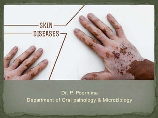
Dental, hair, and nail anomalies
12. Symptoms of a reduction in hair follicles vary from sparse scalp hair
(usually short, fine and dry) to a complete absence of hair
Eccrine sweat glands may be absent or sparse and rudimentary,
particularly in those with hypohidrotic EDS.
In some cases, mucous glands are absent in the upper respiratory tract
and in the bronchi, esophagus, and duodenum.
The mouth may be dry from hypoplasia of the salivary glands; lacrimal
glands also may be deficient.
Teeth show abnormal morphogenesis or they may be absent.
Nails are often brittle and thin or show abnormal ridging, but they may
be grossly deformed
Other signs and symptoms - deficient hearing or vision, cleft lip and/or
palate and missing fingers or toes are also seen.
13. 1. Hypohidrotic (anhidrotic) ED (Christ-SiemensTouraine
syndrome):
Most common phenotype in this group
Usually inherited as an X-linked recessive trait; autosomal recessive
and autosomal dominant forms have been reported but are rare.
It is characterized by several defects:
Hypohidrosis,
Anomalous Dentition,
Onychodysplasia,
Hypotrichosis.
14. • Typical facies are
characterized by:
• Frontal Bossing
• Sunken Cheeks
• Saddle Nose
• Thick, Everted
Lips
• Wrinkled,
Hyperpigmented
Skin Around The
Eyes
• Large, Low-set
Ears.
15. Because such characteristics are not obvious at birth, clinical clues for
diagnosis in the neonatal period are extensive scaling of the skin and
unexplained pyrexia.
The prevalence of atopic eczema is high.
Other common signs are:
Short stature,
Eye abnormalities,
Decreased flow of tears and
Photophobia.
Intelligence is normal.
16. Oral Manifestations:
Hypodontia or complete anodontia
Conical or pegged teeth
Delayed eruption of permanent teeth.
Even when complete anodontia exists, the growth of the jaw is not
impaired.
Since the alveolar process does not develop in the absence of teeth,
there is a reduction from the normal vertical dimension resulting in the
protuberant lips.
High palatal arch and a cleft palate may be present
17. Salivary glands, including the intraoral accessory glands, are
sometimes hypoplastic - xerostomia
Protuberant lips may be dry and cracked with pseudorhagades
formation.
There may be hypoplasia of the nasal and pharyngeal mucous glands
which leads to chronic rhinitis and/or pharyngitis
pseudorhagades
18. 1. Hidrotic ED (Clouston syndrome):
Autosomal dominant manner
Clinical features include:
Nail dystrophy - Nails are thickened and discolored; persistent
paronychial infections are frequent.
Hair defects - Scalp hair is very sparse, fine, and brittle. Eyebrows are
thinned or absent.
Palmoplantar dyskeratosis.
Patients have normal facies, normal sweating and
No specific dental defect is seen.
19. Histologic Findings:
Skin histopathology - reduction in the number of sweat glands, hair
follicles, and sebaceous glands
In hypohidrotic EDS, the epidermis is thin and flattened. Eccrine sweat
glands are few or poorly developed or are very rudimentary.
Salivary glands may show ectasia of ducts and inflammatory changes.
Treatment:
There is no treatment for the condition
Affected individuals with dental defects could be subjected to early
dental evaluation and intervention beginning with dentures as early as
two years.
20.
21. Though this disease is not classified as a dermatologic disease but is
discussed here because of the similarity of certain of its features to
hereditary anhidrotic ectodermal dysplasia.
Clinical Features:
Chondroectodermal dysplasia is characterized by a number of
ectodermal disturbances, including:
Involvement of the nails and teeth
Chondrodysplasia,
Polydactyly.
The nails are generally hypoplastic with marked koilonychia.
22. The sweat mechanism has been reported to be normal in contrast to
that in hereditary anhidrotic ectodermal dysplasia.
The arms and legs are shortened and thickened.
The bilateral polydactyly affects the hands and occasionally the feet.
23. Oral manifestations:
The most constant oral finding is a fusion of the middle portion of the upper
lip to the maxillary gingival margin.
Thus, the middle portion of the upper lip appears hypoplastic.
Natal teeth, prematurely erupted deciduous teeth, frequently occur as well
as congenital absence of teeth
Tooth eruption is often delayed and those erupted are commonly defective,
being small, cone-shaped, irregularly spaced and demonstrating enamel
hypoplasia.
Supernumerary teeth are also reported.
Treatment:
No treatment for the disease.
Some patients die in early childhood.
24.
25. Noncontagious skin disorder
Most commonly appears as inflamed, edematous skin lesions covered
with a silvery white scale.
The most common type of psoriasis is plaque psoriasis and is
characterized by patches on the scalp, trunk, and limbs.
Etiology:
The cause of psoriasis is unknown.
Genetic predisposition - strong association with HLA Cw6 and B57
region.
The trigger event may be unknown in most cases but is likely to be an
immunologic event.
Some evidence suggest that psoriasis is an autoimmune disease
26. Lesions of psoriasis are associated with increased activity of T-cells in
underlying skin.
Perceived stress can cause exacerbation of psoriasis.
Some authors suggest that psoriasis is a stress-related disease
The pathogenesis of psoriatic lesions is due to an increase in the
turnover rate of dermal cells, from the normal turnover duration of 23
days to three to five days in affected areas
28. Clinical Features:
Lesions on the skin is characterized by the occurrence of small,
sharply delineated, dry papules, each covered by a delicate silvery
scale which has been described as resembling a thin layer of mica.
If the deep scales are removed, one or more tiny bleeding points are
disclosed - Auspitz’s sign.
After removal of the scale the surface of the skin is red and dusky in
appearance.
29. The cutaneous lesions are painless and seldom pruritic
The disease commences with the appearance of a few small papules,
which gradually increase in size.
The papules enlarge at the periphery and tend to become slightly
infiltrating and elevated, smaller lesions coalescing to form large
plaques of irregular outline.
They are roughly symmetrical and are most frequently grouped on the
extensor surfaces of the extremities, particularly the elbows and
knees, the scalp, back and chest, face and abdomen.
30. The disease may remain static for a long time, progresses slowly
The disease is more severe in the winter and less severe in the
summer as a result of increased exposure to ultraviolet light
Mental anxiety or stress - increase the severity of the disease
Psoriasis is uncommon in children
It most frequently arises in the second and third decades of life. The
median age at onset is 28 years.
Psoriasis is slightly more common in women.
31. Oral Manifestations:
Psoriatic involvement of oral mucosa extremely rare
lesions have been reported on the lips, buccal mucosa, palate, gingiva
and floor of the mouth
Clinically, they are described as gray or yellowish-white plaques; as
silvery white, scaly lesions with an erythematous base
Oral lesions have exhibited all histologic features of psoriasis and in
some instances have been identical with the coexisting skin lesions.
33. Histologic Features:
Parakeratosis
Absence of the stratum granulosum
Elongation and clubbing of the rete pegs
The epithelium over the connective tissue papillae is thinned, and it is
from these points that bleeding occurs when the scales are peeled off.
Tortuous, dilated capillaries extending high in the papillae are
prominent.
Intraepithelial microabscesses (Monro’s abscesses)
Treatment:
UV-A light, psoralen plus UV-A light (PUVA), retinoids (e.g.,
isotretinoin, acitretin), methotrexate (particularly for arthritis)
37. Pityriasis denotes fine scales, and rosea implies rose-colored or pink
It resembles Secondary syphilis. It is not contagious.
Etiology:
Viral: picornavirus and parvovirus B19, HHV7 – but their role is still
controversial.
Lesions - increased in individuals with high stress levels.
Clinical Features:
The disease is more common in hot, dry climate countries like
Australia, Malaysia and India.
More common in women than in men
Commonly develops in children and young adults
more common in the spring and autumn than at other time
38. Pityriasis rosea is characterized by the appearance of superficial light
red macules or papules, either generalized over most of the skin
surface, with the usual exception of the face and hands, or localized to
certain areas such as the trunk, thighs, axillae or groin.
This generalized outbreak is frequently preceded by the appearance of
a ‘primary lesion’ or ‘herald spot’ 7–10 days previously.
This spot is brighter red and larger (3–4 cm in diameter) than the
multiple eruptions which follow its appearance.
The individual exanthematous lesions are commonly ovoid, with the
long axis parallel to the natural lines of cleavage of the skin, and are
covered by a thin silvery scale.
lesions often manifest mild itching sometimes accompanied by
headache and low-grade fever, cervical lymphadenopathy
40. Oral manifestations:
The oral lesions appear either concomitantly with or subsequent to the
skin manifestations, they are not present throughout the clinical course
of the disease, but are usually prominent during its most severe phase.
The oral lesions usually occur only on the buccal mucosa
They appear as erythematous macules with or without a central area
of grayish desquamation.
The lesions may be single or multiple, are irregular in shape,
occasionally show a raised border and vary in size from a few
millimeters to 1 or 2 cm in diameter.
These lesions are asymptomatic and clear simultaneously with the
skin lesions.
42. Treatment:
Pruritus - topical steroids, oral antihistamines, topical menthol-phenol
lotions, and oatmeal baths.
Systemic steroids are not recommended - they may prolong or
exacerbate the disease.
Ultraviolet B (UV-B) light therapy may rapidly relieve pruritus in
resistant cases.
Prognosis for PR is excellent.
43.
44. It is an acute self-limiting febrile illness in a large number of Japanese
children was first described in 1967 by Dr Tomisaku Kawasaki.
Mucocutaneous lymph node syndrome is a systemic vasculitis of
unknown etiology and the most common cause of acquired heart
disease in children in Japan and the United States.
The hallmarks of KD:
Fever of unknown origin for more than five days
Generalized erythema and desquamation of skin
Cervical nonsuppurative lymphadenopathy, and
Swelling of the hands and the feet.
45. Etiology:
Unknown.
Evidence suggests an abnormal inflammatory response triggered by a
neoantigen or a conventional antigen from one or more etiologic
agents - Epstein-Barr virus; retroviruses; Streptococcus pyogenes;
Streptococcus viridans; Staphylococcus species; Chlamydia infections;
Propionibacterium, and Pseudomonas species – but this is not
confirmed.
Other postulated etiologic agents are immunization; medications; and
environmental agents, such as house dust mites.
46. Clinical Features:
Majority occur in children between 3 months and 12 years of age
Boys > Girls, with a ratio of about 1.4 : 1.
The most frequent symptoms of the disease are: in addition to
hallmarks:
Bilateral congestion of ocular conjunctiva.
Dryness of mouth, redness and fissuring of lips, strawberry-like
reddening and swelling of tongue papillae and diffuse reddening of oral
and pharyngeal mucosa
Acute, nonpurulent swelling of cervical lymph nodes of 1.5 cm or more.
47.
48. Common complication: Cardiac abnormality
Treatment:
First we have to reduce fever and aim at reducing inflammation of the
myocardium and coronary artery wall to prevent subsequent cardiac
sequelae.
In acute phase - intravenously administered gammaglobulin (IVGG).
49.
50. Pachy = thick, Onchia = nails
It is a rare form of hereditary palmoplantar keratoderma, extremely
uncommon in occurrence.
Currently two distinct syndromes of PC are recognized:
PC-1 (the Jadassohn-Lewandowsky type)
PC-2 (the Jackson-Lawler type).
Etiology:
Mutations in the genes encoding epidermal keratinocyte
keratins, specifically K6a, K6b, K16, and K17.
51. Clinical Features:
The skin lesions usually occur shortly after birth
Both genders are affected equally
Dystrophic changes in the fingernails and toenails
Hyperkeratotic calluses of the palms and soles
Follicular keratosis about the knees and elbows
Hyperhidrosis or excessive sweating of the hands and feet.
Marked thickening of nail often causing the nail to project upward at
free edge.
52. Oral Manifestations:
Focal or generalized, white, opaque thickening of the mucosa
involving the buccal mucosa, tongue or lips.
These leukoplakic oral lesions should not be confused clinically
with lichen planus.
Histologic Findings:
Show acanthosis, hyperkeratosis, and parakeratosis.
Treatment: Currently, there is no treatment for this disease,
which is not considered to be a serious condition
53.
54. characterized by hyperkeratotic papules in seborrheic regions
and various nail abnormalities.
The disease was first reported independently by Darier and
White in 1889.
Etiology:
Abnormal cell-cell adhesion and aberrant epidermal
keratinization are the primary features of DD.
Mutations in the gene ATP2A2 (located in band 12q23-24.1)
were found in patients with DD.
55. Clinical Features:
Keratosis follicularis is usually manifested during childhood or
adolescence and has an equal gender distribution.
The cutaneous lesions appear as small, firm papules (red when
they first appear) → grayish brown or even purple, ulcerate and
crust over
Especially in the skin folds, the lesions tend to coalesce and
produce verrucous or vegetating macerated, foul-smelling
masses.
They are generally distributed on the forehead, scalp, neck and
over the shoulders, but often spread to the limbs, chest and
genitalia.
56.
57. Palmar and plantar keratotic thickening may be so severe as to
interfere with function.
In severe cases, all the intertriginous areas are involved.
Characteristic nail changes are also seen consisting of
splintering, fissuring, longitudinal streaking and subungual
keratosis.
58. Oral Manifestations:
The oral mucosa is probably more commonly involved
Other mucosal surfaces such as vulva, pharynx and larynx have
also been reported as sites of the disease.
The oral lesions appear as minute, whitish papules which feel
rough upon palpation - rough, pebbly areas with verrucous white
plaques or as having a cobblestone appearance
These are most frequently found on the gingiva, tongue, hard
and soft palates, buccal mucosa and even the pharynx
59.
60. Histologic Features: Central keratin plug that overlies epithelium with
test tube reteridges.
Corps ronds –
larger than normal squamous cells and have round, homogenous,
basophilic nucleus with a dark eosinophilic cytoplasm and a
distinct cell membrane.
seen in granular and superficial spinous layer
Grains –
small, elongated parakeratotic cells with cigar shaped nucleus
Situated in keratin layer
Both Corps ronds and Grains represent partially keratinized cells – seen
in intraepithelial vesicle at suprabasal level
Treatment: Oral Retinoids
63. Is an X-linked dominant singlegene disorder with:
Neurologic, Ophthalmologic, Dental involvement and cutaneous
findings
Bloch and Sulzberger defined the condition as a clinical syndrome with
a constellation of unique features, which include typical cutaneous
lesions.
Etiology:
The patchy distribution of the skin lesions is thought to be the result of
tissue mosaicism due to random X-inactivation.
Normal X chromosomes are active in unaffected skin, and mutated X
chromosomes are active in skin affected with IP.
64. Clinical Features:
The disease generally appears shortly after birth.
More than 95% occur in females
Characterized by the appearance of erythematous and
vesiculobullous lesions on the trunk and extremities which
frequently disappear, then reappear.
These are gradually replaced by white keratotic, lichenoid,
papillary or verrucous lesions which then persist for some
months.
The third type of characteristic skin lesions in these infants are
brownish-gray macules in a streaked, patchy distribution over
the trunk and extremities
65. This pigmentation begins to fade within a few years.
It is the heavy melanin pigmentation of the epithelium, dropping
down into clusters of chromatophores in the upper dermis
(incontinence), which gives the disease its name and is
considered the hallmark of the syndrome.
66. A variety of associated defects - local or generalized baldness;
ophthalmologic lesions including cataracts, optic atrophy, central
nervous system involvement and lesions of the skeletal system.
Oral Manifestations:
Both the deciduous and permanent dentitions may be affected.
Delayed tooth eruption, peg or cone-shaped tooth crowns,
congenitally missing teeth, malformed teeth and additional
cusps.
The cone-shaped teeth are very similar to those seen in
hereditary ectodermal dysplasia.
67. Treatment:
Treatment of the cutaneous lesions is usually not required.
The vesicles of the inflammatory stage should be left intact, and
the skin should be kept clean to prevent secondary bacterial
infection.
Oral hygiene and regular dental care is necessary, and dental
restoration may be indicated.
The prognosis is generally good.
68.
69. Was first described in 1893 by Vittorio Mibelli
Uncommon genokeratosis characterized by faulty keratinization
of the skin followed by atrophy.
It appears to be inherited as a simple dominant characteristic,
although many sporadic cases are known.
There is no adequate documentation that the lesions of
porokeratosis, despite the name of the disease, have their origin
in the epidermal pores of sweat glands
Clinical Features:
Begin in early childhood but the progression of lesions is slow.
Males > Females
70. Lesions start as crateriform keratotic papules which gradually
enlarge to form elevated plaques ranging in size from a few
millimeters to several centimeters.
The lesions have a predilection for the extremities, particularly
the hands and feet, as well as the shoulders, face and neck,
and the genitalia.
The nails commonly become thickened and ridged.
The central portion of the lesions ultimately becomes atrophic,
leaving permanent scarring.
Epidermoid carcinoma has been reported developing in this
atrophic skin.
71.
72. Histologic Features:
The elevated horny margin of the lesion exhibits hyperkeratosis
and acanthosis with a deep groove filled with parakeratin and a
characteristic absence of the usual underlying granular layer
- This constitutes the ‘cornoid lamella’ which is characteristic of
the lesion.
The central portion of the lesion shows epithelial atrophy and
occasionally dyskeratosis.
The connective tissue beneath the cornoid lamella may exhibit a
lymphocytic infiltrate.
Treatment: There is no treatment for the disease except for
removal of individual lesions.
73.
74.
75. It is a rare genodermatosis characterized by cutaneous
reticulated hyperpigmentation, nail dystrophy, premalignant
leukoplakia of the oral mucosa, and progressive pancytopenia.
The importance of the syndrome lies in the high incidence of
oral cancer
Etiology:
Mutations in DKC1gene encoding for an enzyme that
modifies ribosomal RNA (rRNA) - X-linked form of DKC.
The inheritance pattern of most cases of DKC is X-linked
recessive, but autosomal dominant and recessive patterns have
been reported.
76. Clinical Features:
Because this disorder is primarily X-linked recessive, M:F is
approximately 10:1.
The nail changes are usually the first manifestation of the
disease, becoming dystrophic and shedding some time after the
age of five years.
The grayish-brown skin pigmentation appears at the same time
or a few years later and is distributed over the trunk, neck, and
thighs.
The skin may become atrophic and telangiectatic and the face
appears red.
77.
78. other minor manifestations including a frail skeleton, mental
retardation, small sella turcica, dysphagia, transparent tympanic
membranes, deafness, epiphora and eyelid infections, urethral
anomalies, small testis, dental abnormalities and, commonly,
hyperhidrosis of the palm and soles.
Oral Manifestations:
Mucosal leukoplakia typically occurs on the buccal mucosa and
can affect the tongue and oropharynx.
The leukoplakia may become verrucous, and ulceration may
occur.
79. Other mucosal sites may be involved (e.g. esophagus, urethral
meatus, glans penis, lacrimal duct, conjunctiva, vagina, anus).
Constriction and stenosis can occur, with the development of
dysphagia, dysuria, phimosis, and epiphora.
Patients have an increased incidence of malignant neoplasms,
particularly squamous cell carcinoma of the skin, mouth,
nasopharynx, esophagus, rectum, vagina, and cervix.
These often occur within sites of leukoplakia.
Patients also may have an increased incidence and severity of
dental caries and tooth loss.
80. Histologic Findings:
Areas of reticulated pigmentation typically show mild
hyperkeratosis, epidermal atrophy, telangiectasia of the
superficial blood vessels, and melanophages in the papillary
dermis.
Interface changes have also been reported, with mild basal
layer vacuolization and a lymphocytic inflammatory infiltrate in
the upper dermis.
Oral lesions - leukoplakic lesions appear to be nonspecific
hyperparakeratosis or hyperorthokeratosis and acanthosis.
Depending on the stage of the disease, the epithelium may
show dysplasia.
81. Laboratory Findings:
Due to Bone marrow failure - hematologic changes including
anemia, leukopenia, thrombocytopenia and pancytopenia can
be seen.
Treatment
Bone marrow failure
Short-term treatment options - erythropoietin and granulocyte
colony-stimulating factor
Long-term, the only curative option - allogenic bone marrow
transfer.
The high frequency of malignant transformation of oral lesions
would necessitate careful periodic examination of the patient
82.
83. Is a relatively uncommon condition of the oral mucosa described
by Cannon in 1935.
The disease appears to follow a hereditary pattern as an
autosomal dominant trait.
Clinical Features:
This mucosal abnormality is congenital in many instances.
The oral lesions may be widespread, often involving the cheeks,
palate, gingiva, floor of the mouth and portions of the tongue.
The mucosa appears thickened and folded or corrugated with a
soft or spongy texture and a peculiar white opalescent hue
84. There is sometimes a minimal amount of folding present
Ragged white areas may also be present which can be removed
sometimes by gentle rubbing without any ensuing bleeding
The lesions tare almost invariably asymptomatic.
85. Histologic Features:
The epithelium is generally thickened, showing both
hyperparakeratosis and acanthosis, and the basal layer is intact.
The cells of the entire spinous layer, continuing to the very
surface, exhibit intracellular edema
These vacuolated cells may show pyknotic nuclei.
Parakeratin plugs running deep into the spinous layer are
typically found.
The submucosa may show a mild inflammatory cell infiltration
Treatment and Prognosis: There is no treatment for the
condition, but since it is perfectly benign, the prognosis is
excellent.
86.
87.
88.
89. Is an unusual dermatosis, and is divided into two broad
categories, benign and malignant.
Patients with the benign form experience very few complications
and Malignant acanthosis nigricans is associated with significant
complications because the underlying malignancy is often an
aggressive tumor (e.g. adenocarcinomas of various internal
organs, particularly the stomach or malignant lymphomas).
The average survival time of patients with signs of malignant
acanthosis nigricans is two years.
90. Etiology:
Most likely caused by factors that stimulate epidermal
keratinocyte and dermal fibroblast proliferation.
In the benign form of acanthosis nigricans, the factor is probably
insulin or an insulin-like growth factor (IGF) that stimulate the
epidermal cells.
In malignant acanthosis nigricans, the stimulating factor is
hypothesized to be a substance secreted by the tumor
91. Clinical Features:
More common in people with darker skin.
M = F.
Benign acanthosis nigricans - present at any age
Malignant AN occurs more frequently in elderly persons
Generally, the skin lesions vary from a symmetric, mild
hyperpigmentation and mild papillary hypertrophy of only small
patchy areas to heavily pigmented, aggressively verrucous
lesions involving much of the skin, especially the axillae, palms
and soles, and face and neck
92.
93. Oral manifestations:
The tongue and lips appear to be most frequently involved
There is hypertrophy of the filiform papillae producing a shaggy,
papillomatous surface to the dorsal tongue.
The lips may be enlarged and covered by papillomatous
growths, particularly at the angles of the mouth.
The buccal mucosa shows a velvety white appearance with
occasional papillary lesions.
Gingival enlargement has been reported, clinically resembling
idiopathic fibromatosis.
94.
95. Histologic Findings:
Hyperkeratosis, papillomatosis, and slight irregular acanthosis
with minimal or no hyperpigmentation.
The dermal papillae project upward as finger-like projections,
with occasional thinning of the adjacent epidermis
Pseudohorn cysts may be present
Clinical discoloration is secondary to the hyperkeratosis and not
to increased melanocytes or increased melanin deposition.
96.
97. Treatment:
The goal of therapy is to correct the underlying disease process.
Correction of hyperinsulinemia often reduces the burden of
hyperkeratotic lesions.
The prognosis for patients with malignant AN is often poor.
98.
99.
100. Vesiculobullous (VB) diseases are a distinct group of oral
disorders characterized by the formation of vesicles or
bullae. It is uncommon to see vesicles or bullae intraorally,
as they soon rupture, leaving erosions or ulcers.
Fitzpatrick classification - Based on specific separation
according to the anatomical plane
105. Erythema multiforme (EM) is an acute self-limiting dermatitis
characterized iris or target lesion.
EM may present with a wide spectrum of severity:
1. EM minor - localized eruption of the skin with mild or no
mucosal involvement.
2. EM major – severe form, mucosal erosions of raised atypical
target lesions. These are usually located on the extremities
and/or on the face
3. Stevens-Johnson syndrome (SJS) - severe form, mucosal
erosions + widespread distribution of flat atypical targets. The
lesions may be present on the trunk, the face, and on the
extremities.
106. Etiology:
Consequence of immune-complex mechanisms that target
small blood vessels in skin and mucosa.
Most common cause – infection
Infection – Herpes simplex virus followed by
Mycoplasma pnuemoniae
Drugs – Sulfa drugs
Clinical Features:
Occurs chiefly in young adults - the highest incidence is in
the second to fourth decades of life
M > F
107. Characterized by the occurrence of asymptomatic,
erythematous discrete macules, papules or occasionally
vesicles and bullae distributed in a rather symmetrical pattern
most commonly over the hands and arms, feet and legs, face
and neck.
A concentric ringlike appearance of the lesions, resulting from
the varying shades of erythema, - ‘target’, ‘iris’, or ‘bull’s eye’
In oral cavity - tongue, palate, buccal mucosa and gingiva are
commonly involved
Occasionally, mucous membrane lesions occur before the
cutaneous manifestations
108.
109. Steven-Johnson syndrome:
Very severe bullous form of erythema multiforme with
widespread involvement typically including the skin, oral cavity,
eyes and genitalia.
Cutaneous lesions - are similar to those of erythema
multiforme and they are commonly hemorrhagic
Oral mucosa lesions - extremely severe, so painful that
mastication is impossible. Mucosal vesicles - rupture and leave
surfaces covered with a thick white or yellow exudate. Erosions
of the pharynx are also common. The lips may exhibit ulceration
with bloody crusting. The mucosal involvement in SJS is more
severe and extensive than in EM major.
110. Eye lesions – photophobia, corneal ulceration and
Keratoconjunctivitis sicca finally leading to Blindness
Genital lesions - nonspecific urethritis, balanitis
Other reported complications are related to respiratory tract
involvement
Histologic Features:
Epithelial hyperplasia, spongiosis.
Subepithelial (skin lesions) / Intraepithelial vesiculation (oral
lesions) with necrotic basal keratinocytes,
Mixed inflammatory infiltrate arranged in perivascular
orientation.
113. Toxic epidermal necrolysis (TEN):
Toxic epidermal necrolysis is a very serious, often fatal, bullous
drug eruption, so severe that large sheets of skin peel off, giving
the appearance of a widespread scalding burn.
It is now considered to be a confluent form of Stevens-Johnson
syndrome.
Toxic epidermal necrolysis must be differentiated from the
staphylococcal scalded skin syndrome, which appears clinically
similar even though the latter is a milder disease with a better
prognosis.
116. Treatment:
Identification of the cause should be made if possible.
If a drug is suspected, it must be withdrawn.
Infections should be appropriately treated after cultures and/or
serologic tests have been performed.
For all forms of EM, symptomatic treatment - oral
antihistamines, analgesics, Topical steroids may be considered.
Oral antacids may be helpful for discrete oral ulcers. The use of
liquid antiseptics, such as 0.05% chlorhexidine, during bathing
is preferable. Systemic corticosteroids - may predispose to
complications.
117.
118. Is a common mucocutaneous disease.
It was first described by Wilson in 1869
Can affect either the skin or mucosa or both.
It can cause bilateral white striations, papules, or plaques on the
buccal mucosa, tongue, and gingivae.
The involvement of the oral mucous membrane is so frequent
and accompanies or precedes the appearance of lesions on the
skin and genital mucous membrane.
Epidemiology: The overall prevalence of oral lichen planus
among Indians was 1.5%. The relative risk for oral lichen planus
was highest (13.7) among those who smoked and chewed
tobacco.
119. Etiology:
It is a T-cell-mediated autoimmune disease in which cytotoxic
CD8+ T-cells trigger the apoptosis of oral epithelial cells.
The precise cause of OLP is unknown.
CD8+ lesional T-cells may recognize an antigen associated with the
MHC class I on keratinocytes.
↓
After antigen recognition and activation, CD8+ cytotoxic T-cells may
trigger keratinocyte apoptosis.
↓
Activated CD8+ T-cells may release cytokines that attract additional
lymphocytes into the developing lesion.
120. The lichen planus antigen is unknown, although it may be a
selfpeptide.
The expression or unmasking of the lichen planus antigen:
May be induced by drugs (lichenoid drug reaction),
Contact allergens in dental restorative materials or
toothpastes (contact hypersensitivity reaction),
Mechanical trauma (Koebner phenomenon),
Viral infection,
The course of the disease is long, from months to several years,
frequently undergoing periods of remission followed by
exacerbations
122. Clinical Features:
F : M - 1.4:1.
It predominantly occurs in adults older than 40 years
Skin lesions - appear as small, angular, flat-topped papules only
a few millimeter in diameter - gradually coalesce into larger
plaques, each of which is covered by a fine, glistening scale.
Early in the course of the disease the lesions appear red →
purple or violaceous hue → brownish color develops
Its surface is covered by characteristic, very fine grayish-white
lines, called Wickham’s striae.
123. The lesions usually are distributed in a bilaterally symmetrical
pattern, most often on the flexor surfaces of the wrist and
forearms, the inner aspect of the knees and thighs, and the
trunk, especially the sacral area.
The face frequently remains uninvolved. In chronic cases,
hypertrophic plaques may develop, especially over the shins.
The primary symptom of lichen planus is a severe pruritus that
may be intolerable.
In patients with OLP, scalp involvement (lichen planopilaris) and
nail involvement is rare.
124. Oral Manifestations:
In the oral cavity - lesions consisting of radiating white or gray,
velvety, thread-like papules in a linear, annular or retiform
arrangement forming typical lacy, reticular patches, rings and
streaks over the buccal mucosa
A tiny white elevated dot is frequently present at the intersection
of the white lines, known here also as the striae of Wickham.
Buccal mucosa – most common site followed by tongue, lips,
gingiva, floor of mouth and palate.
Patients will complain of a burning sensation in the involved
areas.
127. Reticular:
Most common
Presents as a series of fine, radiant, white striae known as
‘Wickham Striae’
Buccal mucosa is the most common site
Striae are typically bilateral and symmetrical
Wickham striae
128. Papular:
Presents as small pinpoint papules about 0.5 mm in size
Rare
Papules
129. Plaque-like:
Resembles leukoplakia
Presents as homogenous white patches
Common on dorsum of tongue and buccal mucosa
More common among tobacco smokers
White patch
130. Erosive :
Second most common type
Lesions are irregular in shape and covered with fibrinous plaque
or pseudomembrane and the periphery is surrounded by
reticular or radiating keratotic striae.
Painful and has a greater potential for malignant transformation
Erosion
131. Atrophic :
Lesions are diffuse, red and usually show white striae around
the lesion.
Commonly involves attached gingiva – referred as ‘Chronic
Desquamative Gingivitis’
Lingual gingiva is less severely involved
Burning sensation – on contact with spicy foods
132. Bullous:
Appear as small bullae or vesicles that tend to rupture easily
leaving an ulcerated, painful surface.
Size ranges from few millimeters to several centimeters
Very rare
Commonly involves buccal mucosa followed by lateral margins
of tongue
Vesicle
134. Showing hyperkeratosis, saw-toothed
reteridges and band like subepithelial
infiltrate of lymphocytes
Showing migration of lymphocytes
into epithelium with degeneration of
basal cell layer
135. Malignant Transformation: The overall frequency of malignant
transformation is low, varying between 0.3 and 3%
The forms that more commonly undergo malignant
transformation are the erosive and atrophic forms.
Treatment:
At present there is no cure, although various agents have been
tried.
As it is an autoimmune mediated condition, corticosteroids are
recommended.
136.
137. Is a serious chronic skin disease characterized by the
appearance of vesicles and bullae that develop in cycles.
Pemphigus is derived from the Greek word pemphix meaning
bubble or blister.
Pemphigus includes a group of autoimmune blistering diseases
characterized histologically by intradermal blisters and
immunologically by the finding of circulating immunoglobulin G
(IgG) antibody directed against the cell surface of keratinocytes.
The three primary subsets of pemphigus include:
Pemphigus vulgaris (PV) – 70% of cases
Pemphigus foliaceus, and
Paraneoplastic pemphigus.
138.
139. It is an
Autoimmune
Intraepithelial blistering disease affecting the skin and mucous
membranes
mediated by circulating autoantibodies directed against
keratinocyte cell surfaces.
IgG autoantibodies to keratinocyte cell surface molecules (Dsg1 &
Dsg3)
↓
These antibodies bind to keratinocyte desmosomes of the
keratinocyte cell membrane.
↓
The binding of autoantibodies results in a loss of cell-cell adhesion
↓
Blister
144. Clinical Features:
The mean age of onset is approximately 50–60 years
M = F
Characterized by the rapid appearance of vesicles and bullae,
varying in diameter from a few millimeters to several
centimeters
Blisters are filled initially with thin, watery fluid - become
purulent
When the bullae rupture, they leave a raw eroded surface
145. The loss of epithelium occasioned by rubbing apparently
unaffected skin is termed Nikolsky’s sign.
It is a characteristic feature of pemphigus
The course of pemphigus vulgaris is a variable one, the disease
terminating in death or recovery within a few days or weeks, or
being prolonged over a period of months or even years.
146. Pemphigus vegetans is an uncommon variant of pemphigus
vulgaris.
It occurs in 1–2% of pemphigus vulgaris cases.
The median age of onset is 40–50 years.
Two clinical subtypes of pemphigus vegetans exist,
characterized initially by:
Flaccid bullae and erosions (Neumann) or
Pustules (Hallopeau).
Both subtypes subsequently develop into hyperpigmented
vegetative plaques with pustules and hypertrophic granulation
tissue at the periphery.
147. Lesions are typically located at intertriginous areas and the oral
mucosa.
A characteristic feature of pemphigus vegetans is the
cerebriform tongue, characterized by a pattern of sulci and
gyri on the dorsum of the tongue.
148. Oral Manifestations:
Oral lesions are ‘the first to show, and the last to go’
So. they precede skin lesions
Intact bullae are rare in the mouth.
Patients have ill-defined, irregularly shaped, gingival, buccal or
palatine erosions, which are painful and slow to heal
Erosions may be seen on any part of the oral cavity often
extensive.
The patient is often unable to eat or drink
Other mucosal surfaces may be involved - conjunctiva,
esophagus, labia, vagina, cervix, penis, urethra, and anus.
149.
150. Intraepithelial split at suprabasal level
Sometimes superficial layers stripped away – leaving only
basal cells – “row of tombstones”
Cells in spinous layer – acantholysis – cells are round with
eosinophilic cytoplasm and pyknotic nuclei – Tzanck cells –
in cytologic smear- Tzanck test
Mild to moderate chronic inflammatory cell infiltrate in
underlying connective tissue
151.
152. Immunofluorescence:
Direct immunofluorescence - predominantly IgG but
sometimes in combination with C3, IgA and IgM, in the
intercellular spaces or intercellular substance in epithelium –
done on biopsy specimen (either frozen section or one specially
fixed in Michel solution) – Chicken wire configuration
Indirect immunofluorescence - by incubating normal animal or
human mucosa with serum from the patient suspected of having
the disease, presence of circulating immunoglobulin antibodies.
153.
154. Differential Diagnosis:
Dermatitis herpetiformis
Erythema multiforme bullosum,
Bullous lichen planus,
Epidermolysis bullosa
Bullous pemphigoid and
Cicatricial pemphigoid.
Treatment. Aim of treatment in PV is the same as in other
autoimmune bullous diseases, which is to decrease blister
formation, promote healing of blisters and erosions, and
determine the minimal dose of medication necessary to control
the disease process
155.
156. Is a benign variety of pemphigus.
It is an autoimmune skin disorder characterized by the loss of
intercellular adhesion of keratinocytes in the upper parts of the
epidermis (acantholysis) - superficial blisters.
Pemphigus foliaceus - chronic course, with little or no
involvement of the mucous membranes.
Autoantibodies directed against a cell adhesion molecule,
desmoglein 1 (Dsg1), expressed mainly in the granular layer of
the epidermis.
Precipitating factors include medications and ultraviolet light
radiation.
157. Clinical Features:
Early bullous lesions which rapidly rupture and dry to leave
masses of flakes or scales suggestive of an exfoliative
dermatitis or eczema.
It is a relatively mild form of pemphigus, which is most common
in older adults
Brazilian pemphigus (fogo selvagem or Brazilian wildfire) is a
mild endemic form of pemphigus foliaceus found in tropical
regions, particularly in Brazil, that often occurs in children and
frequently in family groups.
The course of the disease is similar to that of pemphigus
foliaceus.
Oral lesions in pemphigus foliaceus are rare
158.
159. Histologic Findings:
It begins as acantholysis of the upper epidermis.
It usually enlarges and detaches without bullae formation,
More established lesions may have acanthosis and mild-to-
moderate papillomatosis.
Hyperkeratosis and parakeratosis may also be evident, with
dyskeratotic cells within the granular layer.
A mild dermal lymphocytic infiltrate occurs, often with the
presence of eosinophils.
160. Treatment:
Therapy for PF is usually less aggressive than that for
pemphigus vulgaris
Mestinon may be used to slow down progression of the disease
and to treat mild cases with chronic lesions on limited areas.
161.
162. Anhalt et al, first described paraneoplastic pemphigus in 1990.
A summary of criteria for the diagnosis of paraneoplastic
pemphigus includes the following:
Painful mucosal erosions
Histopathologic changes of acantholysis, keratinocyte necrosis,
and interface dermatitis.
Direct immunofluorescence- reveals IgG & complement (C3)
within the epidermal intercellular spaces & at basement
membrane.
Indirect immunofluorescence - circulating antibodies specific for
stratified squamous epithelia is found.
Immunoprecipitation of a complex of proteins
163. Autoimmune response to intercellular adhesins (plakins).
This autoantibody response leads to blistering in mucosa and
other epithelia.
Paraneoplastic pemphigus is often fatal.
Causes of death include sepsis, with resultant multiorgan failure
and respiratory failure due to the direct effects of the disease on
the respiratory epithelium.
The susceptibility to infection caused by the loss of skin integrity
is exacerbated by the potent immunosuppressive medications
used to treat the condition.
164.
165. Clinical Features:
The mean age at onset is 60 years.
M = F
All patients with paraneoplastic pemphigus have had tumors,
most of which have been malignant.
The most common malignancy associated is non-Hodgkin
lymphoma.
Other associated malignancies include chronic lymphocytic
leukemia, Castleman tumor, giant cell lymphoma, Waldenstrom
macroglobulinemia, poorly differentiated sarcoma, bronchogenic
squamous cell carcinoma etc
166. Oral Manifestations:
Most patients present oral erosions or ulcerations.
The erosions can occur anywhere in the mouth, including the
buccal, the labial, the gingival, and the lingual mucosa.
Erosions and subsequent crusting on the vermilion of the lips
are typical and similar to that seen in Stevens-Johnson
syndrome.
The nose, the pharynx, and the tonsils can be affected, as can
the genital mucosal surfaces.
Nasal ulcers may cause epistaxis.
167. Histologic Findings:
Oral and cutaneous lesions show variable epidermal necrosis,
suprabasal acantholysis, dyskeratotic keratinocytes, vacuolar
interface dermatitis, and lymphocytic infiltration.
Oral mucosal lesions show the greatest acantholysis, while
some skin lesions may not have any acantholysis at all.
A distinctive feature of paraneoplastic pemphigus is
dyskeratosis - constant feature
The presence of dyskeratosis in a suprabasal acantholytic
bullous disorder is a clue to the presence of paraneoplastic
pemphigus.
168.
169. Treatment:
Initial care is aimed at treating superinfection, if present.
Warm compresses, nonadherent wound dressings, and topical
antibiotic ointments are helpful.
Potent immunosuppressive agents are required to decrease
blistering, but they are often ineffective.
In general, skin lesions are more responsive to therapy than
mucosal lesions.
Other therapeutic options include plasmapheresis and
immunopheresis.
For solid neoplasms, curative resection
The prognosis of paraneoplastic pemphigus is poor.
170.
171. Described by Hailey brothers in 1939.
It is a chronic autosomal dominant disorder
A history of multiple relapses and remissions is characteristic.
It is hypothesized to result from a genetic defect in a calcium
pump protein. The pump mutation is in ATP2C1, a gene
localized on chromosome 3.
This gene defect is similar to the genetic defect in Darier
disease (ATP2A2) , which also is a calcium pump defect.
The contributing factors like heat, friction, and infection are
known to exacerbate the disease.
172. Clinical Features:
Common in adolescence or young adult life
No gender predilection
The lesions themselves develop as small groups of vesicles
appearing on normal or erythematous skin, which soon rupture
to leave eroded, crusted areas.
These lesions then appear to enlarge peripherally but heal in
the center.
Nikolsky’s sign is present.
173. Heat – exacerbates and cool weather - remission
The lesions themselves develop most commonly on those areas
of skin which are exposed to friction, e.g. flexure surfaces of the
axillae and groin, the neck and the genital area.
174. Oral Manifestations:
The lesions develop as crops of vesicles which rapidly rupture
leaving raw eroded areas.
Histologic Features:
The histologic appearance bears remarkable similarity to that
seen in pemphigus vulgaris and in keratosis follicularis or
Darier’s disease.
However, in familial benign chronic pemphigus there is generally
more extensive acantholysis than in pemphigus vulgaris and
there is usually less damage to the acantholytic cells.
175. One of the characteristic features of this disease is that
occasional intercellular bridges persist so that adjacent epithelial
cells still adhere to each other and are not entirely acantholytic.
This appearance has been given the classic description of the
dilapidated brick wall effect.
176. Treatment:
Familial benign pemphigus waxes and wanes in intensity.
Soothing compresses (aluminum acetate) followed by
intermittent use of mild corticosteroid preparations and topical
antibiotics (clindamycin or erythromycin) result in transient
improvement.
More widespread flares may require systemic antibiotics to
suppress protease activation and acantholysis.
Erythromycin and tetracycline are favored.
Bacterial culture and sensitivity can help guide appropriate
therapy.
177.
178. Cicatricial pemphigoid (CP) is an autoimmune blistering disease
that predominately affects the mucous membranes,
including the mouth and the oropharynx, the conjunctiva, the
nares, and the genitalia.
Patients with cutaneous involvement present with tense blisters
and erosions, often on the head and the neck or at sites of
trauma.
Blisters heal with scarring and pigmentation.
Sequelae of mucosal involvement include decreased vision,
blindness, and supraglottic stenosis with hoarseness or airway
obstruction.
179. Etiology:
Autoantibodies directed against basement membrane zone
target antigens (IgG subclass, particularly IgG4)
The two major antigens associated with CP are bullous
pemphigoid antigen 2(BPAG2 0r BP180) and epiligrin
(laminin-5).
Lamina lucida
• Bullous pemphigoid
• Cicatricial pemphigoid
• Epidermolysis bullosa
junctional
• Dermatitis herpetiformis
180. Clinical Features:
peak age of involvement - between 40 and 50 years.
F : M – 2 : 1
Vesiculobullous lesions occur on the oral mucous membranes
and conjunctiva.
Lesions also occur on the skin, particularly around the genitalia
and near the body orifices in 25% of cases
Typically, these lesions heal by scar formation, particularly on
the conjunctiva.
Other mucous membrane surfaces may be involved such as the
nose, larynx, pharynx, esophagus, vulva, vagina, penis and
anus.
181. The ocular involvement is probably the most serious
complication of this disease.
Initial conjunctivitis
↓
Adhesions develop between the palpebral and bulbar conjunctivae
↓
Obliteration of the palpebral fissurewith opacity of the cornea
↓
Complete blindness.
182. Oral Manifestations:
Gingiva is most commonly involved
The mucosal lesions are also vesiculobullous in nature but
appear to be relatively thick-walled, and for this reason, may
persist for 24– 48 hours before rupturing and desquamating.
Eventually their rupture does occur leaving a raw, eroded,
bleeding surface.
These oral lesions rarely scar.
In the past, this disease has often been diagnosed as ‘chronic
desquamative gingivitis,’ a term now used only in the descriptive
sense and not as a specific disease entity.
183. Histologic Features:
The vesicles and bullae are subepidermal rather than
suprabasilar
No evidence of acantholysis
The basement membrane structure appears to detach with the
epithelium from the underlying connective tissue
There is a nonspecific chronic inflammatory infiltrate in the
connective tissue, chiefly lymphocytes, plasma cells and
eosinophils.
184.
185. Immunofluorescence:
Direct Immunofluoroscence: linear basement membrane
zone pattern of IgG, IgA, IgM, and C3
Indirect Immunofluoroscence: show positivity only for IgG;
all others negative)
Differential Diagnosis:
Pemphigus vulgaris
Bullous pemphigoid
Erosive lichen planus, and
Bullous erythema multiforme.
Direct Immunofluoroscence
186. Treatment:
The goal of treatment is to suppress extensive blister formation,
to promote healing, and to prevent scarring.
This disorder is extremely difficult to treat.
Even with optimum control, blisters may continue to develop in
some patients.
Complications of CP include visual loss or blindness, airway
stenosis, esophageal stricture, or cutaneous blistering with
scarring and milia formation.
187.
188. Chronic, autoimmune, subepidermal, blistering skin disease
that rarely involves mucous membranes.
Characterized by the presence of immunoglobulin G (IgG)
autoantibodies specific for the hemidesmosomal bullous
pemphigoid antigens BP230 (BPAg1) and BP180 (BPAg2).
Lamina lucida
• Bullous pemphigoid
• Cicatricial pemphigoid
• Epidermolysis bullosa
junctional
• Dermatitis herpetiformis
189. IgG autoantibodies bind to the skin basement membrane
↓
activate complement and inflammatory mediators
↓
attracts inflammatory cells to the basement membrane
↓
Recriuted Inflammatory cells release proteases
↓
Degrade hemidesmosomal proteins
↓
lead to blister formation.
Eosinophils are characteristically present, although their
presence is not an absolute diagnostic criteria.
190.
191. Clinical Features:
Disease of elderly persons, approximately 80% of patients being
over 60 years of age.
No gender predilection.
The cutaneous lesions begin as a generalized nonspecific rash,
commonly on the limbs, which appears urticarial or eczematous
→ vesicles and bullae
In addition to the limbs, the abdomen is frequently affected.
Vesicles and bullae are relatively thick-walled and may remain
intact for some days.
Rupture does not always occur, if occurs – erosion – heals
rapidly
192. Oral Manifestations:
Oral lesions occur far less frequently in bullous pemphigoid than
in cicatricial pemphigoid – if they occur - vesicles and areas of
erosion and ulceration.
An important feature of the oral involvement is the similarity of
gingival lesions to those of cicatricial pemphigoid.
The gingival tissues appear extremely erythematous and may
desquamate as the result of even minor frictional trauma and is
exceedingly painful.
The vesicles and ultimate erosions may develop not only on the
gingival tissues but in any other area such as the buccal
mucosa, palate, floor of the mouth and tongue.
193. Histologic Features:
The vesicles and bullae are subepidermal
No evidence of acantholysis of epithelial cells; in fact, the
epithelium appears relatively normal.
The vesicles contain a fibrinous exudate admixed with
occasional inflammatory cells.
Electron microscopic studies have shown that, in contrast to
cicatricial pemphigoid, the basement membrane remains
attached to the connective tissue rather than to the
overlying separated epithelium.
194. Treatment:
The goal of therapy is to decrease blister formation, to promote
healing of blisters and erosions.
Most patients affected with BP require therapy for 6–60 months,
after which many patients experience long-term remission of the
disease.
However, some patients have long-standing disease requiring
treatment for years.
195.
196. Epidermolysis bullosa (EB) is a group of inherited bullous
disorders characterized by blister formation in response to
mechanical trauma.
Epidermolysis bullosa is classified into three major categories
including:
Epidermolysis bullosa simplex (EBS) (intraepidermal skin
separation)
Junctional epidermolysis bullosa (skin separation in lamina
lucida or central basement membrane zone)
Dystrophic epidermolysis bullosa (sublamina densa
basement membrane zone separation).
200. Two forms:
Generalized and
Localized (Weber-Cockayne syndrome)
Manifests itself at birth or shortly thereafter
Characterized by the formation of vesicles and bullae, chiefly on
the hands and feet at sites of friction or trauma
Tend to exacerbate in hot weather
There is no scarring upon healing.
Oral lesions are rare
201. Histopathology:
Split is due to destruction of basal and suprabasal cells
PAS positive basement membrane is on dermal side
202.
203. It was thought that this is simply an extremely severe from of the
dystrophic recessive form which is incompatible with prolonged
survival.
However, recent studies have proven that the two are distinctly
different disorders.
Clinical Features. Three criteria have been established for the
diagnosis of this form of the disease. They are:
Onset at birth
Absence of scarring, milia or pigmentation
Death within three months of age.
204. Oral manifestations:
Extreme fragility oral bullae are extensive feeding
problems
Similar lesions in upper respiratory tract, bronchioles and
esophagus
Severe disturbances in enamel and dentin
Histopathology:
Similar to DEB. Can be differentiated only with EM
appearance
208. Blister formation below lamina densa
Also called ‘Dermolytic EB’
Blisters tend to ‘heal with scarring’ and development of
contractures – Hallmark
Major forms are:
Dominant DEB – Mild form
Severe generalized recessive – severe form
209. Clinical features:
Vesicles or bullae in areas exposed to low-grade chronic
trauma like knuckles or knees bullae ruptures
ulcerations heals with scarring fingernails may be lost
Oral manifestations:
Mild with some gingival erythema and tenderness
Gingival recession
Reduction in depth of buccal vestibule
210. Clinical features:
More debilitating disease
Vesicles and bullae form even with minor trauma
As large surfaces are involved more secondary infections
Mitten like deformity of hands
Oral manifestations:
Even friction due to food
vesicle or bulla
repeated cycles of scarring – (Microstomia, ankyloglossia)
213. Treatment:
Therapy is chiefly symptomatic.
The simplex form of the disease requires little treatment; the
lethal form will terminate fatally in most cases regardless of
management. In the dystrophic forms, prevention of trauma may
reduce the incidence of bulla formation, but this is almost
impossible to achieve.
Antibiotics are useful in controlling secondary infection and
corticosteroids have sometimes been found effective.
214.
215. Rare, benign, chronic, recurrent, immune-mediated blistering
dermatologic disease with an associated, most often
asymptomatic, gluten-sensitive enteropathy (GSE).
The pathogenesis of DH is associated with the presence of
GSE:
An increased expression of HLA-A1, HLA-B8, HLA-DR3, and
HLA-DQ2 haplotypes; and granular deposition of IgA at the
dermal-epidermal junction of the skin.
216.
217. Between 20 and 55 years of age
M > F
The first manifestations of the disease - pruritus and severe
burning, followed by the development of erythematous papules,
vesicles, bullae or pustules.
Occur most frequently on the extremities, trunk and buttocks as
well as on the face, scalp and sometimes oral cavity.
The vesicle is the most common and characteristic lesion,
usually occurring symmetrically and in groups.
Pigmentation of involved areas of skin develops
Increased disease severity in summer months
218. Histologic Features:
The lesions begin by accumulation of neutrophils and
eosinophils in the dermal papillae producing a microabscess
The connective tissue becomes necrotic and the overlying
epithelium separates, usually forming a subepithelial vesicle
with destruction of basement membrane.
Direct Immunofluorescence:
Granular IgA deposits at dermo-epidermal junction
If deposits are linear instead of granular, that variant is termed
as Linear IgA Disease
219.
220. Treatment:
Skin lesions can be treated with dapsone, with relief of
symptoms within 24–48 hours of the start of therapy.
Alternatively, many patients can control the skin disease with a
gluten-free diet, often without medication.
Linear IgA Disease
Dermatitis Herpetiformis
222. Systemic lupus erythematosus (SLE) is an autoimmune disease
characterized by autoantibodies, immune complex
formation, and immune dysregulation resulting in damage to
essentially any organ, including the kidney, skin, blood cells, and
the CNS.
The natural history of this illness is unpredictable
patients may present with many years of symptoms or with
acute life-threatening disease.
Because of its protean manifestations, lupus must be
considered in the differential diagnosis of many problems,
including fevers of unknown origin, arthralgia, anemia, nephritis
etc.
225. Clinical Features:
Serious cutaneous-systemic disorder which characteristically
manifests repeated remissions and exacerbations.
This disease has its peak age of onset at about 30 years in
females but about 40 years in males.
F : M - 2 : 1 before puberty, and 4 : 1 - after puberty.
The cutaneous lesions - erythematous patches on the face
which coalesce to form a roughly symmetrical pattern over the
cheeks and across the bridge of the nose - butterfly
distribution
226.
227. Also involved are the neck, upper arms, shoulders and fingers.
These lesions may present itching or burning sensations as well
as areas of hyperpigmentation.
Severity of rash is intensified by exposure to sunlight.
The widespread tissue involvement and the nature of the
lesions have led to the inclusion of this disease in that group
known as the ‘collagen diseases,’ which also includes rheumatic
fever, rheumatoid arthritis, polyarteritis nodosa, scleroderma
and dermatomyositis.
228.
229. Oral manifestations:
Oral lesions are similar to Discoid LE, but more severe here
Oral ulcers with erythematous borders
Greater tendency for bleeding, petechiae
Damage to salivary glands - xerostomia
230. Histologic features:
Both SLE and DLE show similar histological features
In SLE, degenerative changes are more than DLE and
inflammatory changes are less than DLE
Common fetures are:
Hyperkeratosis with keratin plugging
Atrophy of reteridges
Liquefaction degeneration of basal cell layer
Perivascular infiltration of lymphocytes
Subepithellial split
232. Direct Immunofluoroscence:
In SLE - Reveal presence of Immunoglobulins – band like
deposition of IgG, IgM and IgA at basement membrane zone.
Also seen are deposition of C3 and fibrinogen
In DLE – deposition is ‘particulate’ (or ‘speckled’) pattern.
SLE DLE
233. Laboratory Findings:
Routine clinical tests which suggest that the person has an
active systemic disease include:
1. Elevated ESR and CRP
2. Serum protein electrophoresis which may reveal increased
gammaglobulin and decreased albumin.
3. Routine blood counts - reveal anemia and low platelet and WBC
4. Routine blood chemistry which may reveal:
Kidney involvement by increases in serum BUN and creatinine
Abnormalities of liver function tests
Increased muscle enzymes (such as CPK) if muscle involvement is
present.
234. Commonly used blood tests in the diagnosis of SLE are:
Antinuclear antibody test (ANA) to determine if autoantibodies to cell
nuclei are present in the blood
Anti-DNA antibody test to determine if there are antibodies to the
genetic material in the cell
Anti-Sm antibody test to determine if there are antibodies to Sm, which
is a ribonucleoprotein found in the cell nucleus
Serum (blood) complement test to examine the total level of a group of
proteins which can be consumed in immune reactions – low levels
Complement proteins C3 and C4 test to examine specific levels.
235. Positive ANA:
A positive ANA test, by itself, is not proof of lupus since the test may
also be positive
In other connective tissue diseases - scleroderma, Sjogren’s
syndrome, rheumatoid arthritis, thyroid diseases, liver diseases,
juvenile arthritis
In individuals being treated with certain drugs like procainamide,
hydralazine, isoniazid, chlorpromazine, etc.
In viral illnesses - infectious mononucleosis, in other chronic infections
such as, hepatitis, lepromatous leprosy, subacute bacterial
endocarditis, malaria, etc.
In other autoimmune diseases, including thyroiditis, multiple sclerosis.
A positive ANA does not equate to having a disease.
236. Disease activity correlates with
CBC, Complement factors,
Serum Albumin
CRP, ESR, Anti- DNA, Liver
and kidney function tests,
CPK, Urine protein casts
237. Treatment:
The most important - frequent clinical and laboratory
evaluation to provide prompt recognition and treatment of
disease flare, which is the cornerstone of successful
intervention.
Lupus is a lifelong illness, and patients must be monitored
indefinitely.
SLE is a high-risk disease with the possibility of end-organ
damage to any organ, vital or otherwise.
This damage can severely affect organ function and can
lead to decreased quality of life.
238.
239. Chronic, Scarring, atrophy producing, photosensitive
dermatosis.
Patients with DLE rarely have clinically significant systemic
disease.
Lesions may produce scarring or atrophy.
The pathophysiology of DLE is not well understood.
It has been suggested that a heat shock protein is induced in
the keratinocyte following ultraviolet (UV) light exposure or
stress.
This protein may act as a target for T-cell-mediated epidermal
cell cytotoxicity
240. Clinical Features:
Occurs predominantly in the third and fourth decades.
F > M
Although any skin area may be involved by the discoid form of
lupus erythematosus, the most common sites are the face, oral
mucous membranes, chest, back and extremities.
The typical cutaneous lesions are slightly elevated red or purple
macules that are often covered by gray or yellow adherent
scales.
Forceful removal of the scale reveals numerous ‘carpet tack’
extensions which had dipped into enlarged pilosebaceous
canals.
243. The oral lesions in the discoid form begin as erythematous
areas, sometimes slightly elevated but more often depressed,
usually without induration and typically with white spots.
The margins of the lesions are not sharply demarcated but
frequently show the formation of a narrow zone of keratinization.
Often, fine white striae radiate out from the margins. Central
healing may result in depressed scarring.
Most common on the buccal mucosa, palate and tongue
Malignant transformation of these lip lesions occurs with some
frequency
244.
245. Treatment:
Goals - to control existing lesions and limit scarring, and to
prevent the development of further lesions.
The prognosis of patients with chronic DLE is favorable
Many patients continue to experience pain in their lesions or
may experience disfigurement from the scars or atrophy that
can develop.
246.
247. Systemic sclerosis (SSc) is a systemic connective tissue disease,
characterized by:
Vasomotor disturbances;
Fibrosis;
Subsequent atrophy of the skin, subcutaneous tissue, muscles, and
internal organs (e.g. alimentary tract, lungs, heart, kidney, CNS);
Associated immunologic disturbances.
Etiology:
Genetic factors
Environmental factors
Vascular Factors
Increased collagen deposition in tissues is a characteristic
feature of SSc.
248.
249. Clinical Features:
Common between 30 – 50 yrs of age
F > M
Usually begins on the face, hands or trunk.
Starts as edeme – erythema - skin becomes hardened and
atrophic and cannot be wrinkled or picked up because of its firm
fixation to the deep connective tissue.
This contracture of the skin gives a mask-like appearance to
the face and a claw-like appearance to the hands
Sometimes deposition of calcium in affected areas is also found.
250.
251.
252. Progressive diffuse systemic sclerosis may ultimately involve
many internal organs by fibrosis, loss of smooth muscle and
loss of visceral function.
One variant of systemic sclerosis is the CREST syndrome, an
acronym of the five major findings:
1. Calcinosis cutis,
2. Raynaud’s phenomenon,
3. Esophageal dysfunction,
4. Sclerodactyly and
5. Telangiectasia.
253.
254. Circumscribed scleroderma, commonly termed morphea:
Manifested by the appearance of slightly elevated or depressed
cutaneous patches, which are white or yellowish and are
surrounded by a violaceous halo.
The lesions commonly occur on the sides of the chest and the
thighs.
Linear scleroderma:
Lesions occur as linear bands or ribbons on the face, particularly
the forehead, on the chest and trunk or on an extremity.
Coup de sabre appearance resembles the mark produced by the
blow of a saber
255.
256. Oral Manifestations:
The tongue, soft palate and larynx are usually involved
The tongue often becomes stiff and board like, causing the
patient difficulty in eating and speaking
The gingival tissues are pale and unusually firm.
The lips become thin, rigid and partially fixed, producing
microstomia.
Dysphagia
Limitation of mouth opening
257. Radiographic Features:
Extreme widening of the periodontal ligament, two to four
times normal thickness
One additional radiographic feature reported has been partial or
complete resorption of condyles and/or coronoid processes of
the mandible.
Histologic features:
Thickening and hyalinization of the collagen fibers in the skin
Loss of dermal appendages, particularly the sweat glands
Atrophy of the epithelium with loss of rete pegs and
Increased melanin pigmentation.
258.
259.
260. Treatment:
There is no adequate treatment for progressive diffuse systemic
sclerosis, although partial remissions have been reported
following cortisone therapy.
Circumscribed scleroderma has an excellent prognosis, since
spontaneous remission usually occurs.
261.
262.
263. Ehlers-Danlos syndrome (EDS) - group of >10 different inherited
disorders; all involving a genetic defect in collagen and
connective-tissue synthesis and structure.
EDS can affect the skin, joints, and blood vessels.
Underlying collagen abnormality is different for each type.
Clinical recognition of the types of EDS is important.
One type, type IV, is associated with arterial rupture and visceral
perforation, with possible life-threatening consequences.
Recently in 2017 international classification for Ehlers Danlos
syndrome recognized 13 subtypes of EDS
264. Clinical Features:
Characteristic clinical features of this disease are:
Hyperelasticity of skin,
Hyperextensibility of the joints, and
Fragility of the skin and blood vessels resulting in excessive
bruising as well as defective healing of skin wounds
However, there may be considerable variation in the clinical
manifestations depending upon the type of the syndrome
present in the patient
Among all subtypes – Type IV or Ecchymotic or Vascular EDS is
most fatal since rupture of even large arteries as well as the
intestine often occurs, producing a life-threatening situation
265. The facies - hypertelorism, a wide nasal bridge and epicanthic
folds being common features.
Protruding ears and frontal bossing are often present.
Freely movable subcutaneous nodules are frequently found,
and these appear to represent fibrosed lobules of fat.
The scarring of the skin following wound healing in these
patients is unusual - scars tend to spread rather than
contract in time.
266.
267. Oral Manifestations:
Oral mucosa was of normal color but was excessively fragile
and bruised easily.
No remarkable hyperextensibility of mucous membrane
The gingival tissues appeared fragile and bled after
toothbrushing, gingival hyperplasia
Hypermobility of the temporomandibular joint, resulting in
repeated dislocations of the jaw
lack of normal scalloping of DEJ
Passage of many dentinal tubules into the enamel, the
formation of much irregular dentin and
Increased tendency to form pulp stones.
268. Histologic Features:
Histologic fails to reveal any characteristic or diagnostic
abnormality.
Ultrastructural changes in collagen can be seen
Treatment:
No known treatment for the disease.
Surgical procedures should be carried out with care because
difficulty in suturing and healing problems may exist.
With the exception of EDS type IV, all the other variants of this
syndrome are not too dangerous.
269. References:
Rajendran DA, Sundaram DS, Rajendran R. Shafer’s textbook of oral pathology. 7th ed. New Delhi,
India: Elsevier; 2012.
Neville B. Oral and maxillofacial pathology. 7th ed. St. Louis, Mo.: Saunders/Elsevier; 2009
Fitzpatrick T, Freedberg I. Fitzpatrick's dermatology in general medicine. New York: McGraw-Hill,
Medical Pub. Division; 2003.
Regezi J, Sciubba J, Jordan R. Oral pathology. St. Louis, Mo.: Saunders; 2003.
Cawson RA, Odell EW. Cawson’s essentials of Oral pathology and Oral medicine. 8th edition.
Rastogi V, Sharma R, Misra SR, Yadav L. Diagnostic procedures for autoimmune vesiculobullous
diseases: A review. Journal of Oral and Maxillofacial Pathology. 2014; 18(3): 390 – 7.
Roopashree MR, Gondhalekar RV, Shashikanth MV, George J, Thippeswamy SH, Shukla A.
pathogenesis of oral lichen planus – a review. J oral path med. 2010; 39: 729 – 34.
Google Images