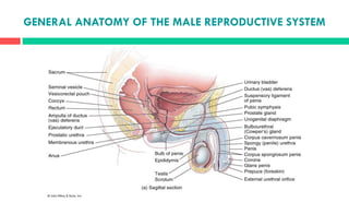
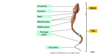
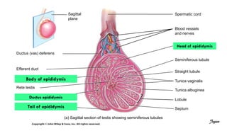
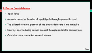
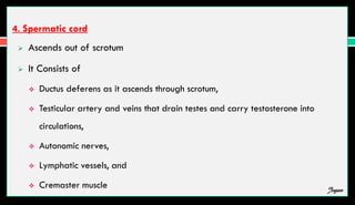
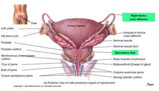
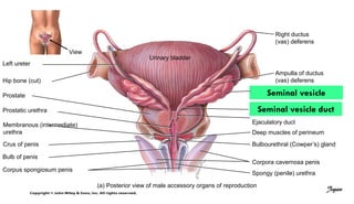
The male reproductive system includes the testes, duct system, and accessory sex glands. The testes produce sperm and testosterone. Sperm mature as they move through the duct system, which includes the epididymis and vas deferens. The ductus deferens joins the seminal vesicles and prostate to form the ejaculatory ducts, which empty into the urethra. During ejaculation, the seminal vesicles and prostate gland secrete fluids that combine with sperm to form semen. The scrotum and penis are supporting structures that regulate temperature and allow delivery of semen during intercourse.
2. The organs of male reproductive system include
1. Testes
2. A system of ducts
The epididymis,
Ductus deferens,
Ejaculatory ducts, and
Urethra
3. Accessory sex glands
Seminal vesicles,
Prostate, and
Bulbourethral glands
4. Supporting structures
Scrotum
Penis Jegan
5. SCROTUM
The scrotum the supporting structure for the testes consists of loose skin hanging from the
root (attached portion) of the penis
Externally, the scrotum looks like a single pouch of skin separated into lateral portions by a
median ridge called the raphe
Internally, the scrotal septum divides the scrotum into two sacs, each containing a single
testis
The septum is made up of a subcutaneous layer and muscle tissue called the dartos muscle
Jegan
6. Associated with each testis in the scrotum is the cremaster muscle, a series of
small bands of skeletal muscle that descend through the spermatic cord to
surround the testes.
The location of the scrotum and the contraction of its muscle fibers regulate
the temperature of the testes
Normal sperm production requires a temperature about 2–3 C below core
body temperature
Jegan
7. Anterior view of scrotum and testes and transverse section of penis
Cremaster muscle
Spermatic cord
Scrotal septum
Cremaster muscle
Dartos muscle
Skin of scrotum
Raphe
Jegan
8. PENIS
The penis contains the urethra and is a passageway for the ejaculation of
semen and excretion of urine
It is cylindrical in shape and consists of
a body,
glans penis, and
a root
Jegan
9. (a) Frontal section
Internal urethral orifice
External urethral orifice
Prostatic urethra
Bulbourethral (Cowper’s) gland
Deep muscles of perineum
BODY OF PENIS:
Corpora cavernosa penis
Corpus spongiosum penis
Spongy (penile) urethra
Urinary bladder
Prostate
Orifice of ejaculatory duct
Membranous urethra
ROOT OF PENIS:
Bulb of penis
Crus of penis
Frontal
plane
Corona
GLANS PENIS
Prepuce (foreskin)
Jegan
10. 1. THE BODY OF THE PENIS
The body of the penis is composed of three cylindrical masses of tissue
Tunica Albuginea
Corpora Cavernosa Penis
Corpus Spongiosum Penis
Jegan
11. (a) Frontal section
BODY OF PENIS:
Frontal
plane
Corpus spongiosum penis
Corpora cavernosa penis
Jegan
12.
13. 2. GLAN PENIS
The distal end of the corpus spongiosum penis is a slightly enlarged, acorn-
shaped region called the glans penis
Its margin is the corona
The distal urethra enlarges within the glans penis and forms a terminal
slitlike opening, the external urethral orifice.
Covering the glans in an uncircumcised penis is the loosely fitting prepuce
or foreskin.
Jegan
15. 3. THE ROOT OF THE PENIS
It is the attached proximal portion
It consists of the
Bulb of the penis
Crura of the penis
The bulb of the penis is attached to the inferior surface of the deep
muscles of the perineum and is enclosed by the bulbospongiosum muscle, a
muscle that aids ejaculation
Jegan
16. (a) Frontal section
Prostatic urethra
Bulbourethral (Cowper’s) gland
Deep muscles of perineum
Membranous urethra
ROOT OF PENIS:
Bulb of penis
Crus of penis
Frontal
plane
Jegan
18. TESTES OR TESTICLES
Paired oval glands in the scrotum
Develops near kidney and descends through inguinal canals near 7th month of fetal
development
Tunica vaginalis partially covers testes
Tunica albuginea – internal to tunica vaginalis
Tunica albuginea extends inward forming septa that divide testis into many lobules
Each of 200-300 lobules contains 1-3 seminiferous tubules
Sperm are produced here through spermatogenesis Jegan
19. (a) Sagittal section of testis showing seminiferous tubules
Blood vessels
and nerves
Head of epididymis
Seminiferous tubule
Straight tubule
Tunica vaginalis
Tunica albuginea
Lobule
Septum
Sagittal
plane
Jegan
20. SEMINIFEROUS TUBULE
The seminiferous tubules contain 3 types of cells:
1. Spermatogenic cells – sperm-forming cells
Spermatagonia (stem cell) develop from primordial germ cells that arise in yolk sac
and enter testes in 5th week of development
Primary spermatocytes → secondary spermatocytes → spermatids → sperm cells →
lumen
2. Sertoli cells or sustentacular cells– support cells
Tight junction form blood-testis barrier – prevents immune response against sperm cell
surface antigens
Jegan
21. Nourish spermatocytes, spermatids and sperm,
Phagocytize excess spermatid cytoplasm,
Control movements of spermatogenic cells, release sperm into lumen,
Produce fluid for sperm transport,
Secrete inhibin, regulate effects of testosterone and follicle-stimulating
hormone (FSH)
3. Leydig (interstitial) cells
It is found in spaces between seminiferous tubules
Secrete testosterone
Jegan
22. (b) Transverse section of part of seminiferous tubule
SPERMATOGENIC CELLS:
Spermatogonium (2n)
(stem cell)
Primary spermatocyte (2n)
Spermatid (n)
Sperm cell or
spermatozoon (n)
Secondary spermatocyte (n)
Leydig cell
Blood capillary
Basement membrane
Blood–testis barrier
(tight junction)
Lumen of seminiferous tubule
Sertoli cell nucleus
Jegan
23. SPERMATOGENESIS
Takes 65-75 days
Begins with spermatogonia – diploid (2n)
Spermatogonia are type of Stem cells which undergo mitosis to replace themselves
(reservior for future) and some continue development
Primary spermatocytes – diploid (2n)
Each duplicates its DNA and meiosis begins
Jegan
24. Meiosis I – homologous pairs line up, crossing over occurs
Secondary spermatocytes (haploid or n)
2 cells at end of Meiosis I
Each chromosome made up of 2 chromatids attached at centromere
Meiosis II – 2 chromatids separate
Spermatids – 4 haploid cells at end of meiosis II
Cells remain attached to each other by cytoplasmic bridges
Jegan
27. Spermatids Sperm cells
Secondary
spermatocyte
Second meiotic
division
First meiotic
division
(23 chromosomes, each
with 2 chromatids)
Paired homologous
chromosomes
(46 chromosomes,
each with 2 chromatids)
(23 chromosomes, each
with 2 chromatids) (23 chromosomes,
each chromatid now an
independent chromosome)
Primary
spermatocyte
Jegan
29. SPERMIOGENESIS
Development of spermatids into sperm
Spherical spermatids transform into elongated sperm
Acrosome and flagella form, mitochondria multiply
Sertoli cells dispose of excess cytoplasm
SPERMIATION – release from connections to Sertoli cells
Not yet able to swim
Jegan
30. SPERM
Each day about 300 million sperm complete spermatogenesis
The major part of sperm are head and tail
Head
4-5µm long
Nucleus with 23 chromosomes (haploid or n)
Acrosome – vesicle filled with oocyte penetrating enzymes
Jegan
31. Tail
Neck – contains centrioles forming microtubules that comprise remainder of tail
Middle piece – contains mitochondria
Principal piece – longest portion of tail
End piece – terminal, tapering portion of tail
Once ejaculated,
sperm do not
survive more than
48 hours in female
reproductive tract
Jegan
35. HORMONAL CONTROL OF TESTES
At puberty, secretion of gonadotropin-releasing hormone (GnRH) increases
GnRH Stimulates anterior pituitary to increase secretion of luteinizing hormone
(LH) and follicle-stimulating hormone (FSH)
LH stimulates Leydig cells to secrete testosterone
Synthesized from cholesterol mainly in testes
Suppresses secretion of LH and GnRH via negative feedback
Enzyme 5-alpha-reductase converts testosterone into dihydrotestosterone (DHT) in
external genitals and prostate
Jegan
36. FSH acts indirectly to stimulate spermatogenesis
FSH and testosterone act on Sertoli cells to stimulate secretion of androgen-binding
protein (ABP)
ABP binds testosterone keeping concentration high
Testosterone stimulates spermatogenesis
Sertoli cells release inhibin which inhibits FSH
Jegan
37. Hypothalamus
GnRH
Testosterone decreases
release of GnRH and LH
Anterior pituitary
Gonadotroph
LH stimulates
testosterone secretion
Together with
testosterone, FSH
stimulates
spermatogenesis
Inhibin decreases
release of FSH
Inhibin
ABP
Spermatogenic
cells
Sertoli cells secrete
androgenbinding
protein (ABP)
Dihydrotestosterone
(DHT)
Leydig cells secrete
testosterone
FSH LH
LH
LH receptor
FSH
FSH receptor
Testosterone
Androgen
receptor
Key:
• Male pattern of development (before birth)
• Enlargement of male sex organs and expression of
male secondary sex characteristics (starting at
puberty)
• Anabolism (protein synthesis)
Testosterone
Jegan
39. REPRODUCTIVE SYSTEM DUCTS IN MALES
1. Ducts of testis
Pressure generated by fluid produced by Sertoli cells push sperm along
seminiferous tubules into straight tubules
rete testis
efferent ducts in epididymis
ductus epididymis
Jegan
41. 2. Epididymis
Consists of tightly coiled ductus epididymis
Has Head, body, tail
Tail continues as ductus (vas) deferens
Stereocilia are microvilli that reabsorb degenerated sperm
It is site of sperm maturation – acquire motility and ability to fertilize
It can store sperm for several months
Jegan
42. (a) Sagittal section of testis showing seminiferous tubules
Spermatic cord
Blood vessels
and nerves
Head of epididymis
Seminiferous tubule
Straight tubule
Tunica vaginalis
Tunica albuginea
Lobule
Septum
Sagittal
plane
Ductus (vas) deferens
Efferent duct
Rete testis
Body of epididymis
Tail of epididymis
Ductus epididymis
Jegan
43. 3. Ductus (vas) deferens
45cm long
Ascends posterior border of epididymis through spermatic cord
The dilated terminal portion of the ductus deferens is the ampulla
Conveys sperm during sexual arousal through peristaltic contractions
Can also store sperm for several months
Jegan
44. 4. Spermatic cord
Ascends out of scrotum
It Consists of
Ductus deferens as it ascends through scrotum,
Testicular artery and veins that drain testes and carry testosterone into
circulations,
Autonomic nerves,
Lymphatic vessels, and
Cremaster muscle Jegan
45. View
Left ureter
Urinary bladder
Hip bone (cut)
Prostate
Prostatic urethra
Membranous (intermediate)
urethra
Crus of penis
Bulb of penis
Corpus spongiosum penis
Right ductus
(vas) deferens
Ampulla of ductus
(vas) deferens
Seminal vesicle
Seminal vesicle duct
Ejaculatory duct
Deep muscles of perineum
Bulbourethral (Cowper’s) gland
Corpora cavernosa penis
Spongy (penile) urethra
(a) Posterior view of male accessory organs of reproduction
Jegan
46. 5. Urethra
20cm long
Shared terminal duct of reproductive and urinary systems
Subdivided into
Prostatic urethra. 2-3 cm long
Membranous urethra. 1 cm long
Spongy (penile) urethra. 15-20 cm long
Ends at external urethral orifice
Jegan
47. View
Urinary bladder
Spongy (penile) urethra
(a) Posterior view of male accessory organs of reproduction
Prostatic urethra
Membranous
(intermediate) urethra
Jegan
49. ACCESSORY SEX GLANDS
Accessory sex glands – secrete most of liquid portion of semen
1. Seminal vesicles
The paired seminal vesicles or seminal glands are convoluted pouchlike structures
About 5 cm (2 in.) in length
Lying posterior to the base of the urinary bladder and anterior to the rectum
Secrete alkaline, viscous fluid containing fructose, prostaglandins, and clotting proteins
(different from blood)
About 60% of semen volume Jegan
50. View
Left ureter
Urinary bladder
Hip bone (cut)
Prostate
Prostatic urethra
Membranous (intermediate)
urethra
Crus of penis
Bulb of penis
Corpus spongiosum penis
Right ductus
(vas) deferens
Ampulla of ductus
(vas) deferens
Seminal vesicle
Seminal vesicle duct
Ejaculatory duct
Deep muscles of perineum
Bulbourethral (Cowper’s) gland
Corpora cavernosa penis
Spongy (penile) urethra
(a) Posterior view of male accessory organs of reproduction
Jegan
51. 2. Prostate
The prostate is a single, doughnut-shape gland
It measures about 4 cm from side to side, about 3 cm from top to bottom.
It is inferior to the urinary bladder and surrounds the prostatic urethra
Secretes milky, slightly acidic fluid containing
Citric acid,
Several proteolytic enzymes ( lysozyme, amylase, hyaluronidase),
Acid phosphatase,
Seminalplasmin (antibiotic)
About 25% of semen volume
Jegan
52. View
Left ureter
Urinary bladder
Hip bone (cut)
Prostatic urethra
Membranous (intermediate)
urethra
Crus of penis
Bulb of penis
Corpus spongiosum penis
Right ductus
(vas) deferens
Ampulla of ductus
(vas) deferens
Seminal vesicle
Seminal vesicle duct
Ejaculatory duct
Deep muscles of perineum
Bulbourethral (Cowper’s) gland
Corpora cavernosa penis
Spongy (penile) urethra
(a) Posterior view of male accessory organs of reproduction
Prostate
Jegan
53. 3. Bulbourethral glands
The paired bulbourethral glands are also known as cowpers gland
They are located inferior to the prostate
During sexual arousal, the bulbourethral glands secrete an alkaline fluid into the urethra
that protects the passing sperm by neutralizing acids from urine in the urethra
They also secrete mucus that lubricates the end of the penis and the lining of the
urethra, decreasing the number of sperm damaged during ejaculation
Jegan
54. View
Left ureter
Urinary bladder
Hip bone (cut)
Prostate
Prostatic urethra
Membranous (intermediate)
urethra
Crus of penis
Bulb of penis
Corpus spongiosum penis
Right ductus
(vas) deferens
Ampulla of ductus
(vas) deferens
Seminal vesicle
Seminal vesicle duct
Ejaculatory duct
Deep muscles of perineum
Bulbourethral (Cowper’s)
gland
Corpora cavernosa penis
Spongy (penile) urethra
(a) Posterior view of male accessory organs of reproduction
Jegan
55. SEMEN
Mixture of sperm and seminal fluid
Typical volume 2.5-5 mL with 50-150 million sperm/mL
Slightly alkaline pH of 7.2-7.7 due to seminal vesicle secretions
Provides transport medium, nutrients, and protection
Coagulates within 5 mins after ejaculation due to clotting proteins
After about 10 to 20 minutes, semen reliquefies because prostate-specific antigen
(PSA) and other proteolytic enzymes produced by the prostate break down the clot
Jegan