Course: BBT221
Human Physiology
ArifulIslam, Ph.D.
Assistant Professor
Department of Biochemistry & Microbiology
North South University, Dhaka, Bangladesh
E-mail: islam.ariful02@northsouth.edu
Office: SAC832A
Lecture: 20
Reproductive system
2.
Reproductive system
➢ ReproductiveSystem
– It is not needed for the survival of the individual
– Its needed for the survival of species
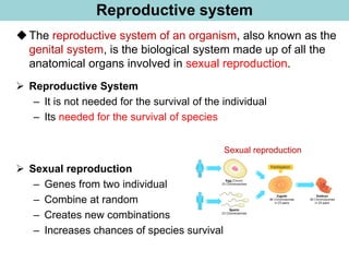
◆The reproductive system of an organism, also known as the
genital system, is the biological system made up of all the
anatomical organs involved in sexual reproduction.
Sexual reproduction
➢ Sexual reproduction
– Genes from two individual
– Combine at random
– Creates new combinations
– Increases chances of species survival
3.
Sexual Reproduction
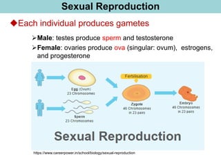
◆Each individualproduces gametes
➢Male: testes produce sperm and testosterone
➢Female: ovaries produce ova (singular: ovum), estrogens,
and progesterone
https://www.careerpower.in/school/biology/sexual-reproduction
4.
Male Reproduction system
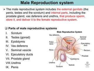
➢The male reproductive system includes the external genitals (the
penis, testes and the scrotum) and internal parts, including the
prostate gland, vas deferens and urethra, that produce sperm,
store it, and deliver it to the female reproductive system.
❑ Parts of male reproductive systems
I. Scrotum
II. Testes (gonad)
III. Epididymis
IV. Vas deferens
V. Seminal vesicle
VI. Ejaculatory ducts
VII. Prostate gland
VIII.Urethra
IX. Penis
5.
Parts of MaleReproduction system
I. Scrotum: this is a loose bag of skin that hangs behind the penis.
It holds the testes in place.
II. Testes: they sit in the scrotum, and produce sperm and
testosterone.
III. Epididymis: it is highly coiled tube that lies at the back of the
testes. Sperm from the testes must pass through the epididymis,
where they mature and start to 'swim'.
Continue…
IV. Vas deferens: this is a thick-walled tube that carries sperm from
the epididymis up to the prostate glands and urethra.
V. Seminal vesicle: these are 2
small glands above the prostate
gland that make up much of the
fluid in semen.
https://www.macmillan.org.uk/cancer-information-
and-support/testicular-cancer/the-testicles
6.
Parts of MaleReproduction system
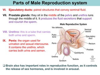
VI. Ejaculatory ducts: paired structures that convey seminal fluid.
VII. Prostate glands: they sit in the middle of the pelvis and urethra runs
through the middle of it. It produces the fluid secretions that support
and nourish the sperm.
❑ Brain also has important roles in reproductive function, as it controls
the release of sex hormones, and is involved in arousal.
VIII. Urethra: this is a tube that carries
both urine and sperm.
IX. Penis: the organ used for
urination and sexual intercourse.
It contains the urethra, which
carries both urine and semen.
7.
Male Reproduction system:Testes
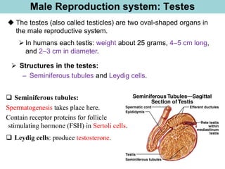
➢ Structures in the testes:
– Seminiferous tubules and Leydig cells.
◆ The testes (also called testicles) are two oval-shaped organs in
the male reproductive system.
➢ In humans each testis: weight about 25 grams, 4–5 cm long,
and 2–3 cm in diameter.
❑ Seminiferous tubules:
Spermatogenesis takes place here.
Contain receptor proteins for follicle
stimulating hormone (FSH) in Sertoli cells.
❑ Leydig cells: produce testosterone.
8.
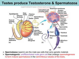
Testes produce Testosterone& Spermatozoa
➢ Spermatozoa (sperm) are the male sex cells that carry genetic material.
➢ Spermatogonia: undifferentiated male germ cell that undergo spermatogenesis
to form mature spermatozoa in the seminiferous tubules of the testis.
9.
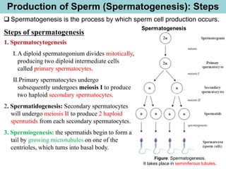
Production of Sperm(Spermatogenesis): Steps
Steps of spermatogenesis
1. Spermatocytogenesis
I.A diploid spermatogonium divides mitotically,
producing two diploid intermediate cells
called primary spermatocytes.
II.Primary spermatocytes undergo
subsequently undergoes meiosis I to produce
two haploid secondary spermatocytes.
2. Spermatidogenesis: Secondary spermatocytes
will undergo meiosis II to produce 2 haploid
spermatids from each secondary spermatocytes.
3. Spermiogenesis: the spermatids begin to form a
tail by growing microtubules on one of the
centrioles, which turns into basal body.
Figure: Spermatogenesis.
It takes place in seminiferous tubules.
❑ Spermatogenesis is the process by which sperm cell production occurs.
10.
Spermatogenesis in seminiferoustubules
(Visualizing Human Biology_Kathleen_3rd edition, Page: 490)
Transverse section of several
seminiferous tubules
Spermatogenesis in
seminiferous tubules
[No question from this
slide in the final exam]
11.
Structure of Spermatozoa(sperm)
◆The three anatomical features of a sperm cell are:
1. Head
2. Mid-piece
3. Tail or the flagellum
1. Head: it contains the genetic material and acrosome. Acrosome contains
digestive enzymes (including hyaluronidase and acrosin). These enzymes
break down the outer membrane of the ovum, called the zona pellucida,
allowing the haploid nucleus in the sperm cell to join with the haploid
nucleus in the ovum.
2. Mid piece: it contains the mitochondria. It provides motility, and hence
is called the powerhouse of the sperm.
3. Tail: it flagellum gives movement to the spermatozoa.
12.
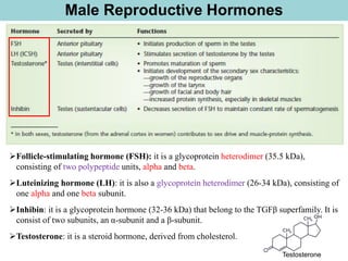
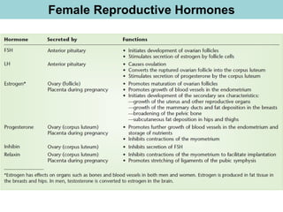
Male Reproductive Hormones
➢Follicle-stimulatinghormone (FSH): it is a glycoprotein heterodimer (35.5 kDa),
consisting of two polypeptide units, alpha and beta.
➢Luteinizing hormone (LH): it is also a glycoprotein heterodimer (26-34 kDa), consisting of
one alpha and one beta subunit.
➢Inhibin: it is a glycoprotein hormone (32-36 kDa) that belong to the TGFβ superfamily. It is
consist of two subunits, an α-subunit and a β-subunit.
➢Testosterone: it is a steroid hormone, derived from cholesterol.
Testosterone
13.
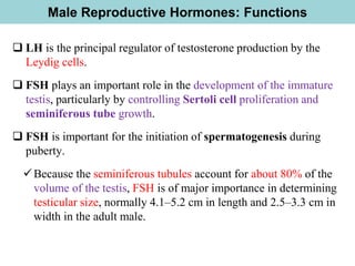
Male Reproductive Hormones:Functions
❑ LH is the principal regulator of testosterone production by the
Leydig cells.
❑ FSH plays an important role in the development of the immature
testis, particularly by controlling Sertoli cell proliferation and
seminiferous tube growth.
❑ FSH is important for the initiation of spermatogenesis during
puberty.
✓Because the seminiferous tubules account for about 80% of the
volume of the testis, FSH is of major importance in determining
testicular size, normally 4.1–5.2 cm in length and 2.5–3.3 cm in
width in the adult male.
14.
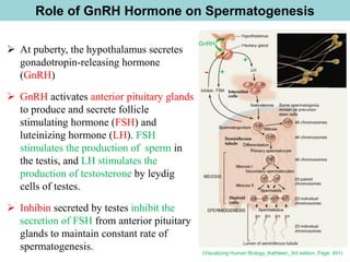
Role of GnRHHormone on Spermatogenesis
(Visualizing Human Biology_Kathleen_3rd edition, Page: 491)
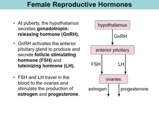
➢ At puberty, the hypothalamus secretes
gonadotropin-releasing hormone
(GnRH)
➢ GnRH activates anterior pituitary glands
to produce and secrete follicle
stimulating hormone (FSH) and
luteinizing hormone (LH). FSH
stimulates the production of sperm in
the testis, and LH stimulates the
production of testosterone by leydig
cells of testes.
➢ Inhibin secreted by testes inhibit the
secretion of FSH from anterior pituitary
glands to maintain constant rate of
spermatogenesis.
- +
+
GnRH
15.
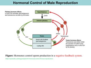
Hormonal Control ofMale Reproduction
Figure: Hormones control sperm production in a negative feedback system.
(https://opentextbc.ca/biology/chapter/24-4-hormonal-control-of-human-reproduction/)
16.
Female Reproductive Systems
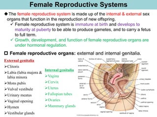
Internalgenitalia
➢Vagina
➢Cervix
➢Uterus
➢Fallopian tubes
➢Ovaries
➢Mammary glands
◆The female reproductive system is made up of the internal & external sex
organs that function in the reproduction of new offspring.
✓ Female reproductive system is immature at birth and develops to
maturity at puberty to be able to produce gametes, and to carry a fetus
to full term.
✓ Growth, development, and function of female reproductive organs are
under hormonal regulation.
Female reproductive organs: external and internal genitalia.
External genitalia
➢Clitoris
➢Labia (labia majora &
labia minora
➢Mons pubis
➢Vulval vestibule
➢Urinary meatus
➢Vaginal opening
➢Hymen
➢Vestibular glands
17.
Parts of TheFemale Reproductive Systems
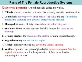
❑ External genitalia: Are collectively called the vulva.
1. Clitoris: a small, sensitive protrusion that’s is very sensitive to stimulation.
2. Labia: labia majora protect other parts of the vulva and the labia minora
protects the vestibule from dryness, infections and irritation.
3. Mons pubis: a mass of fatty tissue where the pubic hair grows.
4. Vulval vestibule: an area between the labia minora that contains the
openings.
5. Urinary meatus: the opening of the urethra for urine to pass through.
6. Vaginal opening: entrance to the vagina.
7. Hymen: connective tissue that covers the vaginal opening.
8. Vestibular glands: two pairs of glands that produce a mucous fluid for
vaginal lubrication, and for the ejaculation of fluid as well as for
lubricating the meatus.
18.
Parts of TheFemale Reproductive Systems
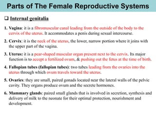
❑ Internal genitalia
1. Vagina: it is a fibromuscular canal leading from the outside of the body to the
cervix of the uterus. It accommodates a penis during sexual intercourse.
2. Cervix: it is the neck of the uterus, the lower, narrow portion where it joins with
the upper part of the vagina.
3. Uterus: it is a pear-shaped muscular organ present next to the cervix. Its major
function is to accept a fertilized ovum, & pushing out the fetus at the time of birth.
4. Fallopian tubes (fallopian tubes): two tubes leading from the ovaries into the
uterus through which ovum travels toward the uterus.
5. Ovaries: they are small, paired gonads located near the lateral walls of the pelvic
cavity. They organs produce ovum and the secrete hormones.
6. Mammary glands: paired small glands that is involved in secretion, synthesis and
delivery of milk to the neonate for their optimal protection, nourishment and
development.
19.
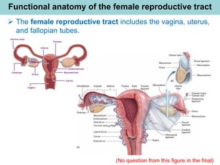
Functional anatomy ofthe female reproductive tract
➢ The female reproductive tract includes the vagina, uterus,
and fallopian tubes.
(No question from this figure in the final)
20.
Female reproductive organs:Ovaries
◆The principal female reproductive organs are ovaries.
✓ They are small, oval-shaped glands located on either side of uterus. Each
ovary has thousands of ovarian follicles (small sacs in the ovaries that
hold immature eggs).
✓ They produce and store eggs (ovum) and make hormones that control
menstrual cycle and pregnancy.
✓ During ovulation, one ovary releases an egg.
✓ Ovaries continue to release an egg each menstrual cycle until menopause.
❑ The ovaries consist of 2 layers;
A.An outer cortex layer
B.The inner medulla
21.
Female reproductive organs:Ovaries
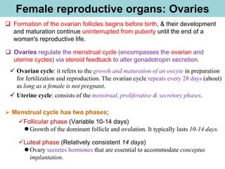
❑ Formation of the ovarian follicles begins before birth, & their development
and maturation continue uninterrupted from puberty until the end of a
woman's reproductive life.
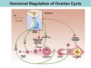
❑ Ovaries regulate the menstrual cycle (encompasses the ovarian and
uterine cycles) via steroid feedback to alter gonadotropin secretion.
✓ Ovarian cycle: it refers to the growth and maturation of an oocyte in preparation
for fertilization and reproduction. The ovarian cycle repeats every 28 days (about)
as long as a female is not pregnant.
✓ Uterine cycle: consists of the menstrual, proliferative & secretory phases.
➢ Menstrual cycle has two phases;
✓Follicular phase (Variable 10-14 days)
⚫ Growth of the dominant follicle and ovulation. It typically lasts 10-14 days.
✓Luteal phase (Relatively consistent 14 days)
⚫ Ovary secretes hormones that are essential to accommodate conceptus
implantation.
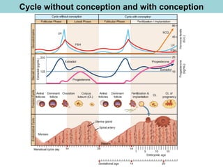
22.
Menstrual cycle: Phasesof ovarian cycle
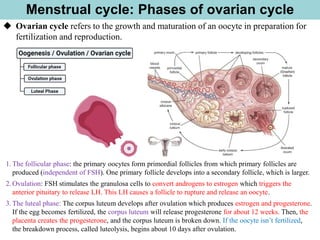
◆ Ovarian cycle refers to the growth and maturation of an oocyte in preparation for
fertilization and reproduction.
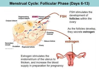
1.The follicular phase: the primary oocytes form primordial follicles from which primary follicles are
produced (independent of FSH). One primary follicle develops into a secondary follicle, which is larger.
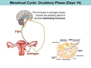
2.Ovulation: FSH stimulates the granulosa cells to convert androgens to estrogen which triggers the
anterior pituitary to release LH. This LH causes a follicle to rupture and release an oocyte.
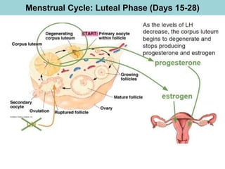
3.The luteal phase: The corpus luteum develops after ovulation which produces estrogen and progesterone.
If the egg becomes fertilized, the corpus luteum will release progesterone for about 12 weeks. Then, the
placenta creates the progesterone, and the corpus luteum is broken down. If the oocyte isn’t fertilized,
the breakdown process, called luteolysis, begins about 10 days after ovulation.
23.
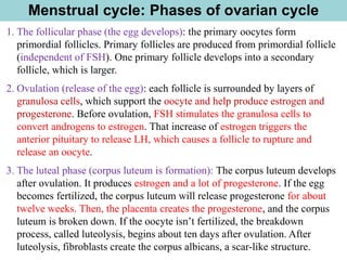
1. The follicularphase (the egg develops): the primary oocytes form
primordial follicles. Primary follicles are produced from primordial follicle
(independent of FSH). One primary follicle develops into a secondary
follicle, which is larger.
2. Ovulation (release of the egg): each follicle is surrounded by layers of
granulosa cells, which support the oocyte and help produce estrogen and
progesterone. Before ovulation, FSH stimulates the granulosa cells to
convert androgens to estrogen. That increase of estrogen triggers the
anterior pituitary to release LH, which causes a follicle to rupture and
release an oocyte.
3. The luteal phase (corpus luteum is formation): The corpus luteum develops
after ovulation. It produces estrogen and a lot of progesterone. If the egg
becomes fertilized, the corpus luteum will release progesterone for about
twelve weeks. Then, the placenta creates the progesterone, and the corpus
luteum is broken down. If the oocyte isn’t fertilized, the breakdown
process, called luteolysis, begins about ten days after ovulation. After
luteolysis, fibroblasts create the corpus albicans, a scar-like structure.
Menstrual cycle: Phases of ovarian cycle
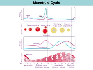
Gonadotropin Regulation ofOvarian Function
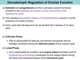
❑ Estradiol and progesterone are the 2 principal ovarian hormones
involved in the regulation of ovarian function and control of the
reproductive cycle.
❑ The variations in pulsatile release of the gonadotropins result in a cyclic
response of ovarian function.
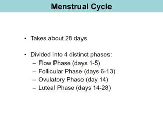
❑ Each cycle lasts 28 days and can be divided into 2 phases of 14 days
each.
❑ Follicular Phase
➢ FSH is responsible for follicular recruitment and growth and for
estrogen synthesis during the follicular phase of the ovarian cycle
❑ Luteal Phase
➢ LH is responsible for ovulation and corpus luteum formation and for
progesterone and estrogen production by the corpus luteum during
the early and mid-luteal phases of the menstrual cycle.
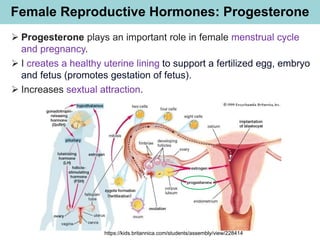
Female Reproductive Hormones:Progesterone
➢ Progesterone plays an important role in female menstrual cycle
and pregnancy.
➢ I creates a healthy uterine lining to support a fertilized egg, embryo
and fetus (promotes gestation of fetus).
➢ Increases sextual attraction.
https://kids.britannica.com/students/assembly/view/228414


![Spermatogenesis in seminiferous tubules
(Visualizing Human Biology_Kathleen_3rd edition, Page: 490)
Transverse section of several
seminiferous tubules
Spermatogenesis in
seminiferous tubules
[No question from this
slide in the final exam]](https://image.slidesharecdn.com/lecture20reproductivesystemarim250321-250424132241-4661571a/85/Lecture-20_Reproductive-System_ArIm_250321-pdf-10-320.jpg)


![ONFH[AVN HIP] -TRIPLE REGIME -A NOVAL SURGICAL CONCEPT .pptx](https://cdn.slidesharecdn.com/ss_thumbnails/onfhavnhip2026koaconcalicutdrgokuldevdrmashraf-260210064517-213ec005-thumbnail.jpg?width=640&height=640&fit=bounds)


![PERI-PROSTHETIC FRACTURE NAIL-PLATE CONSTRUCT [NPC].pptx](https://cdn.slidesharecdn.com/ss_thumbnails/drarunkumardrmohamedashrafperiprostheticfrasturenail-plateconstructnpc-260209164459-7e9d15a1-thumbnail.jpg?width=640&height=640&fit=bounds)
