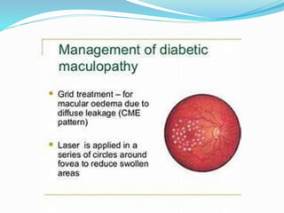
Dr. Prabhakara Sastry,
M.D. (Ophthal), AIIMS.
Professor, Andhra Medical College, Visakhapatnam
Symptoms of Macular Edema
 It’s a painless condition and usually
asymptomatic in the early stages. Patients
may later develop
 Blurred or wavy central vision
 Colors may appear different
 May experience difficulty in reading
Causes of Macular Edema
Diabetes:
 High blood sugar levels due to Diabetes causes leaky
blood vessels at the macula.
Age related Macular Degeneration:
 Here abnormal blood vessels leak fluid and cause
macular swelling.
Retinal Vein oclussions:
 When the veins in the retina become blocked, blood
and fluid then leak out into the macula.
Causes of Macular Edema
Vitreomacular traction(VMT)
Genetic/Hereditary disorders:
 such as Retinoschisis or Retinitis Pigmentosa.
Inflammatory eye diseases:
 Conditions like uveitis, where the body attacks its own
tissues, can damage retinal blood vessels and cause
swelling of the macula.
Medication:
 Certain drugs have side effects that can lead to
macular edema.
Causes of Macular Edema
Eye Malignancies:
 Both benign and malignant tumors can lead to
macular edema.
Eye surgery:
 It’s not common, but sometimes after glaucome,
retinal or cataract surgery, you can get macular edema.
Injuries:
 Trauma to the eye.
Macular Edema Risk Factors
 Metabolic conditions (diabetes)
 Blood vessel diseases (vein occlusion/blockage)
 Aging (macular degeneration)
 Hereditary diseases (retinitis pigmentosa)
 Traction on the macula (macular hole, macular
pucker, and vitreomacular traction)
 Inflammatory conditions (sarcoidosis, uveitis)
Macular Edema Risk Factors
 Toxicity
 Neoplastic conditions (eye tumors)
 Trauma
 Surgical causes (following eye surgery)
 Unknown (idiopathic) causes
Macular Edema Prevention
 Anyone with diabetes should have their eyes
checked annually, at the least.
 People with family history or underlying genetic
condition can have a yearly eye examination.
Macular Edema Diagnosis
 A routine dilated fundus examination by
the ophthalmologist aids in the diagnosis. Further
tests can be ordered to document and measure the
thickness of the macula.
Optical Coherence Tomography (OCT):
 It scans the retina and provides very detailed
images of its thickness. This helps your doctor find
leakage and measure swelling of the macula.It can
also be used to follow the response to treatment
Macular Edema Diagnosis
Fundus Fluorescein Angiography(FFA):
 For this test, fluorescein dye is injected into a
peripheral vein in the hand or forearm. A series of
photographs is taken of the retina as the dye passes
through its blood vessels
Macular Edema Treatment
 First and foremost is addressing the underlying
cause of the macular edema, and the related
leakage and retinal swelling.
 Treatment can include:
Topical NSAIDS:
 Non Steroidal anti inflammatory drugs can be
given as eye drops to cure the swelling.
Macular Edema Treatment
Steroid treatment:
 When macular edema is caused by inflammation,
steroids can be given either as drops,tablets or as
injections into the eye.
Intravitreal injections:
 Anti Vascular Endothelial Growth Factor( Anti-
VEGF)medicines given as intravitreal injections
into the eye helps to reduce the growth of
abnormal blood vessels in the retina, and also
decreases leaking from blood vessels.
Macular Edema Treatment
Laser treatment:
 With this tiny laser pulses are applied to the areas
of fluid leakage around the macula. The goal is to
stabilize vision by sealing off leaking blood vessels
Vitrectomy surgery:
 When macular edema is caused by vitreous pulling
on the macula, a procedure called a vitrectomy
may be needed to restore the macula to its normal
(lying flat) shape.
THANK
YOU

Macular Edema.pptx

  • 1.
    Dr. Prabhakara Sastry, M.D.(Ophthal), AIIMS. Professor, Andhra Medical College, Visakhapatnam
  • 2.
    Symptoms of MacularEdema  It’s a painless condition and usually asymptomatic in the early stages. Patients may later develop  Blurred or wavy central vision  Colors may appear different  May experience difficulty in reading
  • 3.
    Causes of MacularEdema Diabetes:  High blood sugar levels due to Diabetes causes leaky blood vessels at the macula. Age related Macular Degeneration:  Here abnormal blood vessels leak fluid and cause macular swelling. Retinal Vein oclussions:  When the veins in the retina become blocked, blood and fluid then leak out into the macula.
  • 4.
    Causes of MacularEdema Vitreomacular traction(VMT) Genetic/Hereditary disorders:  such as Retinoschisis or Retinitis Pigmentosa. Inflammatory eye diseases:  Conditions like uveitis, where the body attacks its own tissues, can damage retinal blood vessels and cause swelling of the macula. Medication:  Certain drugs have side effects that can lead to macular edema.
  • 5.
    Causes of MacularEdema Eye Malignancies:  Both benign and malignant tumors can lead to macular edema. Eye surgery:  It’s not common, but sometimes after glaucome, retinal or cataract surgery, you can get macular edema. Injuries:  Trauma to the eye.
  • 6.
    Macular Edema RiskFactors  Metabolic conditions (diabetes)  Blood vessel diseases (vein occlusion/blockage)  Aging (macular degeneration)  Hereditary diseases (retinitis pigmentosa)  Traction on the macula (macular hole, macular pucker, and vitreomacular traction)  Inflammatory conditions (sarcoidosis, uveitis)
  • 7.
    Macular Edema RiskFactors  Toxicity  Neoplastic conditions (eye tumors)  Trauma  Surgical causes (following eye surgery)  Unknown (idiopathic) causes
  • 8.
    Macular Edema Prevention Anyone with diabetes should have their eyes checked annually, at the least.  People with family history or underlying genetic condition can have a yearly eye examination.
  • 9.
    Macular Edema Diagnosis A routine dilated fundus examination by the ophthalmologist aids in the diagnosis. Further tests can be ordered to document and measure the thickness of the macula. Optical Coherence Tomography (OCT):  It scans the retina and provides very detailed images of its thickness. This helps your doctor find leakage and measure swelling of the macula.It can also be used to follow the response to treatment
  • 10.
    Macular Edema Diagnosis FundusFluorescein Angiography(FFA):  For this test, fluorescein dye is injected into a peripheral vein in the hand or forearm. A series of photographs is taken of the retina as the dye passes through its blood vessels
  • 11.
    Macular Edema Treatment First and foremost is addressing the underlying cause of the macular edema, and the related leakage and retinal swelling.  Treatment can include: Topical NSAIDS:  Non Steroidal anti inflammatory drugs can be given as eye drops to cure the swelling.
  • 12.
    Macular Edema Treatment Steroidtreatment:  When macular edema is caused by inflammation, steroids can be given either as drops,tablets or as injections into the eye. Intravitreal injections:  Anti Vascular Endothelial Growth Factor( Anti- VEGF)medicines given as intravitreal injections into the eye helps to reduce the growth of abnormal blood vessels in the retina, and also decreases leaking from blood vessels.
  • 13.
    Macular Edema Treatment Lasertreatment:  With this tiny laser pulses are applied to the areas of fluid leakage around the macula. The goal is to stabilize vision by sealing off leaking blood vessels Vitrectomy surgery:  When macular edema is caused by vitreous pulling on the macula, a procedure called a vitrectomy may be needed to restore the macula to its normal (lying flat) shape.
  • 19.