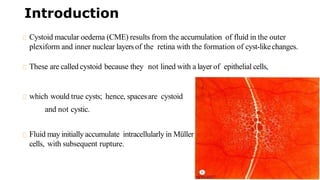
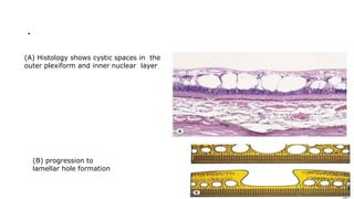
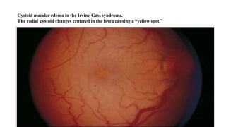
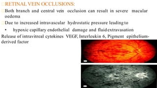
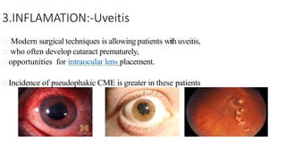
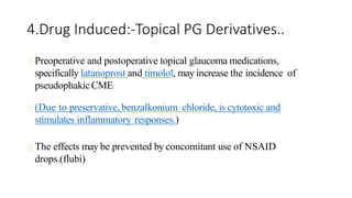
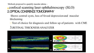
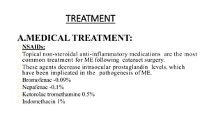
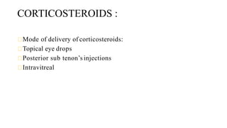
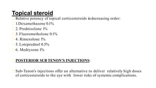
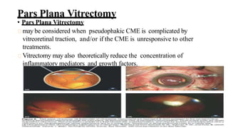
Cystoid Macula
Edema-Causes &Manegment
By:-Dr.Rajesh Kumar Shakya
Deptt of Ophthalmology
Junior resident –second year
M.L.B Mediacal College Jhansi
u.p
Introduction
Cystoid macular oedema (CME) results from the accumulation of fluid in the outer
plexiform and inner nuclear layersof the retina with the formation of cyst-likechanges.
These are called cystoid because they not lined with a layer of epithelial cells,
which would true cysts; hence, spacesare cystoid
and not cystic.
Fluid may initially accumulate intracellularly in Müller
cells, with subsequent rupture.
.
(A) Histology shows cystic spaces in the
outer plexiform and inner nuclear layer
(B) progression to
lamellar hole formation
Pathogenesis & Etiology
Intraretinal fluid may accumulate into CME by:
• Breakdown of normal anatomical barriers:
a) inner blood retinal barrier
b) outer blood retinal barrier
• inflammatory mediators such as
• prostaglandins, leukotrienes, protein kinase C, nitric oxide, vascular endothelial
• growth factor, various other cytokines.
Pathophysiology:-
INFLAMMATION:
Ischaemia and inflammation
Intravitreal cytokine release
Increased permeability of retinal vascular endothelium
Development of Cystoid Macular Oedema
1. (Irvine-GassSyndrome)
POSTOPERATIVE:
Most commonly after cataractsurgery
Highest risk after inadvertent rupture of posterior capsule and/or
persistent traction to anterior segment structures
Also seen after penetrating keratoplasty, scleral buckling, laser
iridotomy, cryotherapy forretinal break, panretinal photocoagulation.
Angiographic CME after intracapsular cataract
extraction: As high as 60%*
Angiographic CME after extracapsular cataract
extraction: 15% to 30%
Clinical CME after small incision phacoemulsification:
0.1% to 2.35%
OCT evidence of CME after small incision
phacoemulsification: 4% to 11%, but also reported to be
as high as 41%
Most patients with CME found via angiography or OCT willnot
have visual changes.
Furthermore, most patients with clinical CME will experience
spontaneous improvement by 3 to 12 months
Cystoid macular edema in the Irvine-Gass syndrome.
The radial cystoid changes centered in the fovea causing a “yellow spot.”
2.Retinal Vascular Disease:-DR,RVD.
DIABETIC RETINOPATHY:
Due to vascular compromise
CME occurs along with diffuse macular oedema
Associated microaneurysms and hard exudates are often evident
RETINAL VEIN OCCLUSIONS:
Both branch and central vein occlusion can result in severe macular
oedema
Due to increased intravascular hydrostatic pressure leading to
• hypoxic capillary endothelial damage and fluid extravasation
Release of intravitreal cytokines VEGF, Interleukin 6, Pigment epithelium-
derived factor
3.INFLAMATION:-Uveitis
Modern surgical techniques is allowing patients with uveitis,
who often develop cataract prematurely,
opportunities for intraocular lens placement.
Incidence of pseudophakic CME is greater in these patients
4.Drug Induced:-Topical PG Derivatives..
Preoperative and postoperative topical glaucoma medications,
specifically latanoprost and timolol, may increase the incidence of
pseudophakicCME.
(Due to preservative, benzalkonium chloride, is cytotoxic and
stimulates inflammatory responses.)
The effects may be prevented by concomitant use of NSAID
drops.(flubi)
5.CNV
CHOROIDAL NEOVASCULAR MEMBRANE:
Exudation of fluid from the capillaries of CNVM.
6.Others causes:-
• Retinal dystrophies:- Retinitis pigmentosa.
• Conditions involving vitreomacular traction:-ERM
• Fundus tumours :-Retinal capillary haemangioma.
• Systemic disease:-Chronic renal failure.
Management:- (DIAGNOSIS & Treatment)
• History and Clinical Presentation
Develop approximately 4-6 weeks postoperatively.
Blurry vision .
Others central scotomas, metamorphopsia, and mild photophobia.
On biomicroscopy, retinal thickening and loss of the foveal depression is
usually appreciated.
findings are best observed with fundus contact lens, red-free light may aide
in demonstrating cystic changes.
In severe or chronic cases, optic disc swelling and/or a lamellar
• hole may also be seen.
Countinue…..
• On fluorescein angiography, pseudophakic CME is characterized by
retinal telangiectasis, capillary dilatation, and leakage from perifoveal
capillaries in the early phase frames, and perifoveal hyperfluorescent
spots classically described as a “petaloid” pattern in late phase frames,
representing fluorescein accumulation in cystic spaces.
• FAis the gold standard in diagnosing pseudophakic CME, but treatment
responses would be more conveniently monitored by biomicroscopy,
visual acuity and OCT.
Methods proposed to quantify macular edema -
1.confocal scanning laser ophthalmoscopy (SLO)
2..OPTICALCOHERENCETOMOGRAPHY
Shows central cysts, loss of foveal depressionand macular
thickening
Test of choice for diagnosis and follow up of patients with CME
3.RETINAL THICKNESS ANALYZER
TREATMENT
A.MEDICAL TREATMENT:
NSAIDs:
Topical non-steroidal anti-inflammatory medications are the most
common treatment for ME following cataract surgery.
These agents decrease intraocular prostaglandin levels, which
have been implicated in the pathogenesis of ME.
Bromofenac -0.09%
Nepafenac -0.1%
Ketorolac tromethamine 0.5%
Indomethacin 1%
CORTICOSTEROIDS :
Mode of delivery of corticosteroids:
Topical eye drops
Posterior sub tenon’s injections
Intravitreal
Topical steroid
Relative potency of topical corticosteroids in decreasing order:
1.Dexamethasone 0.1%
2. Prednisolone 1%
3. Fluorometholone 0.1%
4. Rimexolone 1%
5. Loteprednol 0.5%
6. Medrysone 1%
POSTERIOR SUB TENON’S INJECTIONS:
Sub-Tenon's injections offer an alternative to deliver relatively high doses
of corticosteroids to the eye with lower risks of systemic complications.
INTRAVITREAL TRIAMCINOLONE
ACETONIDE:
The use of intravitreal injection of triamcinolone acetonide
(Tricort) has increased due to its potent ability to ameliorate
refractory ME secondary to diabetes mellitus, retinal venous
occlusions, inflammation, and otheridiopathic causes.
.The effect is temporary and typically lasts for three to six months,
the ME usually responds to re-injection.
Systemic corticosteroids:-
• Sustained drug delivery systems (DDS) have been developed to address
this limitation of intravitreal corticosteroid injections.
• Ozurdex (Allergan, Irvine, CA) is an injectable, biodegradable intravitreal DDS
that provides sustained release of preservative- free dexamethasone, a
potent corticosteroid
Anti-VEGF Treatments
• Bevacizumab (Avastin, Genentech, South San Francisco, CA) is a humanized
• monoclonal antibody that inhibits VEGF-A.
• “Triple therapy” with intravitreal triamcinolone, intravitreal bevacizumab and
topical NSAIDs has been shown to be effective as well,although the effects of
the intravitreal medications were transient.
ACETAZOLAMIDE:
CAIs improve the pumping action of the retinal pigment
epithelium, to decrease intraretinal fluid.
initial dose of 250mg daily,increased to 500mg daily if no effect
• is apparent.
Immunomodulatory Therapy
• Recent small pilot studies have started to examine interferon alpha (IFN-a,
Imgenex, San Diego, CA) and intravitreal infliximab (Remicade, Centocor Ortho Biotech,
Horsham, PA) with mixed results.
HYPERBARIC
OXYGEN
Improvement in aphakic CME with hyperbaric oxygen therapy was
reported by Ploff and Thom*.
The dosage given was 2.2atm oxygen for 1.5h twice a day for 7 days,
followed by 2h daily for 14 days.The mechanism was hypothesized to be
macular capillary contraction.
B.Surgical Treatment
• Neodymium:YAG laser anterior vitreolysis can release vitreous
incarceration in the cataract incision wounds that complicate
pseudophakic CME.
Pars Plana Vitrectomy
• Pars Plana Vitrectomy
may be considered when pseudophakic CME is complicated by
vitreoretinal traction, and/or if the CME is unresponsive to other
treatments.
Vitrectomy may also theoretically reduce the concentration of
inflammatory mediators and growth factors.
DOMINANT CYSTOID MACULAR
EDEMA
• Extremely rare autosomal dominant disease mapped to chromosome 7q.
• Interestingly, autosomal dominant retinitis pigmentosa, known as RP9,
also maps to this region.
Unique macular disorder-
• the inner nuclear layer of retina is the site primarily affected.
Muller's cells are the specific cellular constituents thought to be
• Involved.
TREATMENT, COURSE, AND OUTCOME
Patients first begin to notice decreasing visual acuity at about 30 years of age, with
slowly progressive worsening to a moderate or severe level years later.
Advanced cases have maculae of atrophic appearance, with window defects seen on
fluorescein angiography.
As in allthe macular dystrophies, no known effective treatment.
…….
• …….

C M E ophthalmology book assessment .pptx

  • 1.
    Cystoid Macula Edema-Causes &Manegment By:-Dr.RajeshKumar Shakya Deptt of Ophthalmology Junior resident –second year M.L.B Mediacal College Jhansi u.p
  • 3.
    Introduction Cystoid macular oedema(CME) results from the accumulation of fluid in the outer plexiform and inner nuclear layersof the retina with the formation of cyst-likechanges. These are called cystoid because they not lined with a layer of epithelial cells, which would true cysts; hence, spacesare cystoid and not cystic. Fluid may initially accumulate intracellularly in Müller cells, with subsequent rupture.
  • 4.
    . (A) Histology showscystic spaces in the outer plexiform and inner nuclear layer (B) progression to lamellar hole formation
  • 5.
    Pathogenesis & Etiology Intraretinalfluid may accumulate into CME by: • Breakdown of normal anatomical barriers: a) inner blood retinal barrier b) outer blood retinal barrier • inflammatory mediators such as • prostaglandins, leukotrienes, protein kinase C, nitric oxide, vascular endothelial • growth factor, various other cytokines.
  • 6.
    Pathophysiology:- INFLAMMATION: Ischaemia and inflammation Intravitrealcytokine release Increased permeability of retinal vascular endothelium Development of Cystoid Macular Oedema
  • 7.
    1. (Irvine-GassSyndrome) POSTOPERATIVE: Most commonlyafter cataractsurgery Highest risk after inadvertent rupture of posterior capsule and/or persistent traction to anterior segment structures Also seen after penetrating keratoplasty, scleral buckling, laser iridotomy, cryotherapy forretinal break, panretinal photocoagulation.
  • 8.
    Angiographic CME afterintracapsular cataract extraction: As high as 60%* Angiographic CME after extracapsular cataract extraction: 15% to 30% Clinical CME after small incision phacoemulsification: 0.1% to 2.35% OCT evidence of CME after small incision phacoemulsification: 4% to 11%, but also reported to be as high as 41% Most patients with CME found via angiography or OCT willnot have visual changes. Furthermore, most patients with clinical CME will experience spontaneous improvement by 3 to 12 months
  • 9.
    Cystoid macular edemain the Irvine-Gass syndrome. The radial cystoid changes centered in the fovea causing a “yellow spot.”
  • 10.
    2.Retinal Vascular Disease:-DR,RVD. DIABETICRETINOPATHY: Due to vascular compromise CME occurs along with diffuse macular oedema Associated microaneurysms and hard exudates are often evident
  • 11.
    RETINAL VEIN OCCLUSIONS: Bothbranch and central vein occlusion can result in severe macular oedema Due to increased intravascular hydrostatic pressure leading to • hypoxic capillary endothelial damage and fluid extravasation Release of intravitreal cytokines VEGF, Interleukin 6, Pigment epithelium- derived factor
  • 12.
    3.INFLAMATION:-Uveitis Modern surgical techniquesis allowing patients with uveitis, who often develop cataract prematurely, opportunities for intraocular lens placement. Incidence of pseudophakic CME is greater in these patients
  • 13.
    4.Drug Induced:-Topical PGDerivatives.. Preoperative and postoperative topical glaucoma medications, specifically latanoprost and timolol, may increase the incidence of pseudophakicCME. (Due to preservative, benzalkonium chloride, is cytotoxic and stimulates inflammatory responses.) The effects may be prevented by concomitant use of NSAID drops.(flubi)
  • 14.
    5.CNV CHOROIDAL NEOVASCULAR MEMBRANE: Exudationof fluid from the capillaries of CNVM.
  • 15.
    6.Others causes:- • Retinaldystrophies:- Retinitis pigmentosa. • Conditions involving vitreomacular traction:-ERM • Fundus tumours :-Retinal capillary haemangioma. • Systemic disease:-Chronic renal failure.
  • 16.
    Management:- (DIAGNOSIS &Treatment) • History and Clinical Presentation Develop approximately 4-6 weeks postoperatively. Blurry vision . Others central scotomas, metamorphopsia, and mild photophobia. On biomicroscopy, retinal thickening and loss of the foveal depression is usually appreciated. findings are best observed with fundus contact lens, red-free light may aide in demonstrating cystic changes. In severe or chronic cases, optic disc swelling and/or a lamellar • hole may also be seen.
  • 17.
    Countinue….. • On fluoresceinangiography, pseudophakic CME is characterized by retinal telangiectasis, capillary dilatation, and leakage from perifoveal capillaries in the early phase frames, and perifoveal hyperfluorescent spots classically described as a “petaloid” pattern in late phase frames, representing fluorescein accumulation in cystic spaces. • FAis the gold standard in diagnosing pseudophakic CME, but treatment responses would be more conveniently monitored by biomicroscopy, visual acuity and OCT.
  • 18.
    Methods proposed toquantify macular edema - 1.confocal scanning laser ophthalmoscopy (SLO) 2..OPTICALCOHERENCETOMOGRAPHY Shows central cysts, loss of foveal depressionand macular thickening Test of choice for diagnosis and follow up of patients with CME 3.RETINAL THICKNESS ANALYZER
  • 19.
    TREATMENT A.MEDICAL TREATMENT: NSAIDs: Topical non-steroidalanti-inflammatory medications are the most common treatment for ME following cataract surgery. These agents decrease intraocular prostaglandin levels, which have been implicated in the pathogenesis of ME. Bromofenac -0.09% Nepafenac -0.1% Ketorolac tromethamine 0.5% Indomethacin 1%
  • 20.
    CORTICOSTEROIDS : Mode ofdelivery of corticosteroids: Topical eye drops Posterior sub tenon’s injections Intravitreal
  • 21.
    Topical steroid Relative potencyof topical corticosteroids in decreasing order: 1.Dexamethasone 0.1% 2. Prednisolone 1% 3. Fluorometholone 0.1% 4. Rimexolone 1% 5. Loteprednol 0.5% 6. Medrysone 1% POSTERIOR SUB TENON’S INJECTIONS: Sub-Tenon's injections offer an alternative to deliver relatively high doses of corticosteroids to the eye with lower risks of systemic complications.
  • 22.
    INTRAVITREAL TRIAMCINOLONE ACETONIDE: The useof intravitreal injection of triamcinolone acetonide (Tricort) has increased due to its potent ability to ameliorate refractory ME secondary to diabetes mellitus, retinal venous occlusions, inflammation, and otheridiopathic causes. .The effect is temporary and typically lasts for three to six months, the ME usually responds to re-injection.
  • 23.
    Systemic corticosteroids:- • Sustaineddrug delivery systems (DDS) have been developed to address this limitation of intravitreal corticosteroid injections. • Ozurdex (Allergan, Irvine, CA) is an injectable, biodegradable intravitreal DDS that provides sustained release of preservative- free dexamethasone, a potent corticosteroid
  • 24.
    Anti-VEGF Treatments • Bevacizumab(Avastin, Genentech, South San Francisco, CA) is a humanized • monoclonal antibody that inhibits VEGF-A. • “Triple therapy” with intravitreal triamcinolone, intravitreal bevacizumab and topical NSAIDs has been shown to be effective as well,although the effects of the intravitreal medications were transient.
  • 25.
    ACETAZOLAMIDE: CAIs improve thepumping action of the retinal pigment epithelium, to decrease intraretinal fluid. initial dose of 250mg daily,increased to 500mg daily if no effect • is apparent.
  • 26.
    Immunomodulatory Therapy • Recentsmall pilot studies have started to examine interferon alpha (IFN-a, Imgenex, San Diego, CA) and intravitreal infliximab (Remicade, Centocor Ortho Biotech, Horsham, PA) with mixed results.
  • 27.
    HYPERBARIC OXYGEN Improvement in aphakicCME with hyperbaric oxygen therapy was reported by Ploff and Thom*. The dosage given was 2.2atm oxygen for 1.5h twice a day for 7 days, followed by 2h daily for 14 days.The mechanism was hypothesized to be macular capillary contraction.
  • 28.
    B.Surgical Treatment • Neodymium:YAGlaser anterior vitreolysis can release vitreous incarceration in the cataract incision wounds that complicate pseudophakic CME.
  • 29.
    Pars Plana Vitrectomy •Pars Plana Vitrectomy may be considered when pseudophakic CME is complicated by vitreoretinal traction, and/or if the CME is unresponsive to other treatments. Vitrectomy may also theoretically reduce the concentration of inflammatory mediators and growth factors.
  • 30.
    DOMINANT CYSTOID MACULAR EDEMA •Extremely rare autosomal dominant disease mapped to chromosome 7q. • Interestingly, autosomal dominant retinitis pigmentosa, known as RP9, also maps to this region. Unique macular disorder- • the inner nuclear layer of retina is the site primarily affected. Muller's cells are the specific cellular constituents thought to be • Involved.
  • 31.
    TREATMENT, COURSE, ANDOUTCOME Patients first begin to notice decreasing visual acuity at about 30 years of age, with slowly progressive worsening to a moderate or severe level years later. Advanced cases have maculae of atrophic appearance, with window defects seen on fluorescein angiography. As in allthe macular dystrophies, no known effective treatment.
  • 32.