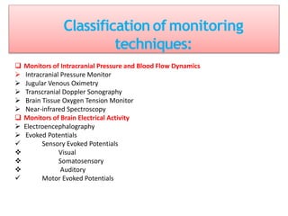
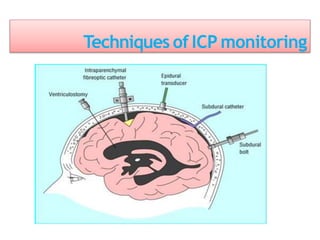
This document discusses various techniques used to monitor brain function during anesthesia, including:
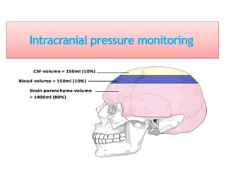
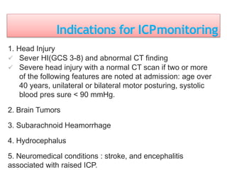
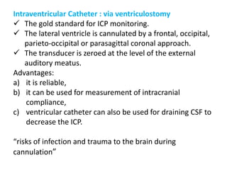
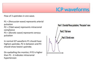
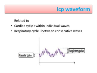
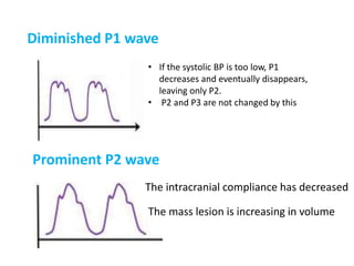
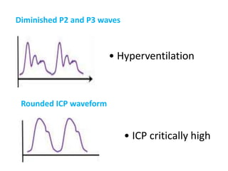
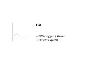
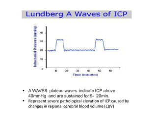
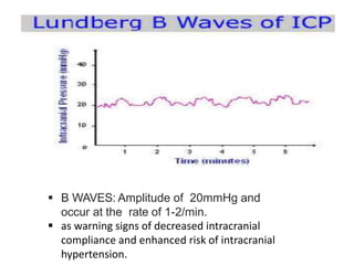
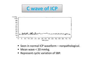
1. Intracranial pressure monitoring via an intraventricular catheter, which is the gold standard. Normal ICP is 10-15 mmHg in adults and waveforms can indicate increased ICP.
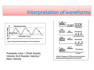
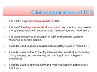
2. Transcranial Doppler measures blood flow velocity in cerebral arteries to monitor for vasospasm.
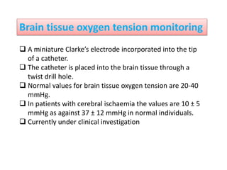
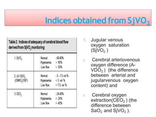
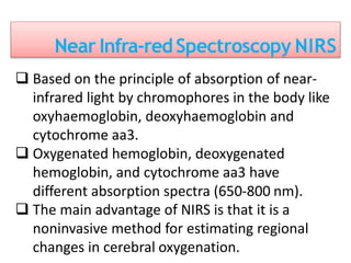
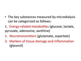
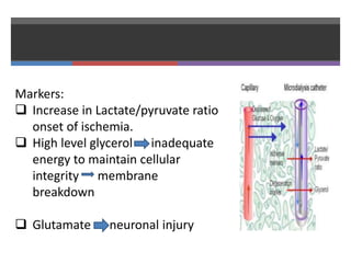
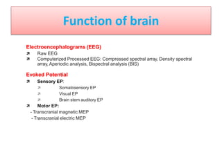
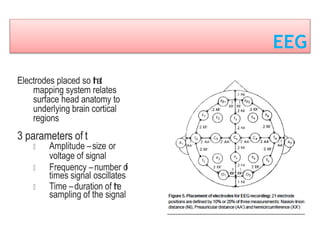
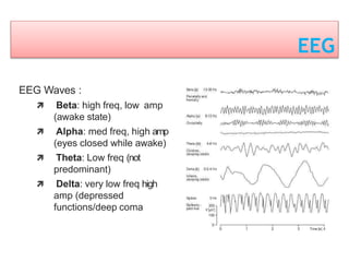
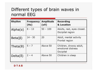
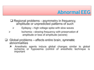
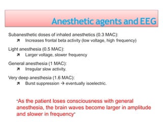
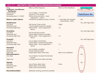
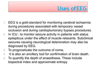
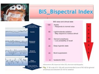
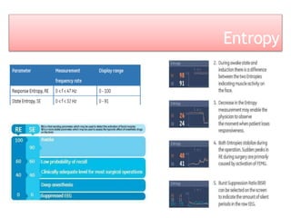
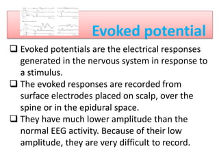
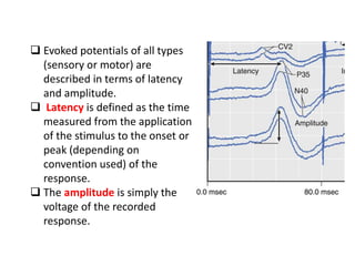
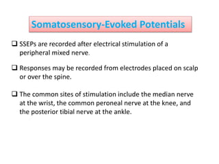
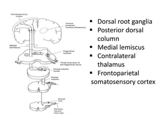
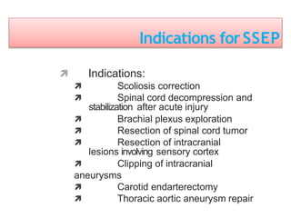
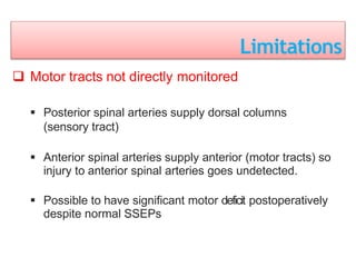
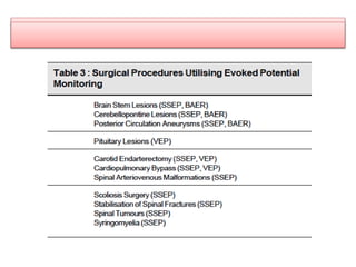
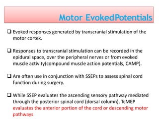
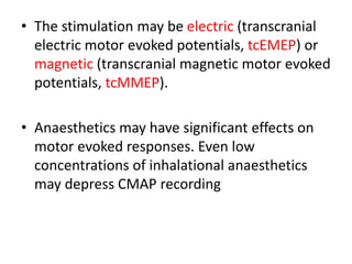
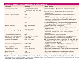
3. EEG, evoked potentials, brain tissue oxygen monitors, microdialysis, and near infrared spectroscopy provide additional insights into brain activity, oxygenation, and metabolism. EEG changes with anesthesia and can detect ischemia. Bispectral index monitors depth of anesthesia.
SUMMARY:
- Neurophysiologic monitoring not universally adopted but in many centers has become routine monitor for some surgical procedures
- Ideal neurophysiologic monitoring in the neurosurgical procedure should be: non-invasive (v.s invasive), high sensitivity & specificity, cost effective, easy to use, simple instrumentation, and real time or continous monitoring.
Introduction of organ donation .
Introduction of brain death and pathophysiology following it.
Perioperative problems in organ retrieval .
Goals of management of these patients .
Anesthetic management of the cadaver during organ harvesting.
Acute Promyelocytic Leukemia with Intracerebral Bleed and ARDSMedicalintensivist
A 27-Year-Old Young lady newly diagnosed case of Acute Promyelocytic Leukemia on treatment with ATRA developed Disseminated intravascular coagulation had drop in her sensorium shifted from ward to Medical ICU
Recomendações da OMS sobre cuidados maternos e neonatais para uma experiência pós-natal positiva.
Em consonância com os ODS – Objetivos do Desenvolvimento Sustentável e a Estratégia Global para a Saúde das Mulheres, Crianças e Adolescentes, e aplicando uma abordagem baseada nos direitos humanos, os esforços de cuidados pós-natais devem expandir-se para além da cobertura e da simples sobrevivência, de modo a incluir cuidados de qualidade.
Estas diretrizes visam melhorar a qualidade dos cuidados pós-natais essenciais e de rotina prestados às mulheres e aos recém-nascidos, com o objetivo final de melhorar a saúde e o bem-estar materno e neonatal.
Uma “experiência pós-natal positiva” é um resultado importante para todas as mulheres que dão à luz e para os seus recém-nascidos, estabelecendo as bases para a melhoria da saúde e do bem-estar a curto e longo prazo. Uma experiência pós-natal positiva é definida como aquela em que as mulheres, pessoas que gestam, os recém-nascidos, os casais, os pais, os cuidadores e as famílias recebem informação consistente, garantia e apoio de profissionais de saúde motivados; e onde um sistema de saúde flexível e com recursos reconheça as necessidades das mulheres e dos bebês e respeite o seu contexto cultural.
Estas diretrizes consolidadas apresentam algumas recomendações novas e já bem fundamentadas sobre cuidados pós-natais de rotina para mulheres e neonatos que recebem cuidados no pós-parto em unidades de saúde ou na comunidade, independentemente dos recursos disponíveis.
É fornecido um conjunto abrangente de recomendações para cuidados durante o período puerperal, com ênfase nos cuidados essenciais que todas as mulheres e recém-nascidos devem receber, e com a devida atenção à qualidade dos cuidados; isto é, a entrega e a experiência do cuidado recebido. Estas diretrizes atualizam e ampliam as recomendações da OMS de 2014 sobre cuidados pós-natais da mãe e do recém-nascido e complementam as atuais diretrizes da OMS sobre a gestão de complicações pós-natais.
O estabelecimento da amamentação e o manejo das principais intercorrências é contemplada.
Recomendamos muito.
Vamos discutir essas recomendações no nosso curso de pós-graduação em Aleitamento no Instituto Ciclos.
Esta publicação só está disponível em inglês até o momento.
Prof. Marcus Renato de Carvalho
www.agostodourado.com
Flu Vaccine Alert in Bangalore Karnatakaaddon Scans
As flu season approaches, health officials in Bangalore, Karnataka, are urging residents to get their flu vaccinations. The seasonal flu, while common, can lead to severe health complications, particularly for vulnerable populations such as young children, the elderly, and those with underlying health conditions.
Dr. Vidisha Kumari, a leading epidemiologist in Bangalore, emphasizes the importance of getting vaccinated. "The flu vaccine is our best defense against the influenza virus. It not only protects individuals but also helps prevent the spread of the virus in our communities," he says.
This year, the flu season is expected to coincide with a potential increase in other respiratory illnesses. The Karnataka Health Department has launched an awareness campaign highlighting the significance of flu vaccinations. They have set up multiple vaccination centers across Bangalore, making it convenient for residents to receive their shots.
To encourage widespread vaccination, the government is also collaborating with local schools, workplaces, and community centers to facilitate vaccination drives. Special attention is being given to ensuring that the vaccine is accessible to all, including marginalized communities who may have limited access to healthcare.
Residents are reminded that the flu vaccine is safe and effective. Common side effects are mild and may include soreness at the injection site, mild fever, or muscle aches. These side effects are generally short-lived and far less severe than the flu itself.
Healthcare providers are also stressing the importance of continuing COVID-19 precautions. Wearing masks, practicing good hand hygiene, and maintaining social distancing are still crucial, especially in crowded places.
Protect yourself and your loved ones by getting vaccinated. Together, we can help keep Bangalore healthy and safe this flu season. For more information on vaccination centers and schedules, residents can visit the Karnataka Health Department’s official website or follow their social media pages.
Stay informed, stay safe, and get your flu shot today!
micro teaching on communication m.sc nursing.pdfAnurag Sharma
Microteaching is a unique model of practice teaching. It is a viable instrument for the. desired change in the teaching behavior or the behavior potential which, in specified types of real. classroom situations, tends to facilitate the achievement of specified types of objectives.
Explore natural remedies for syphilis treatment in Singapore. Discover alternative therapies, herbal remedies, and lifestyle changes that may complement conventional treatments. Learn about holistic approaches to managing syphilis symptoms and supporting overall health.
Ethanol (CH3CH2OH), or beverage alcohol, is a two-carbon alcohol
that is rapidly distributed in the body and brain. Ethanol alters many
neurochemical systems and has rewarding and addictive properties. It
is the oldest recreational drug and likely contributes to more morbidity,
mortality, and public health costs than all illicit drugs combined. The
5th edition of the Diagnostic and Statistical Manual of Mental Disorders
(DSM-5) integrates alcohol abuse and alcohol dependence into a single
disorder called alcohol use disorder (AUD), with mild, moderate,
and severe subclassifications (American Psychiatric Association, 2013).
In the DSM-5, all types of substance abuse and dependence have been
combined into a single substance use disorder (SUD) on a continuum
from mild to severe. A diagnosis of AUD requires that at least two of
the 11 DSM-5 behaviors be present within a 12-month period (mild
AUD: 2–3 criteria; moderate AUD: 4–5 criteria; severe AUD: 6–11 criteria).
The four main behavioral effects of AUD are impaired control over
drinking, negative social consequences, risky use, and altered physiological
effects (tolerance, withdrawal). This chapter presents an overview
of the prevalence and harmful consequences of AUD in the U.S.,
the systemic nature of the disease, neurocircuitry and stages of AUD,
comorbidities, fetal alcohol spectrum disorders, genetic risk factors, and
pharmacotherapies for AUD.
ARTIFICIAL INTELLIGENCE IN HEALTHCARE.pdfAnujkumaranit
Artificial intelligence (AI) refers to the simulation of human intelligence processes by machines, especially computer systems. It encompasses tasks such as learning, reasoning, problem-solving, perception, and language understanding. AI technologies are revolutionizing various fields, from healthcare to finance, by enabling machines to perform tasks that typically require human intelligence.
Report Back from SGO 2024: What’s the Latest in Cervical Cancer?bkling
Are you curious about what’s new in cervical cancer research or unsure what the findings mean? Join Dr. Emily Ko, a gynecologic oncologist at Penn Medicine, to learn about the latest updates from the Society of Gynecologic Oncology (SGO) 2024 Annual Meeting on Women’s Cancer. Dr. Ko will discuss what the research presented at the conference means for you and answer your questions about the new developments.
- Video recording of this lecture in English language: https://youtu.be/lK81BzxMqdo
- Video recording of this lecture in Arabic language: https://youtu.be/Ve4P0COk9OI
- Link to download the book free: https://nephrotube.blogspot.com/p/nephrotube-nephrology-books.html
- Link to NephroTube website: www.NephroTube.com
- Link to NephroTube social media accounts: https://nephrotube.blogspot.com/p/join-nephrotube-on-social-media.html
Title: Sense of Taste
Presenter: Dr. Faiza, Assistant Professor of Physiology
Qualifications:
MBBS (Best Graduate, AIMC Lahore)
FCPS Physiology
ICMT, CHPE, DHPE (STMU)
MPH (GC University, Faisalabad)
MBA (Virtual University of Pakistan)
Learning Objectives:
Describe the structure and function of taste buds.
Describe the relationship between the taste threshold and taste index of common substances.
Explain the chemical basis and signal transduction of taste perception for each type of primary taste sensation.
Recognize different abnormalities of taste perception and their causes.
Key Topics:
Significance of Taste Sensation:
Differentiation between pleasant and harmful food
Influence on behavior
Selection of food based on metabolic needs
Receptors of Taste:
Taste buds on the tongue
Influence of sense of smell, texture of food, and pain stimulation (e.g., by pepper)
Primary and Secondary Taste Sensations:
Primary taste sensations: Sweet, Sour, Salty, Bitter, Umami
Chemical basis and signal transduction mechanisms for each taste
Taste Threshold and Index:
Taste threshold values for Sweet (sucrose), Salty (NaCl), Sour (HCl), and Bitter (Quinine)
Taste index relationship: Inversely proportional to taste threshold
Taste Blindness:
Inability to taste certain substances, particularly thiourea compounds
Example: Phenylthiocarbamide
Structure and Function of Taste Buds:
Composition: Epithelial cells, Sustentacular/Supporting cells, Taste cells, Basal cells
Features: Taste pores, Taste hairs/microvilli, and Taste nerve fibers
Location of Taste Buds:
Found in papillae of the tongue (Fungiform, Circumvallate, Foliate)
Also present on the palate, tonsillar pillars, epiglottis, and proximal esophagus
Mechanism of Taste Stimulation:
Interaction of taste substances with receptors on microvilli
Signal transduction pathways for Umami, Sweet, Bitter, Sour, and Salty tastes
Taste Sensitivity and Adaptation:
Decrease in sensitivity with age
Rapid adaptation of taste sensation
Role of Saliva in Taste:
Dissolution of tastants to reach receptors
Washing away the stimulus
Taste Preferences and Aversions:
Mechanisms behind taste preference and aversion
Influence of receptors and neural pathways
Impact of Sensory Nerve Damage:
Degeneration of taste buds if the sensory nerve fiber is cut
Abnormalities of Taste Detection:
Conditions: Ageusia, Hypogeusia, Dysgeusia (parageusia)
Causes: Nerve damage, neurological disorders, infections, poor oral hygiene, adverse drug effects, deficiencies, aging, tobacco use, altered neurotransmitter levels
Neurotransmitters and Taste Threshold:
Effects of serotonin (5-HT) and norepinephrine (NE) on taste sensitivity
Supertasters:
25% of the population with heightened sensitivity to taste, especially bitterness
Increased number of fungiform papillae
TEST BANK for Operations Management, 14th Edition by William J. Stevenson, Ve...kevinkariuki227
TEST BANK for Operations Management, 14th Edition by William J. Stevenson, Verified Chapters 1 - 19, Complete Newest Version.pdf
Pulmonary Thromboembolism - etilogy, types, medical- Surgical and nursing man...VarunMahajani
Disruption of blood supply to lung alveoli due to blockage of one or more pulmonary blood vessels is called as Pulmonary thromboembolism. In this presentation we will discuss its causes, types and its management in depth.
Lung Cancer: Artificial Intelligence, Synergetics, Complex System Analysis, S...Oleg Kshivets
RESULTS: Overall life span (LS) was 2252.1±1742.5 days and cumulative 5-year survival (5YS) reached 73.2%, 10 years – 64.8%, 20 years – 42.5%. 513 LCP lived more than 5 years (LS=3124.6±1525.6 days), 148 LCP – more than 10 years (LS=5054.4±1504.1 days).199 LCP died because of LC (LS=562.7±374.5 days). 5YS of LCP after bi/lobectomies was significantly superior in comparison with LCP after pneumonectomies (78.1% vs.63.7%, P=0.00001 by log-rank test). AT significantly improved 5YS (66.3% vs. 34.8%) (P=0.00000 by log-rank test) only for LCP with N1-2. Cox modeling displayed that 5YS of LCP significantly depended on: phase transition (PT) early-invasive LC in terms of synergetics, PT N0—N12, cell ratio factors (ratio between cancer cells- CC and blood cells subpopulations), G1-3, histology, glucose, AT, blood cell circuit, prothrombin index, heparin tolerance, recalcification time (P=0.000-0.038). Neural networks, genetic algorithm selection and bootstrap simulation revealed relationships between 5YS and PT early-invasive LC (rank=1), PT N0—N12 (rank=2), thrombocytes/CC (3), erythrocytes/CC (4), eosinophils/CC (5), healthy cells/CC (6), lymphocytes/CC (7), segmented neutrophils/CC (8), stick neutrophils/CC (9), monocytes/CC (10); leucocytes/CC (11). Correct prediction of 5YS was 100% by neural networks computing (area under ROC curve=1.0; error=0.0).
CONCLUSIONS: 5YS of LCP after radical procedures significantly depended on: 1) PT early-invasive cancer; 2) PT N0--N12; 3) cell ratio factors; 4) blood cell circuit; 5) biochemical factors; 6) hemostasis system; 7) AT; 8) LC characteristics; 9) LC cell dynamics; 10) surgery type: lobectomy/pneumonectomy; 11) anthropometric data. Optimal diagnosis and treatment strategies for LC are: 1) screening and early detection of LC; 2) availability of experienced thoracic surgeons because of complexity of radical procedures; 3) aggressive en block surgery and adequate lymph node dissection for completeness; 4) precise prediction; 5) adjuvant chemoimmunoradiotherapy for LCP with unfavorable prognosis.
4. Intra-cranial Pressure
The pressure inside the lateral ventricles/lumbar
subarachnoid space in supine position.
The normal value of ICP is 10-15 mm Hg in adults.
It is around 2-4 mmHg in neonates and infants.
5. Indications for ICPmonitoring
1. Head Injury
Sever HI(GCS 3-8) and abnormal CT finding
Severe head injury with a normal CT scan if two or more
of the following features are noted at admission: age over
40 years, unilateral or bilateral motor posturing, systolic
blood pres sure < 90 mmHg.
2. Brain Tumors
3. Subarachnoid Heamorrhage
4. Hydrocephalus
5. Neuromedical conditions : stroke, and encephalitis
associated with raised ICP.
7. Intraventricular Catheter : via ventriculostomy
The gold standard for ICP monitoring.
The lateral ventricle is cannulated by a frontal, occipital,
parieto-occipital or parasagittal coronal approach.
The transducer is zeroed at the level of the external
auditory meatus.
Advantages:
a) it is reliable,
b) it can be used for measurement of intracranial
compliance,
c) ventricular catheter can also be used for draining CSF to
decrease the ICP.
“risks of infection and trauma to the brain during
cannulation”
8. ICP waveforms
Flow of 3 upstrokes in one wave.
P1 = (Percussion wave) represents arterial
pulsation
P2 = (Tidal wave) represents intracranial
compliance
P3 = (Dicrotic wave) represents venous
pulsation
In normal ICP waveform P1 should have
highest upstroke, P2 in between and P3
should show lowest upstroke.
On eyeballing the monitor, if P2 is higher
than P1 - it indicates intracranial
hypertension
10. Abnormalities of ICP wave
Earliest sign of ↑ ICP – Changes in pulsatile components
Prominent P1 wave
The systolic BP is too high
11. Diminished P1 wave
• If the systolic BP is too low, P1
decreases and eventually disappears,
leaving only P2.
• P2 and P3 are not changed by this
Prominent P2 wave
The intracranial compliance has decreased
The mass lesion is increasing in volume
12. Diminished P2 and P3 waves
• Hyperventilation
Rounded ICP waveform
• ICP critically high
14. A WAVES: plateau waves indicate ICP above
40mmHg and are sustained for 5- 20min.
Represent severe pathological elevation of ICP caused by
changes in regional cerebral blood volume (CBV)
15. B WAVES: Amplitude of 20mmHg and
occur at the rate of 1-2/min.
as warning signs of decreased intracranial
compliance and enhanced risk of intracranial
hypertension.
16. C wave of ICP
Seen in normal ICP waveform – nonpathological.
Mean wave < 20 mmhg.
Represent cyclic variation of SBP.
17. Transcranial Doppler
Measures the blood flow velocity in major cerebral blood
vessles.
Examination carried out through the temporal
window, orbital foramen or foramen magnum.
MCA commonly used.
Change in velocity is proportional to change in flow
considering the vessel diameter is constant.
19. Clinicalapplications ofTCD
1. It is useful as a noninvasive monitor of CBF.
2. It is helpful to diagnose cerebral vasospasm and monitor response to
therapy in patients with subarachnoid haemorrhage and head injury.
3. It is used to study autoregulation of CBF and cerebral vascular
response to carbon dioxide.
4. It can be used to assess intracranial circulatory status in raised ICP.
5. It can be a useful tool to identify intraoperative cerebral embolisation
during surgery on carotid artery and cardiopulmonary bypass
procedures.
6. It can be used to optimise CPP and hyperventilationin patients with
head injury.
20. Intravascular tracercompounds
Method originally described by Kety and Schmidt.
Administration of radioactive isotope of xenon-133
Measurement of radioactivity washout with gamma detectors.
Disadvantages: 1.Exposure to radioactivity
2.Cumbersome detector equipment
3.Focal areas of hypoperfusion missed
4.Snapshot of CBF not continuous monitor.
21. Monitoring of cerebral oxygenation and
metabloism
Brain tissue oxygenation
Jugular bulb venous oximetry monitoring
Microdialysis catheter
Near Infrared Spectroscopy (NIRS)
22. Brain tissue oxygen tension monitoring
A miniature Clarke’s electrode incorporated into the tip
of a catheter.
The catheter is placed into the brain tissue through a
twist drill hole.
Normal values for brain tissue oxygen tension are 20-40
mmHg.
In patients with cerebral ischaemia the values are 10 ± 5
mmHg as against 37 ± 12 mmHg in normal individuals.
Currently under clinical investigation
23. Jugular venousoximetry
Jugular bulb venous oxygen saturation (Sjvo2) measures
the degree of oxygen extraction by the brain and represents
the balance between cerebral oxygen supply and demand.
The dominant jugular vein (i.e., the right for most patients)
drains predominantly cortical venous blood, whereas the
contralateral jugular vein drains more of the subcortical
regions.
Any decrease in CBF must be accompanied by a
corresponding decrease in cerebral venous oxygen
saturation if CMRO2 is constant.
24. Jugular venousoximetry
A catheter placed retrograde
through the internal jugular
vein into the jugular bulb.
The tip of the catheter must
be placed above the C1-C2
vertebral bodies to avoid
contamination with blood
coming from the facial vein.
Correct positioning of the
catheter can be confirmed
with a lateral skull x-ray
25. Indices obtained fromSjVO2
1. Jugular venous
oxygen saturation
(SjVO2 )
2. Cerebral arteriovenous
oxygen difference (A-
VDO2 ) (the difference
between arterial and
jugularvenous oxygen
content) and
3. Cerebral oxygen
extraction(CEO2 ) (the
difference between
SaO2 and SjVO2 ).
26.
27. Near Infra-redSpectroscopy NIRS
Based on the principle of absorption of near-
infrared light by chromophores in the body like
oxyhaemoglobin, deoxyhaemoglobin and
cytochrome aa3.
Oxygenated hemoglobin, deoxygenated
hemoglobin, and cytochrome aa3 have
different absorption spectra (650-800 nm).
The main advantage of NIRS is that it is a
noninvasive method for estimating regional
changes in cerebral oxygenation.
28. NIRS limitations
Its clinical use is limited by an inability
to differentiate between intracranial
and extracranial changes in blood
flow and oxygenation.
Currently, there are no studies
providing evidence that NIRS use
alone can influence outcomes in adult
neurocritical care.
29. Cerebral Microdialysis
A technique for sampling the extracellular space of a tissue.
This method is based on the diffusion of water-soluble substances
through a semipermeable membrane. Small molecules (<20,000 D)
from the extracellular fluid can diffuse across the membrane and
enter the perfusate. Conversely, substances that have been added
to the perfusate can diffuse across the membrane to gain entry to
the tissue.
The technique of cerebral microdialysis allows continuous and
online monitoring of changes in brain tissue chemistry.
30. • The key substances measured by microdialysis
can be categorized as follows:
1. Energy-related metabolites (glucose, lactate,
pyruvate, adenosine, xanthine)
2. Neurotransmitters (glutamate, aspartate)
3. Markers of tissue damage and inflammation
(glycerol)
31. Markers:
Increase in Lactate/pyruvate ratio
onset of ischemia.
High level glycerol inadequate
energy to maintain cellular
integrity membrane
breakdown
Glutamate neuronal injury
32. Electroencephalograms (EEG)
Raw EEG
Computerized Processed EEG: Compressed spectral array, Density spectral
array, Aperiodic analysis, Bispectral analysis (BIS)
Evoked Potential
Sensory EP:
Somatosensory EP
Visual EP
Brain stem auditory EP
Motor EP:
- Transcranial magnetic MEP
- Transcranial electric MEP
Function of brain
34. Electroencephalogram – surface recordings of the
summation of excitatory and inhibitory postsynaptic
potentials generated by pyramidal cells in cerebral
cortex
EEG:
Measures electrical function of brain
Indirectly measures blood flow
Measures anesthetic effects
EEG
35. EEG
Three uses perioperatively:
Identify inadequate blood flow to cerebral cortex
caused by surgical/anesthetic-induced reduction in flow
Guide reduction of cerebral metabolism prior to
induced reduction of blood flow
Predict neurologic outcome after brain insult
Other uses: identify consciousness, unconsciousness,
seizure activity, stages of sleep, coma
36. EEG
Electrodes placed so that
mapping system relates
surface head anatomy to
underlying brain cortical
regions
3 parameters of t
Amplitude –size or
voltage of signal
Frequency –number of
times signal oscillates
Time –duration of the
sampling of the signal
37. EEG
EEG Waves :
Beta: high freq, low amp
(awake state)
Alpha: med freq, high amp
(eyes closed while awake)
Theta: Low freq (not
predominant)
Delta: very low freq high
amp (depressed
functions/deep coma
38.
39. Abnormal EEG
Regional problems - asymmetry in frequency,
amplitude or unpredicted patterns of such
Epilepsy – high voltage spike with slow waves
Ischemia – slowing frequency with preservation of
amplitude or loss of amplitude (severe)
Global problems – affects entire brain, symmetric
abnormalities
Anesthetic agents induce global changes similar to global
ischemia or hypoxemia (control of anesthetic technique is
important
40. Anesthetic agents and EEG
Subanesthetic doses of inhaled anesthetics (0.3 MAC):
Increases frontal beta activity (low voltage, high frequency)
Light anesthesia (0.5 MAC):
Larger voltage, slower frequency
General anesthesia (1 MAC):
Irregular slow activity.
Very deep anesthesia (1.6 MAC):
Burst suppression eventually isoelectric.
“As the patient loses consciousness with general
anesthesia, the brain waves become larger in amplitude
and slower in frequency”
42. Non-anestheticFactorsAffectingEEG
• Surgical
1. Cardiopulmonary bypass
2. Occlusion of major cerebral
vessel (carotid cross-clamping,
aneurysm clipping)
3. Retraction on cerebral cortex
4. Surgically induced emboli to
brain
Pathophysiol
ogic Factors
1. Hypoxemia
2. Hypotension
3. Hypothermia
43. Uses ofEEG
1. EEG is a gold-standard for monitoring cerebral ischaemia:
during procedures associated with temporary vessel
occlusion and during cardioplumonary bypass procedures.
2. In ICU : to monitor seizure activity in patients with status
epilepticus under the effect of muscle relaxants. Subclinical
seizures causing neurological deterioration may also be
diagnosed by EEG.
3. To prognosticate the outcome of coma.
4. It is also an ancillary tool for confirmation of brain death.
5. To quantify the depth of anaesthesia. These include
bispectral index and approximate entropy
46. Evoked potential
EVOKED POTENTIALS
Evoked potentials are the electrical responses
generated in the nervous system in response to
a stimulus.
The evoked responses are recorded from
surface electrodes placed on scalp, over the
spine or in the epidural space.
They have much lower amplitude than the
normal EEG activity. Because of their low
amplitude, they are very difficult to record.
47. Evoked potentials of all types
(sensory or motor) are
described in terms of latency
and amplitude.
Latency is defined as the time
measured from the application
of the stimulus to the onset or
peak (depending on
convention used) of the
response.
The amplitude is simply the
voltage of the recorded
response.
48. Clinically significant of evoked potential
Decreases in amplitude of 50% or more from
baseline associated with a less than 10%
prolongation in latency as clinically significant SER
changes.
Intraoperative changes in evoked responses, such
as decreased amplitude, increased latency, or
complete loss of the waveform, may result from
surgical misdeed, such as retractor placement or
ischemia. They may also reflect systemic changes,
such as changes in the anesthetic drugs or doses,
temperature, or hypoperfusion.
49. Sensory-Evoked Responses(SERs)
SERs are electrical CNS responses to electrical,
auditory, or visual stimuli.
SERs are produced by stimulating a sensory
system and recording the resulting electrical
responses at various sites along the sensory
pathway up to and including the cerebral cortex.
It include:
Somatosensory EP
Visual EP
Brain stem auditory EP
50. Somatosensory-Evoked Potentials
SSEPs are recorded after electrical stimulation of a
peripheral mixed nerve.
Responses may be recorded from electrodes placed on scalp
or over the spine.
The common sites of stimulation include the median nerve
at the wrist, the common peroneal nerve at the knee, and
the posterior tibial nerve at the ankle.
52. Indications forSSEP
Indications:
Scoliosis correction
Spinal cord decompression and
stabilization after acute injury
Brachial plexus exploration
Resection of spinal cord tumor
Resection of intracranial
lesions involving sensory cortex
Clipping of intracranial
aneurysms
Carotid endarterectomy
Thoracic aortic aneurysm repair
53. Limitations
Motor tracts not directly monitored
Posterior spinal arteries supply dorsal columns
(sensory tract)
Anterior spinal arteries supply anterior (motor tracts) so
injury to anterior spinal arteries goes undetected.
Possible to have significant motor deficit postoperatively
despite normal SSEPs
54. Brainstem Auditory-Evoked Potentials
Auditory evoked potentials are
generated in response to
stimulation of the tympanic
membrane by audible clicks.
Reflects the VIII nerve &
brainstem“well-being”.
useful for surgical procedures in
the posterior fossa that risk
hearing or structures in the upper
medulla, pons, and midbrain.
Most resistant to anesthetic
drugs
55. Visual-Evoked Potentials
Visual evoked potentials are generated in response to
photic stimulation of the retina by flashes of light
from light-emitting diodes.
VEPs are the least commonly used evoked response
monitoring technique intraoperatively.
Most useful for testing for optic nerve function.
56. Effect of physiologic variables on evoked potentials
Cerebral Blood Flow: Sensory evoked potentials are normal
upto a CBF value of 20 mL100g–1/min. They start
deteriorating when CBF decreases to 18-13 ml/100g/min.
Evoked potentials cannot be obtained when CBF is below 10-
12 mL100g/min.
Systemic Blood Pressure: Hypotension prolongs the
conduction in the central nervous system thereby increasing
the latencies of various peaks.
Intracranial Pressure : Raised ICP has been shown to result in
an increase in the latency and a decrease in the amplitude
57. Oxygen Tension: Deterioration of evoked potentials
decreases to less than 40 mmHg.
Haematocrit: Latencies of VEP and SSEP are increased
at a haematoctrit of 10-15%. Their amplitude is
decreased when the haematoctrit is less than 10%.
Carbon Dioxide Tension: Extreme hypocapnia
(PaCO2 < 25 mmHg) causes deterioration of evoked
potentials.
Temperature: Hypothermia increases the latency.
58. Effects of anaesthetics on evoked potentials
Most anaesthetics decrease the amplitude and increase the
latencies of the various peaks.
Brainstem and spinal potentials are least affected.
The effects of anaesthetics on evoked potentials are dose-related.
59.
60. Motor EvokedPotentials
Evoked responses generated by transcranial stimulation of the
motor cortex.
Responses to transcranial stimulation can be recorded in the
epidural space, over the peripheral nerves or from evoked
muscle activity(compound muscle action potentials, CAMP).
Are often use in conjunction with SSEPs to assess spinal cord
function during surgery.
While SSEP evaluates the ascending sensory pathway mediated
through the posterior spinal cord (dorsal column), TcMEP
evaluates the anterior portion of the cord or descending motor
pathways
61. • The stimulation may be electric (transcranial
electric motor evoked potentials, tcEMEP) or
magnetic (transcranial magnetic motor evoked
potentials, tcMMEP).
• Anaesthetics may have significant effects on
motor evoked responses. Even low
concentrations of inhalational anaesthetics
may depress CMAP recording
62.
63. References
Millers anesthesia 8th edition
Neurological monitoring. Dr. G S Rao IJA
2002;46(4)
2017 Textbook of Critical Care, 7e 39