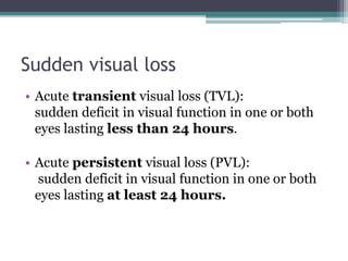
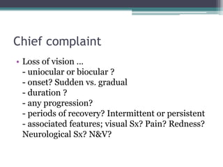
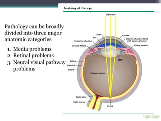
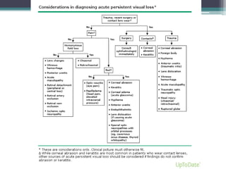
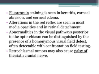
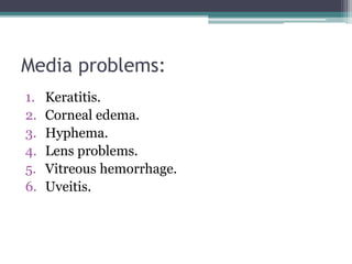
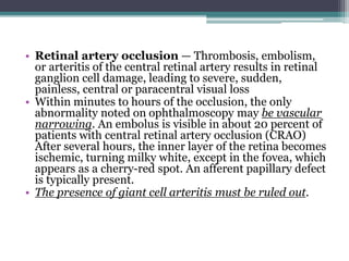
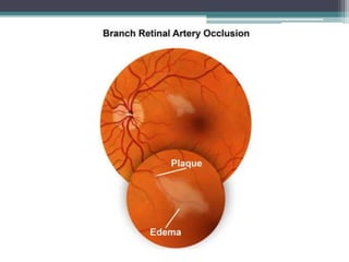
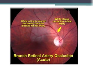
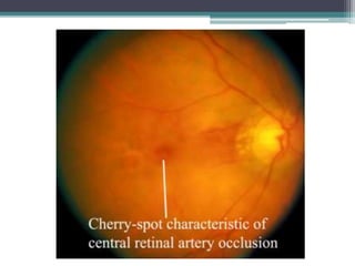
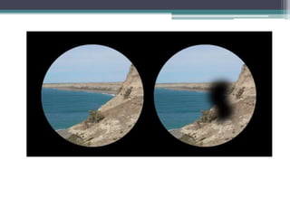
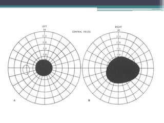
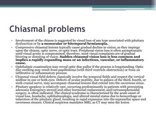
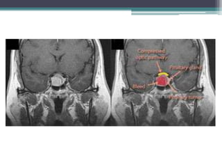
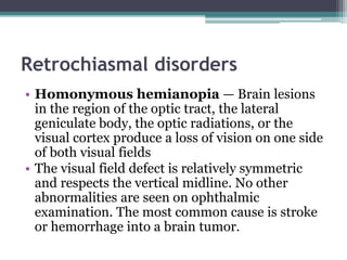
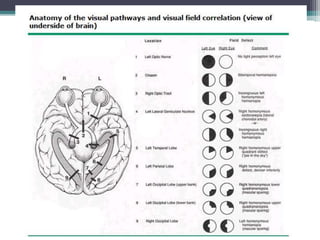
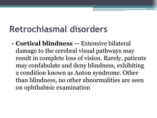
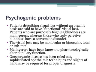
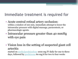
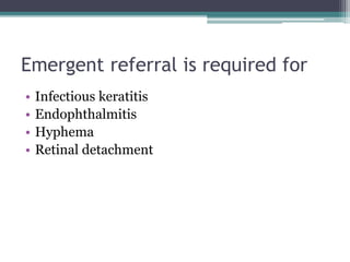
2. Sudden visual loss
• Acute transient visual loss (TVL):
sudden deficit in visual function in one or both
eyes lasting less than 24 hours.
• Acute persistent visual loss (PVL):
sudden deficit in visual function in one or both
eyes lasting at least 24 hours.
3. Chief complaint
• Loss of vision …
- uniocular or biocular ?
- onset? Sudden vs. gradual
- duration ?
- any progression?
- periods of recovery? Intermittent or persistent
- associated features; visual Sx? Pain? Redness?
Neurological Sx? N&V?
4. Afferent pupillary reflex
• demonstrated by shining a light alternately in
one eye and then the other and finding that the
direct response to light is more sluggish in the
affected eye. The room should be dark, and the
patient should fixate on a distant target to
prevent miosis due to accommodation.
• The presence of an afferent pupillary defect is
fairly specific for unilateral optic nerve
pathology.
5.
6.
7. Pathology can be broadly
divided into three major
anatomic categories
1. Media problems
2. Retinal problems
3. Neural visual pathway
problems
8.
9. • Fluorescein staining is seen in keratitis, corneal
abrasion, and corneal edema.
• Alterations in the red reflex are seen in most
media opacities and in retinal detachment.
• Abnormalities in the visual pathways posterior
to the optic chiasm can be distinguished by the
presence of a homonymous visual field defect,
often detectable with confrontation field testing.
• Retrochiasmal tumors may also cause palsy of
the sixth cranial nerve.
11. • Keratitis — Keratitis is inflammation of the cornea due
to trauma, abrasive exposure, allergy, or infection. It is
marked by cloudiness, irregularity, or loss of epithelial or
sub-epithelial corneal tissues. Typically the eye is
tearing, red, and painful or irritated.
• Loss of epithelial cells is demonstrated by corneal uptake
of fluorescein dye, creating a focal or diffuse green glow
under a cobalt blue light. Deeper corneal disease may be
visible as a focal or diffuse white opacity, or by dulling of
the usually distinct reflection of light off of the cornea
(corneal light reflex). The most common causes of
keratitis are infection (viral, bacterial, fungal) and
trauma. Both can also cause corneal edema
12.
13. • Corneal edema — Corneal swelling results in loss of corneal
clarity. Examination may reveal dulling of the corneal light
reflex or a frank grey or white color to the substance of the
cornea.
• A serious cause of sudden corneal edema is acute angle-
closure glaucoma. The patient with severely elevated
intraocular pressure (IOP) typically has nausea, vomiting, and
may see colored halos around lights. The eye is tearing, red,
and extremely painful, often with ipsilateral brow ache. In
angle closure, the pupil may be fixed in a mid-dilated position,
and biomicroscopic (slit lamp) examination may reveal a
shallow anterior chamber. IOP is often dangerously elevated
(usually 40 to 80 mmHg). If formal tonometry is not
available, severely elevated IOP may be suspected by noting
hardness to palpation in comparison to the fellow eye
15. • Hyphema — A hyphema is blood in the
anterior chamber. It may result from blunt
trauma or may occur spontaneously in a handful
of conditions marked by abnormal growth or
fragility of iris blood vessels (often in chronic
poorly controlled diabetes). Biomicroscopic
examination reveals red blood cells
circulating and/or layered in the anterior
chamber. Intraocular pressure may become
dangerously elevated.
17. • Lens changes — Changes in the size, clarity, or
positioning of the crystalline lens may alter the focus
of light onto the retina, resulting in visual
disturbance. Trauma or a variety of congenital
conditions can lead to lens dislocation and resultant
vision loss. Lens clouding (cataract) does not
typically occur acutely, except in the setting of
trauma.
• Elevated blood glucose can cause lens swelling,
altering the refractive error. If the change is great
enough, patients may perceive vision loss. Vision
impairment typically resolves within days to weeks
of normalization of blood glucose.
18. • Vitreous hemorrhage — Bleeding into the
vitreous humor can occur in the setting of trauma,
spontaneous retinal tear, spontaneous vitreous
detachment, or in any condition with retinal
neovascularization (as in poorly controlled
diabetes).
• The reduction in vision is directly proportional to
the amount of blood in the vitreous. If the
hemorrhage is dense enough, there may be a
decreased red reflex, or the retina may not be visible
with ophthalmoscopy
19.
20. • Uveitis — Uveitis is the general term for inflammation inside the
eye. Inflammation of the anterior structures of the eye typically are
associated with red, painful light sensitivity, whereas isolated
inflammation of intermediate and posterior structures may be
associated with normal general appearance of the eye with decrease
in the red reflex and/or patient complaint of new floaters
• The most important cause of acute persistent visual loss in this
category is endophthalmitis. Endophthalmitis is a serious
bacterial or fungal infection of all intraocular tissues, caused by
surface pathogens (usually in the setting of recent ocular surgery) or
blood-borne agents.
• The eye is tearing, red, and painful. Biomicroscopic examination
reveals white blood cells in the anterior chamber, vitreous space, or
both. A layer of pus, called a hypopyon, may collect in the anterior
chamber. Corneal edema and a decreased red reflex may also be
present
23. • Retinal artery occlusion — Thrombosis, embolism,
or arteritis of the central retinal artery results in retinal
ganglion cell damage, leading to severe, sudden,
painless, central or paracentral visual loss
• Within minutes to hours of the occlusion, the only
abnormality noted on ophthalmoscopy may be vascular
narrowing. An embolus is visible in about 20 percent of
patients with central retinal artery occlusion (CRAO)
After several hours, the inner layer of the retina becomes
ischemic, turning milky white, except in the fovea, which
appears as a cherry-red spot. An afferent papillary defect
is typically present.
• The presence of giant cell arteritis must be ruled out.
24.
25.
26.
27.
28.
29. • Retinal vein occlusion — Thrombosis of the central retinal
vein results in venous stasis, leading to disc swelling, diffuse
nerve fiber layer and pre-retinal hemorrhage, and cotton wool
spots that create a dramatic appearance, often called "the
blood and thunder" fundus Depending upon the degree of
ischemia or presence of macular edema, there may be need for
retinal laser to prevent further vision loss or intravitreal
injections of anti-VEGF agents or corticosteroids to recover
vision
• While vision loss may be severe, the onset is typically
subacute in contrast to the sudden visual loss typical of CRAO.
When venous stasis is severe, infarction may occur due to
slowed retinal blood flow on the arterial side. In this setting, a
relative afferent papillary defect is often present
34. • Retinal detachment — Detachment of the neurosensory
retina may occur spontaneously or in the setting of trauma.
The most common form is due to a tear or break in the retina.
Patients may describe sudden onset of new floaters or black
dots in their vision, often accompanied by flashes of light
(photopsias). In its early stages, a detachment may present as
a persistent missing portion of the monocular visual field.
Once the macula (central retina) has become involved, visual
acuity will be severely compromised.
• Retinal detachment is not painful and does not cause a red
eye. There may be a dulling of the red reflex, and
ophthalmoscopic examination may reveal the retina to be
elevated with folds. If the detachment is extensive, there may
be a relative afferent pupillary defect
35.
36.
37.
38. • Acute maculopathy — Conditions that affect the
macula are associated with a central blind spot
(scotoma), blurred vision, or visual distortion.
Alteration of the macula due to fluid leakage,
bleeding, infection, or other causes can occur de
novo or as an acute worsening of a chronic disease
(eg, new edema in previously dry diabetic
retinopathy, or new bleeding in previously dry
macular degeneration). Diagnosis usually requires
detailed ophthalmoscopy with a high magnification
lens through pharmacologically dilated pupils
42. Optic nerve problems
• Optic neuritis is the most common cause of optic
nerve disease in younger adults.
• while ischemic optic neuropathy is the most
common etiology in older patients.
43. • Ischemic optic neuropathy — Ischemic optic
neuropathy is generally categorized as anterior (affecting
the optic disc) or posterior (retrobulbar) and as arteritic
or nonarteritic. Infarction at the optic nerve head due to
thrombosis or transient hypotension leads to a superior
or inferior altitudinal visual field defect, or diffusely
reduced vision.
• An afferent pupillary defect is typically present. In
anterior optic neuropathy, the optic disc is swollen, often
accompanied by splinter hemorrhages. If nerve damage
occurs posterior to the globe, no abnormalities may be
seen on initial ophthalmoscopic exam
• Giant cell arteritis must be ruled out
44.
45. • Optic neuritis — Inflammation of the optic nerve
may be associated with a variety of conditions, most
notably multiple sclerosis. Optic neuritis is the
presenting feature in 15 to 20 percent of patients
with multiple sclerosis, and it occurs at some time
during the course of the disease in 50 percent of
patients
• Affected patients note pain on eye movement,
reduced visual acuity and washed out color vision.
An afferent papillary defect is typically present, and
the optic disc is either normal or swollen
46.
47.
48. • Papilledema — Elevated intracranial pressure
can lead to transient visual obscurations or mild
persistent blurred vision. Examination reveals
bilateral optic nerve swelling without relative
afferent papillary defect.
49.
50.
51. Chiasmal problems
• Involvement of the chiasm is suggested by visual loss of any type associated with pituitary
dysfunction or by a monocular or bitemporal hemianopia.
• Compressive chiasmal lesions typically cause gradual decline in vision, as they impinge
upon the chiasm, optic nerve, or optic tract. Peripheral vision loss is often asymptomatic
until visual acuity is compromised; therefore, most visual complaints are of gradual
blurring or dimming of vision. Sudden chiasmal vision loss is less common and
implies a rapidly expanding mass or an infectious, vascular, or inflammatory
cause.
• Ophthalmic examination may reveal optic disc pallor if the process is longstanding. Optic
disc swelling may result from papilledema (with third ventricle obstruction) or from an
infiltrative or inflammatory process.
• Chiasmal visual field defects classically involve the temporal fields and respect the vertical
midline in one or both eyes. Defects of ocular motility, due to palsies of the third, fourth, or
sixth cranial nerve, may accompany chiasmal lesions that extend into the cavernous sinus.
• Pituitary apoplexy is relatively rare, occurring predominantly in patients with preexisting
adenoma Emergency steroid and other hormonal replacement, and/ortranssphenoidal
surgery, is often indicated. The clinical syndrome is characterized by the acute onset of
visual loss, headache, ophthalmoplegia, and altered mental status due to hemorrhage or
infarction of the pituitary gland, resulting in rapid expansion into the suprasellar space and
cavernous sinuses. Clinical suspicion mandates MRI, as CT may miss the lesion
52.
53. Retrochiasmal disorders
• Homonymous hemianopia — Brain lesions
in the region of the optic tract, the lateral
geniculate body, the optic radiations, or the
visual cortex produce a loss of vision on one side
of both visual fields
• The visual field defect is relatively symmetric
and respects the vertical midline. No other
abnormalities are seen on ophthalmic
examination. The most common cause is stroke
or hemorrhage into a brain tumor.
54.
55. Retrochiasmal disorders
• Cortical blindness — Extensive bilateral
damage to the cerebral visual pathways may
result in complete loss of vision. Rarely, patients
may confabulate and deny blindness, exhibiting
a condition known as Anton syndrome. Other
than blindness, no other abnormalities are seen
on ophthalmic examination
56. Psychogenic problems
• Patients describing visual loss without an organic
basis are said to have "functional" visual loss.
Patients who are purposely feigning blindness are
malingerers, whereas those who truly perceive
blindness have a conversion disorder.
• The visual loss may be monocular or binocular, total
or sub-total.
• Malingerers have been known to pharmacologically
alter pupillary function.
• Once organic disease has been ruled out,
sophisticated ophthalmic techniques and slights of
hand may be required for proper diagnosis
57. Immediate treatment is required for
• Acute central retinal artery occlusion:
within a window of 100 min, immediate attempt to lower the
intraocular pressure with digital massage, paracentesis, or
pharmacologic agents.
• Intraocular pressure greater than 40 mmHg
with eye pain
• Vision loss in the setting of suspected giant cell
arteritis
started on methylprednisolone 1000 mg IV daily for one to three
days, followed by prednisone 60 mg/d for two to four weeks
58. Emergent referral is required for
• Infectious keratitis
• Endophthalmitis
• Hyphema
• Retinal detachment
59. Urgent referral (within 24 to 48 hours)
is appropriate for:
• Non-infectious uveitis
• Vitreous hemorrhage
• Acute maculopathy
• Central retinal vein occlusion
• Optic neuritis
60. • Visual manifestations of intracranial pathology
(stroke, tumor, bleed, or elevated intracranial
pressure) should be treated in the appropriate
neurological or neurosurgical fashion