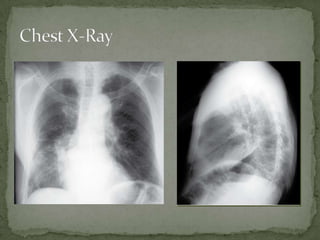
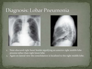
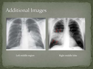
 76 yr old female with pmhx
notable for htn presents
with 3 days worsening
dyspnea on exertion. Pt
endorses cough productive
of “yellow” sputum with
subjective fevers and chills.
She denies outright chest
pain but complains of
weakness and decreased
appetite.
 T 102.7 P 130 BP 130/78
O2 94% (ra), RR 22
 Gen: thin, anxious
 CV:
Tachycardic, RRythm
 Pulm: Lungs –coarse
breath souns bil. with
upper airway
congestion; movement
decreased in the right
mid/lower field.
1. Note obscured right heart border signifying an anterior right middle lobe
process rather than right lower lobe
2. Again on lateral view the consolidation is localized to the right middle lobe.
 Oxygen
 IV Fluids
 Antibiotics
 ABX selection is based on clinical presentation;
community vs inpatient, immunocompetent vs
compromised, severity of illness, previous episodes, etc.
 Admit
 Location based on acuity/respiratory status
 Appearance
 Lobar pneumonia appears as an ill-defined opacity with indistinct
borders on CXR secondary to focal air-space filling .
 Antibiotics
 Antibiotic selection is based on the clinical scenario; the causative
organism is rarely known and radiologic presentation is not
organism specific.
 Populations
 Immunocompromised, asplenic, and patients at the extremes of age
are susceptible to a rapidly progressive illness.
 Chest X-ray Utility
 Pneumonia is a clinical diagnosis; CXR will occasionally appear
normal if the patient presents early or with mild illness.
Left middle region Right middle lobe

Lobar pneumonia Power Point

  • 2.
     76 yrold female with pmhx notable for htn presents with 3 days worsening dyspnea on exertion. Pt endorses cough productive of “yellow” sputum with subjective fevers and chills. She denies outright chest pain but complains of weakness and decreased appetite.  T 102.7 P 130 BP 130/78 O2 94% (ra), RR 22  Gen: thin, anxious  CV: Tachycardic, RRythm  Pulm: Lungs –coarse breath souns bil. with upper airway congestion; movement decreased in the right mid/lower field.
  • 4.
    1. Note obscuredright heart border signifying an anterior right middle lobe process rather than right lower lobe 2. Again on lateral view the consolidation is localized to the right middle lobe.
  • 5.
     Oxygen  IVFluids  Antibiotics  ABX selection is based on clinical presentation; community vs inpatient, immunocompetent vs compromised, severity of illness, previous episodes, etc.  Admit  Location based on acuity/respiratory status
  • 6.
     Appearance  Lobarpneumonia appears as an ill-defined opacity with indistinct borders on CXR secondary to focal air-space filling .  Antibiotics  Antibiotic selection is based on the clinical scenario; the causative organism is rarely known and radiologic presentation is not organism specific.  Populations  Immunocompromised, asplenic, and patients at the extremes of age are susceptible to a rapidly progressive illness.  Chest X-ray Utility  Pneumonia is a clinical diagnosis; CXR will occasionally appear normal if the patient presents early or with mild illness.
  • 7.
    Left middle regionRight middle lobe