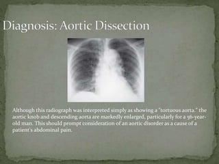
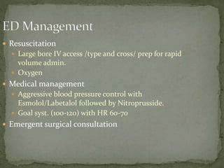
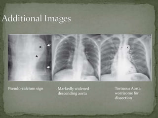
 56 yo wm with a pmhx notable
for hypertension and coronary
artery disease presents with 4
hours of epigastric abdominal
pain he describes as “sharp” or
“stabbing”. He denies any
previus similar episodes. His
pain began at rest and has
remained constant. He denies
any recent illness or injury –
no n/v, cough/congestion,
hematemesis or
hematochezia. At presentation
he is awake/alert in no
respiratory distress.
 T 98.7 P 125 BP 105/60
O2 95% RR 20
 Gen: WDWN, anxious
 CV: Tachycardic,
RRythm, cr approx 3
seconds
 Pulm: Lungs CTA bilat
 Abd: Obese, non-
tender, no pulsatile
mass. Hypoactive
bowel sounds.
 Extremities – cool to
the touch
Although this radiograph was interpreted simply as showing a "tortuous aorta." the
aortic knob and descending aorta are markedly enlarged, particularly for a 56-year-
old man. This should prompt consideration of an aortic disorder as a cause of a
patient's abdominal pain.
 Resuscitation
 Large bore IV access /type and cross/ prep for rapid
volume admin.
 Oxygen
 Medical management
 Aggressive blood pressure control with
Esmolol/Labetalol followed by Nitroprusside.
 Goal syst. (100-120) with HR 60-70
 Emergent surgical consultation
 Presentation
 Abrupt onset is the most common historical clue.
 Pain quality and migration are inconsistent complaints
 A past medical history of hypertension is the most common risk
factor
 Signs of Aortic branch occlusion (neurologic deficits, pulse
deficit/ discrepancy, mesenteric/renal ischemia, murmur of aortic
insufficiency) are present in the minority of cases but are a
hallmark of aortic dissection.
 Do not miss an aortic dissection in the clinical setting of stroke with
chest/abdominal pain.
 8 cm is upper limit of normal for mediastinal width
 CXR
 Findings:
 (1) enlargement of the aorta (the principal sign),
 (2) enlargement of one aortic segment in comparison to the others
(ascending aorta, aortic knob, or descending aorta)
 (3) progressive enlargement of the aorta compared to prior
radiographs
 (4) widening of the mediastinum,
 (5) a pleural effusion (usually left-sided)
 (6) displacement of the calcified intima into the lumen of the
aorta—the “pseudo -calcium sign“
 Utility
 Chest x-ray shows moderate sensitivity/specificity in the diagnosis of aortic
dissection, however in combination with the clinical picture it’s utility
is greatly increased in that it is a rapid bedside study.
 Enlargement of the Aorta on CXR is age-related, making the finding more
useful in diagnosing dissection in a younger individual.
Pseudo-calcium sign Markedly widened
descending aorta
Tortuous Aorta
worrisome for
dissection

Aortic dissection

  • 2.
     56 yowm with a pmhx notable for hypertension and coronary artery disease presents with 4 hours of epigastric abdominal pain he describes as “sharp” or “stabbing”. He denies any previus similar episodes. His pain began at rest and has remained constant. He denies any recent illness or injury – no n/v, cough/congestion, hematemesis or hematochezia. At presentation he is awake/alert in no respiratory distress.  T 98.7 P 125 BP 105/60 O2 95% RR 20  Gen: WDWN, anxious  CV: Tachycardic, RRythm, cr approx 3 seconds  Pulm: Lungs CTA bilat  Abd: Obese, non- tender, no pulsatile mass. Hypoactive bowel sounds.  Extremities – cool to the touch
  • 4.
    Although this radiographwas interpreted simply as showing a "tortuous aorta." the aortic knob and descending aorta are markedly enlarged, particularly for a 56-year- old man. This should prompt consideration of an aortic disorder as a cause of a patient's abdominal pain.
  • 5.
     Resuscitation  Largebore IV access /type and cross/ prep for rapid volume admin.  Oxygen  Medical management  Aggressive blood pressure control with Esmolol/Labetalol followed by Nitroprusside.  Goal syst. (100-120) with HR 60-70  Emergent surgical consultation
  • 6.
     Presentation  Abruptonset is the most common historical clue.  Pain quality and migration are inconsistent complaints  A past medical history of hypertension is the most common risk factor  Signs of Aortic branch occlusion (neurologic deficits, pulse deficit/ discrepancy, mesenteric/renal ischemia, murmur of aortic insufficiency) are present in the minority of cases but are a hallmark of aortic dissection.  Do not miss an aortic dissection in the clinical setting of stroke with chest/abdominal pain.  8 cm is upper limit of normal for mediastinal width
  • 7.
     CXR  Findings: (1) enlargement of the aorta (the principal sign),  (2) enlargement of one aortic segment in comparison to the others (ascending aorta, aortic knob, or descending aorta)  (3) progressive enlargement of the aorta compared to prior radiographs  (4) widening of the mediastinum,  (5) a pleural effusion (usually left-sided)  (6) displacement of the calcified intima into the lumen of the aorta—the “pseudo -calcium sign“  Utility  Chest x-ray shows moderate sensitivity/specificity in the diagnosis of aortic dissection, however in combination with the clinical picture it’s utility is greatly increased in that it is a rapid bedside study.  Enlargement of the Aorta on CXR is age-related, making the finding more useful in diagnosing dissection in a younger individual.
  • 8.
    Pseudo-calcium sign Markedlywidened descending aorta Tortuous Aorta worrisome for dissection