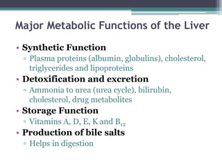
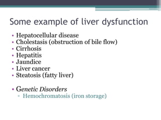
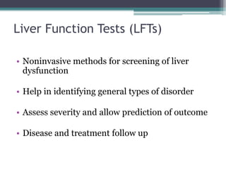
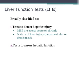
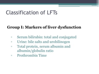
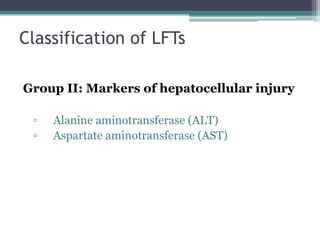
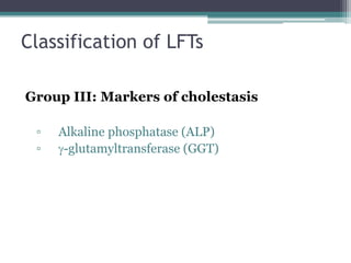
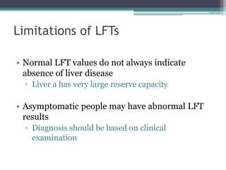
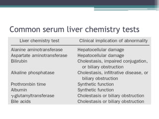
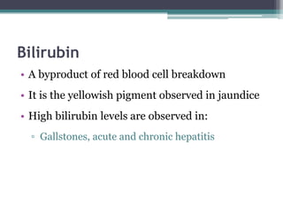
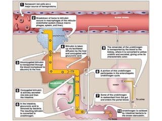
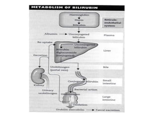
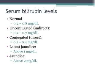
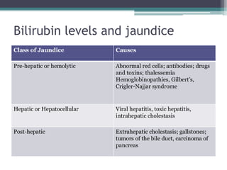
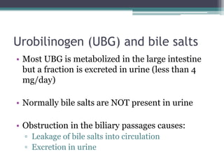
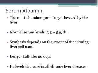
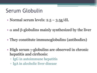
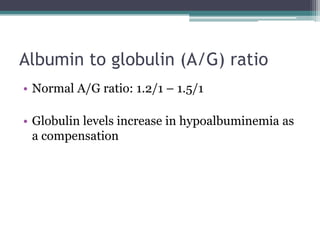
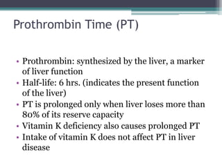
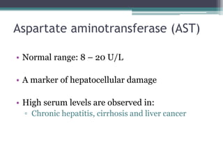
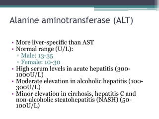
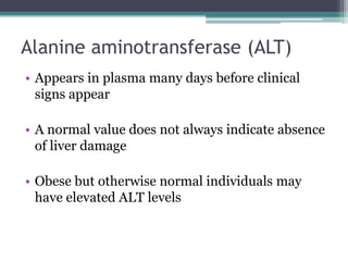
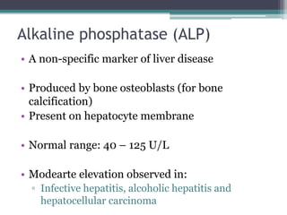
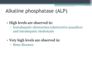
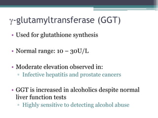
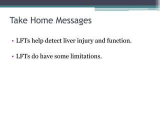
Liver
Function
Tests (LFTs)
GIT/Hematology Block
Objectives
Upon completion of this lecture, the students
should be able to:
• Understand the major metabolic functions of the liver and
causes of liver dysfunction.
• Discuss markers of liver function tests such as liver
enzymes, bilirubin, albumin and prothrombin time that
can diagnose hepatic injury and assess hepatic function.
Major Metabolic Functions of the Liver
• Synthetic Function
▫ Plasma proteins (albumin, globulins), cholesterol,
triglycerides and lipoproteins
• Detoxification and excretion
▫ Ammonia to urea (urea cycle), bilirubin,
cholesterol, drug metabolites
• Storage Function
▫ Vitamins A, D, E, K and B12
• Production of bile salts
▫ Helps in digestion
Some example of liver dysfunction
• Hepatocellular disease
• Cholestasis (obstruction of bile flow)
• Cirrhosis
• Hepatitis
• Jaundice
• Liver cancer
• Steatosis (fatty liver)
• Genetic Disorders
▫ Hemochromatosis (iron storage)
Liver Function Tests (LFTs)
• Noninvasive methods for screening of liver
dysfunction
• Help in identifying general types of disorder
• Assess severity and allow prediction of outcome
• Disease and treatment follow up
Liver Function Tests (LFTs)
Broadly classified as:
1.Tests to detect hepatic injury:
• Mild or severe; acute or chronic
• Nature of liver injury (hepatocellular or
cholestasis)
2.Tests to assess hepatic function
Classification of LFTs
Group I: Markers of liver dysfunction
▫ Serum bilirubin: total and conjugated
▫ Urine: bile salts and urobilinogen
▫ Total protein, serum albumin and
albumin/globulin ratio
▫ Prothrombin Time
Classification of LFTs
Group II: Markers of hepatocellular injury
▫ Alanine aminotransferase (ALT)
▫ Aspartate aminotransferase (AST)
Classification of LFTs
Group III: Markers of cholestasis
▫ Alkaline phosphatase (ALP)
▫ g-glutamyltransferase (GGT)
Limitations of LFTs
• Normal LFT values do not always indicate
absence of liver disease
▫ Liver a has very large reserve capacity
• Asymptomatic people may have abnormal LFT
results
▫ Diagnosis should be based on clinical
examination
Common serum liver chemistry tests
Bilirubin
• A byproduct of red blood cell breakdown
• It is the yellowish pigment observed in jaundice
• High bilirubin levels are observed in:
▫ Gallstones, acute and chronic hepatitis
Serum bilirubin levels
• Normal
▫ 0.2 – 0.8 mg/dL
• Unconjugated (indirect):
▫ 0.2 – 0.7 mg/dL
• Conjugated (direct):
▫ 0.1 – 0.4 mg/dL
• Latent jaundice:
▫ Above 1 mg/dL
• Jaundice:
▫ Above 2 mg/dL
Bilirubin levels and jaundice
Class of Jaundice Causes
Pre-hepatic or hemolytic Abnormal red cells; antibodies; drugs
and toxins; thalessemia
Hemoglobinopathies, Gilbert’s,
Crigler-Najjar syndrome
Hepatic or Hepatocellular Viral hepatitis, toxic hepatitis,
intrahepatic cholestasis
Post-hepatic Extrahepatic cholestasis; gallstones;
tumors of the bile duct, carcinoma of
pancreas
Urobilinogen (UBG) and bile salts
• Most UBG is metabolized in the large intestine
but a fraction is excreted in urine (less than 4
mg/day)
• Normally bile salts are NOT present in urine
• Obstruction in the biliary passages causes:
▫ Leakage of bile salts into circulation
▫ Excretion in urine
Serum Albumin
• The most abundant protein synthesized by the
liver
• Normal serum levels: 3.5 – 5 g/dL
• Synthesis depends on the extent of functioning
liver cell mass
• Longer half-life: 20 days
• Its levels decrease in all chronic liver diseases
Serum Globulin
• Normal serum levels: 2.5 – 3.5g/dL
• a and b-globulins mainly synthesized by the liver
• They constitute immunoglobulins (antibodies)
• High serum g-globulins are observed in chronic
hepatitis and cirrhosis:
▫ IgG in autoimmune hepatitis
▫ IgA in alcoholic liver disease
Albumin to globulin (A/G) ratio
• Normal A/G ratio: 1.2/1 – 1.5/1
• Globulin levels increase in hypoalbuminemia as
a compensation
Prothrombin Time (PT)
• Prothrombin: synthesized by the liver, a marker
of liver function
• Half-life: 6 hrs. (indicates the present function
of the liver)
• PT is prolonged only when liver loses more than
80% of its reserve capacity
• Vitamin K deficiency also causes prolonged PT
• Intake of vitamin K does not affect PT in liver
disease
Aspartate aminotransferase (AST)
• Normal range: 8 – 20 U/L
• A marker of hepatocellular damage
• High serum levels are observed in:
▫ Chronic hepatitis, cirrhosis and liver cancer
Alanine aminotransferase (ALT)
• More liver-specific than AST
• Normal range (U/L):
▫ Male: 13-35
▫ Female: 10-30
• High serum levels in acute hepatitis (300-
1000U/L)
• Moderate elevation in alcoholic hepatitis (100-
300U/L)
• Minor elevation in cirrhosis, hepatitis C and
non-alcoholic steatohepatitis (NASH) (50-
100U/L)
Alanine aminotransferase (ALT)
• Appears in plasma many days before clinical
signs appear
• A normal value does not always indicate absence
of liver damage
• Obese but otherwise normal individuals may
have elevated ALT levels
Alkaline phosphatase (ALP)
• A non-specific marker of liver disease
• Produced by bone osteoblasts (for bone
calcification)
• Present on hepatocyte membrane
• Normal range: 40 – 125 U/L
• Modearte elevation observed in:
▫ Infective hepatitis, alcoholic hepatitis and
hepatocellular carcinoma
Alkaline phosphatase (ALP)
• High levels are observed in:
▫ Extrahepatic obstruction (obstructive jaundice)
and intrahepatic cholestasis
• Very high levels are observed in:
▫ Bone diseases
g-glutamyltransferase (GGT)
• Used for glutathione synthesis
• Normal range: 10 – 30U/L
• Moderate elevation observed in:
▫ Infective hepatitis and prostate cancers
• GGT is increased in alcoholics despite normal
liver function tests
▫ Highly sensitive to detecting alcohol abuse
Take Home Messages
• LFTs help detect liver injury and function.
• LFTs do have some limitations.
References
• Lippincott’s Illustrated Reviews Biochemistry: 6th
edition, Unit IV, Chapter 21, Pages 282 - 285.
• Lecture notes: Clinical Biochemistry: 9th edition, Chapter
13, Pages 174 - 187.
• Clinical Chemistry - Techniques, Principales and
Correlations: 6th edition, Chapter 24, Pages 520 -521.

liver_function_tests.ppt

  • 1.
  • 2.
    Objectives Upon completion ofthis lecture, the students should be able to: • Understand the major metabolic functions of the liver and causes of liver dysfunction. • Discuss markers of liver function tests such as liver enzymes, bilirubin, albumin and prothrombin time that can diagnose hepatic injury and assess hepatic function.
  • 3.
    Major Metabolic Functionsof the Liver • Synthetic Function ▫ Plasma proteins (albumin, globulins), cholesterol, triglycerides and lipoproteins • Detoxification and excretion ▫ Ammonia to urea (urea cycle), bilirubin, cholesterol, drug metabolites • Storage Function ▫ Vitamins A, D, E, K and B12 • Production of bile salts ▫ Helps in digestion
  • 4.
    Some example ofliver dysfunction • Hepatocellular disease • Cholestasis (obstruction of bile flow) • Cirrhosis • Hepatitis • Jaundice • Liver cancer • Steatosis (fatty liver) • Genetic Disorders ▫ Hemochromatosis (iron storage)
  • 5.
    Liver Function Tests(LFTs) • Noninvasive methods for screening of liver dysfunction • Help in identifying general types of disorder • Assess severity and allow prediction of outcome • Disease and treatment follow up
  • 6.
    Liver Function Tests(LFTs) Broadly classified as: 1.Tests to detect hepatic injury: • Mild or severe; acute or chronic • Nature of liver injury (hepatocellular or cholestasis) 2.Tests to assess hepatic function
  • 7.
    Classification of LFTs GroupI: Markers of liver dysfunction ▫ Serum bilirubin: total and conjugated ▫ Urine: bile salts and urobilinogen ▫ Total protein, serum albumin and albumin/globulin ratio ▫ Prothrombin Time
  • 8.
    Classification of LFTs GroupII: Markers of hepatocellular injury ▫ Alanine aminotransferase (ALT) ▫ Aspartate aminotransferase (AST)
  • 9.
    Classification of LFTs GroupIII: Markers of cholestasis ▫ Alkaline phosphatase (ALP) ▫ g-glutamyltransferase (GGT)
  • 10.
    Limitations of LFTs •Normal LFT values do not always indicate absence of liver disease ▫ Liver a has very large reserve capacity • Asymptomatic people may have abnormal LFT results ▫ Diagnosis should be based on clinical examination
  • 11.
    Common serum liverchemistry tests
  • 12.
    Bilirubin • A byproductof red blood cell breakdown • It is the yellowish pigment observed in jaundice • High bilirubin levels are observed in: ▫ Gallstones, acute and chronic hepatitis
  • 15.
    Serum bilirubin levels •Normal ▫ 0.2 – 0.8 mg/dL • Unconjugated (indirect): ▫ 0.2 – 0.7 mg/dL • Conjugated (direct): ▫ 0.1 – 0.4 mg/dL • Latent jaundice: ▫ Above 1 mg/dL • Jaundice: ▫ Above 2 mg/dL
  • 16.
    Bilirubin levels andjaundice Class of Jaundice Causes Pre-hepatic or hemolytic Abnormal red cells; antibodies; drugs and toxins; thalessemia Hemoglobinopathies, Gilbert’s, Crigler-Najjar syndrome Hepatic or Hepatocellular Viral hepatitis, toxic hepatitis, intrahepatic cholestasis Post-hepatic Extrahepatic cholestasis; gallstones; tumors of the bile duct, carcinoma of pancreas
  • 17.
    Urobilinogen (UBG) andbile salts • Most UBG is metabolized in the large intestine but a fraction is excreted in urine (less than 4 mg/day) • Normally bile salts are NOT present in urine • Obstruction in the biliary passages causes: ▫ Leakage of bile salts into circulation ▫ Excretion in urine
  • 18.
    Serum Albumin • Themost abundant protein synthesized by the liver • Normal serum levels: 3.5 – 5 g/dL • Synthesis depends on the extent of functioning liver cell mass • Longer half-life: 20 days • Its levels decrease in all chronic liver diseases
  • 19.
    Serum Globulin • Normalserum levels: 2.5 – 3.5g/dL • a and b-globulins mainly synthesized by the liver • They constitute immunoglobulins (antibodies) • High serum g-globulins are observed in chronic hepatitis and cirrhosis: ▫ IgG in autoimmune hepatitis ▫ IgA in alcoholic liver disease
  • 20.
    Albumin to globulin(A/G) ratio • Normal A/G ratio: 1.2/1 – 1.5/1 • Globulin levels increase in hypoalbuminemia as a compensation
  • 21.
    Prothrombin Time (PT) •Prothrombin: synthesized by the liver, a marker of liver function • Half-life: 6 hrs. (indicates the present function of the liver) • PT is prolonged only when liver loses more than 80% of its reserve capacity • Vitamin K deficiency also causes prolonged PT • Intake of vitamin K does not affect PT in liver disease
  • 22.
    Aspartate aminotransferase (AST) •Normal range: 8 – 20 U/L • A marker of hepatocellular damage • High serum levels are observed in: ▫ Chronic hepatitis, cirrhosis and liver cancer
  • 23.
    Alanine aminotransferase (ALT) •More liver-specific than AST • Normal range (U/L): ▫ Male: 13-35 ▫ Female: 10-30 • High serum levels in acute hepatitis (300- 1000U/L) • Moderate elevation in alcoholic hepatitis (100- 300U/L) • Minor elevation in cirrhosis, hepatitis C and non-alcoholic steatohepatitis (NASH) (50- 100U/L)
  • 24.
    Alanine aminotransferase (ALT) •Appears in plasma many days before clinical signs appear • A normal value does not always indicate absence of liver damage • Obese but otherwise normal individuals may have elevated ALT levels
  • 25.
    Alkaline phosphatase (ALP) •A non-specific marker of liver disease • Produced by bone osteoblasts (for bone calcification) • Present on hepatocyte membrane • Normal range: 40 – 125 U/L • Modearte elevation observed in: ▫ Infective hepatitis, alcoholic hepatitis and hepatocellular carcinoma
  • 26.
    Alkaline phosphatase (ALP) •High levels are observed in: ▫ Extrahepatic obstruction (obstructive jaundice) and intrahepatic cholestasis • Very high levels are observed in: ▫ Bone diseases
  • 27.
    g-glutamyltransferase (GGT) • Usedfor glutathione synthesis • Normal range: 10 – 30U/L • Moderate elevation observed in: ▫ Infective hepatitis and prostate cancers • GGT is increased in alcoholics despite normal liver function tests ▫ Highly sensitive to detecting alcohol abuse
  • 28.
    Take Home Messages •LFTs help detect liver injury and function. • LFTs do have some limitations.
  • 29.
    References • Lippincott’s IllustratedReviews Biochemistry: 6th edition, Unit IV, Chapter 21, Pages 282 - 285. • Lecture notes: Clinical Biochemistry: 9th edition, Chapter 13, Pages 174 - 187. • Clinical Chemistry - Techniques, Principales and Correlations: 6th edition, Chapter 24, Pages 520 -521.

Editor's Notes

  • #18 Obstruction can occur in obstructive jaundice and also in hepatic jaundice due to obstruction of microbiliary channels caused by inflammation