Objectives
Upon completion ofthis lecture, the students
should be able to:
•Understand the major metabolic functions of the liver and
causes of liver dysfunction.
•Discuss markers of liver function tests such as liver
enzymes, bilirubin, albumin and prothrombin time that can
diagnose hepatic injury and assess hepatic function.
3.
Major Metabolic Functionsof the
Liver
• Synthetic Function
▫ Plasma proteins (albumin, globulins), cholesterol,
triglycerides and lipoproteins
• Detoxification and excretion
▫ Ammonia to urea (urea cycle), bilirubin,
cholesterol, drug metabolites
• Storage Function
▫ Vitamins A, D, E, K and B12
• Production of bile salts
▫ Helps in digestion
4.
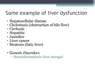
Some example ofliver dysfunction
• Hepatocellular disease
• Cholestasis (obstruction of bile flow)
• Cirrhosis
• Hepatitis
• Jaundice
• Liver cancer
• Steatosis (fatty liver)
• Genetic Disorders
▫ Hemochromatosis (iron storage)
5.
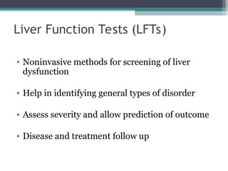
Liver Function Tests(LFTs)
• Noninvasive methods for screening of liver
dysfunction
• Help in identifying general types of disorder
• Assess severity and allow prediction of outcome
• Disease and treatment follow up
6.
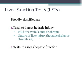
Liver Function Tests(LFTs)
Broadly classified as:
1.Tests to detect hepatic injury:
• Mild or severe; acute or chronic
• Nature of liver injury (hepatocellular or
cholestasis)
2.Tests to assess hepatic function
7.
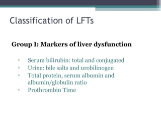
Classification of LFTs
GroupI: Markers of liver dysfunction
▫ Serum bilirubin: total and conjugated
▫ Urine: bile salts and urobilinogen
▫ Total protein, serum albumin and
albumin/globulin ratio
▫ Prothrombin Time
8.
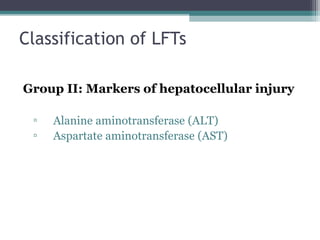
Classification of LFTs
GroupII: Markers of hepatocellular injury
▫ Alanine aminotransferase (ALT)
▫ Aspartate aminotransferase (AST)
9.
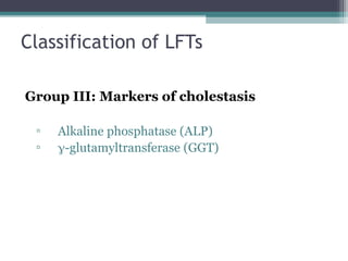
Classification of LFTs
GroupIII: Markers of cholestasis
▫ Alkaline phosphatase (ALP)
▫ -glutamyltransferase (GGT)
10.
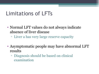
Limitations of LFTs
•Normal LFT values do not always indicate
absence of liver disease
▫ Liver a has very large reserve capacity
• Asymptomatic people may have abnormal LFT
results
▫ Diagnosis should be based on clinical
examination
Bilirubin
• A byproductof red blood cell breakdown
• It is the yellowish pigment observed in jaundice
• High bilirubin levels are observed in:
▫ Gallstones, acute and chronic hepatitis
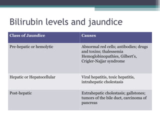
Bilirubin levels andjaundice
Class of Jaundice Causes
Pre-hepatic or hemolytic Abnormal red cells; antibodies; drugs
and toxins; thalessemia
Hemoglobinopathies, Gilbert’s,
Crigler-Najjar syndrome
Hepatic or Hepatocellular Viral hepatitis, toxic hepatitis,
intrahepatic cholestasis
Post-hepatic Extrahepatic cholestasis; gallstones;
tumors of the bile duct, carcinoma of
pancreas
17.
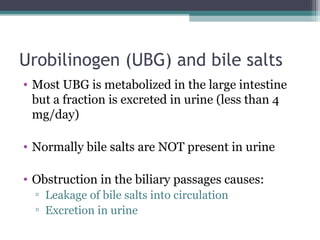
Urobilinogen (UBG) andbile salts
• Most UBG is metabolized in the large intestine
but a fraction is excreted in urine (less than 4
mg/day)
• Normally bile salts are NOT present in urine
• Obstruction in the biliary passages causes:
▫ Leakage of bile salts into circulation
▫ Excretion in urine
18.
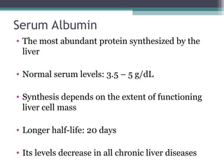
Serum Albumin
• Themost abundant protein synthesized by the
liver
• Normal serum levels: 3.5 – 5 g/dL
• Synthesis depends on the extent of functioning
liver cell mass
• Longer half-life: 20 days
• Its levels decrease in all chronic liver diseases
19.
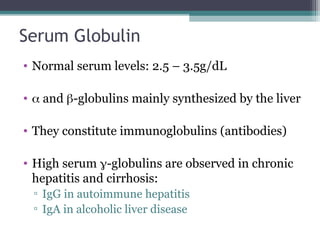
Serum Globulin
• Normalserum levels: 2.5 – 3.5g/dL
• and -globulins mainly synthesized by the liver
• They constitute immunoglobulins (antibodies)
• High serum -globulins are observed in chronic
hepatitis and cirrhosis:
▫ IgG in autoimmune hepatitis
▫ IgA in alcoholic liver disease
20.
Albumin to globulin(A/G) ratio
• Normal A/G ratio: 1.2/1 – 1.5/1
• Globulin levels increase in hypoalbuminemia as
a compensation
21.
Prothrombin Time (PT)
•Prothrombin: synthesized by the liver, a marker
of liver function
• Half-life: 6 hrs. (indicates the present function of
the liver)
• PT is prolonged only when liver loses more than
80% of its reserve capacity
• Vitamin K deficiency also causes prolonged PT
• Intake of vitamin K does not affect PT in liver
disease
22.
Aspartate aminotransferase (AST)
•Normal range: 8 – 20 U/L
• A marker of hepatocellular damage
• High serum levels are observed in:
▫ Chronic hepatitis, cirrhosis and liver cancer
23.
Alanine aminotransferase (ALT)
•More liver-specific than AST
• Normal range (U/L):
▫ Male: 13-35
▫ Female: 10-30
• High serum levels in acute hepatitis (300-
1000U/L)
• Moderate elevation in alcoholic hepatitis (100-
300U/L)
• Minor elevation in cirrhosis, hepatitis C and
non-alcoholic steatohepatitis (NASH) (50-
100U/L)
24.
Alanine aminotransferase (ALT)
•Appears in plasma many days before clinical
signs appear
• A normal value does not always indicate absence
of liver damage
• Obese but otherwise normal individuals may
have elevated ALT levels
25.
Alkaline phosphatase (ALP)
•A non-specific marker of liver disease
• Produced by bone osteoblasts (for bone
calcification)
• Present on hepatocyte membrane
• Normal range: 40 – 125 U/L
• Modearte elevation observed in:
▫ Infective hepatitis, alcoholic hepatitis and
hepatocellular carcinoma
26.
Alkaline phosphatase (ALP)
•High levels are observed in:
▫ Extrahepatic obstruction (obstructive jaundice)
and intrahepatic cholestasis
• Very high levels are observed in:
▫ Bone diseases
27.
-glutamyltransferase (GGT)
• Usedfor glutathione synthesis
• Normal range: 10 – 30U/L
• Moderate elevation observed in:
▫ Infective hepatitis and prostate cancers
• GGT is increased in alcoholics despite normal
liver function tests
▫ Highly sensitive to detecting alcohol abuse
28.
Take Home Messages
•LFTs help detect liver injury and function.
• LFTs do have some limitations.


![L8-Liver_Function_Test[1] SF.pptbwdqdbdwub](https://cdn.slidesharecdn.com/ss_thumbnails/l8-liverfunctiontest1sf-250507164033-55d694e3-thumbnail.jpg?width=640&height=640&fit=bounds)


