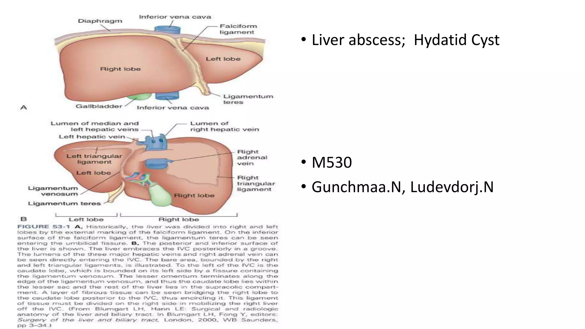
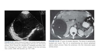
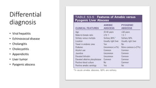
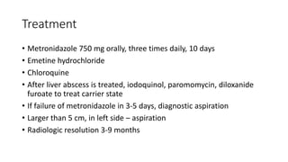
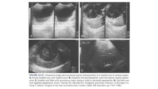
• Liver abscess; Hydatid Cyst
• M530
• Gunchmaa.N, Ludevdorj.N
Pyogenic Abscess
Epidemiology
• Patients in their 50s or 60s
• Often related to biliary tract disease
• Cryptogenic in nature (uncertain in origin)
Pathogenesis
• An inoculum of bacteria exceeds the livers ability to clear it.
• Tissue invasion
• Neutrophil infiltration
• Abscess formation
Biliary tree
• Stone or malignant disease
• Biliary obstruction
• Bile stasis
• Bacterial colonization
• Ascends to liver
• Ascending suppurative cholangitis
Portal vein
• Ascending portal vein infection – pyelophlebitis
• Diverticulitis, appendicitis, pancreatitis, IBD etc
• Malignant disease
Hepatic artery
• Any systemic infection
• Altered immune response
• Direct infection extension
Other causes
• Necrotic liver:
• Trauma
• Hepatic artery embolization
• Thermal ablative procedures
Pathology
• 75% right hemiliver
• 20% left hemiliver
• 5% caudate lobe
• Mostly solitary
• 1 mm – 4 cm
• Multiloculated or in single cavity
Microbiology
• Either polymicrobial or single organism
• Escherichia coli
• Klebsiella pneumoniae
• Staphylococcus aureus
• Enterococcus sp.
Clinical features
• Fever
• Chills
• Jaundice
• RUQ pain
• Tenderness to
palpation
• Hepatomegaly
• Acute or
chronic
Complication
• If diaphragm is involved, cough or dyspnea
• Peritonitis secondary to rupture
• Rupture into pleural space or pericardium
• Endogenous endophthalmitis (Klebsiella)
Lab findings
• Leukocytosis
• Anemia
• LFT abnormality
• ALP elevated /80%/
• Total bilirubin elevated /20-50%/
• Transaminases increase /60%/
• Hypoalbuminemia, increased PT
Radiology
• Chest X-ray
Atelectasis
Right pleural effusion
Elevated right
hemidiaphragm
• Plain abdominal X-ray
- Air fluid levels
- Portal venous gas
Radiology
• Ultrasound – round or oval, less echogenic than surrounding liver
• CT – lesions are of lower attenuation than surrounding liver, multiple
small abscesses
• Both are useful in diagnosing intra-abdominal diseases
Differential diagnosis
• Amebic abscess
• Echinococcal cyst
Treatment
• Immediate broad spectrum IV antibiotics /2 weeks</
• Combination of ampicillin, aminoglycosides, metronidazole
• Aspiration, send for culture.
• Percutaneous catheter drainage
• Contraindications: ascites, coagulopathy, proximity to vital structures
• If larger than 5 cm, surgery is better
• Liver resection if destruction is severe
Amebic Abscess
Epidemiology
• Largely a disease in tropical or developing countries
• Less than 50% are symptomatic
• Mostly Hispanic men
• 20-40 age
• Socioeconomic status
• Alcohol consumption
• Immunosuppression
• Travel history
Pathogenesis
• E.histolytica is a protozoan that exists as trophozoite or cyst
• Fecal-oral route
• When ingested, cysts aren’t degraded in stomach
• Trophozoite released
• Invades colon mucosa
• Enzymatic cellular hydrolysis
Pathogenesis
• Localized necrosis
• Cavity forms
• It contains proteinaceous debris
• Surrounded by rim of trophozoites
• Rapid IgA release
Pathology
• Cavity full of blood, liquefied liver tissue
• The fluid is odorless
• Glisson capsule resistant to hydrolysis
• Early stage, ill-defined
• Right hemiliver
Clinical features
• Last from days to 4 weeks
• Fever
• Chills
• RUQ pain and tenderness
• Hepatomegaly
• Constant, dull abdominal pain
• Diarrhea
• Anorexia
• Jaundice
Clinical features
• Acute <10 days
• High fevers, chills, abdominal tenderness
• Multiple lesions usually
• Chronic > 2 weeks
• Single right-sided lesion
• Response is similar in both groups
Lab findings
• Mild to moderate leukocytosis with no eosinophilia
• Anemia
• LFT abnormality
• Elevated PT-INR
• Enzyme immunoassay
• Detection kit
Radiology
• Plain chest x-ray: elevated right hemidiaphragm, pleural effusion,
atelectasis
• Ultrasound: Round lesions, liver capsule, without significant rim
echoes. Contents are hypoechogenic, nonhomogenous
• CT: more sensitive
• Nuclear studies: to differentiate from pyogenic abscess. No
leukocytes, does not light up
Differential
diagnosis
• Viral hepatitis
• Echinococcal disease
• Cholangitis
• Cholecystitis
• Appendicitis
• Liver tumor
• Pyogenic abscess
Treatment
• Metronidazole 750 mg orally, three times daily, 10 days
• Emetine hydrochloride
• Chloroquine
• After liver abscess is treated, iodoquinol, paromomycin, diloxanide
furoate to treat carrier state
• If failure of metronidazole in 3-5 days, diagnostic aspiration
• Larger than 5 cm, in left side – aspiration
• Radiologic resolution 3-9 months
Hydatid Cyst
• Hydatid disease or echinococcosis is a zoonosis that is common
worldwide because the dog is a definitive host.
• Human contract the disease from dogs, but there is no human to
human transmission
• 3 species that cause hydatid cyst:
• Echinococcus granulosus
• Echinococcus multilocularis
• Echinococcus ligartus
• Humans are end stage to the parasite.
• In the human duodenum, the parasitic embryo releases an
oncosphere containing hooklets that penetrate the mucosa, allowing
access to the bloodstream.
• In the blood, it can reaches the liver or lungs, where the parasite
develops its larval stage
• 3 weeks after infection, a visible hydatid cyst develops, which then
slowly grows in a spherical manner.
• The cyst wall itself has 2 layers, an outer gelatinous membrane
(ectocyst) and an inner germinal membrane (endocyst).
• Brood capsules are small, intracystic cellular masses in which future
worm heads develop into scoleces. In a definitive host, the scoleces
develops into an adult tapeworm.
Hydatid cysts can die with:
• degeneration of the membranes
• Development of cystic vacuoles
• Calcification of the wall
• Calcification of a hydatid cyst, however, doesn’t always imply that the
cyst is dead
• Hydatic cysts are diagnosed in equal numbers of en and woman at an
average age of about 45 years.
• Approximately 75% of hydatid cysts are located in the right liver and
are solitary
• The clinical presentation of a hydatid cyst is largely asymptomatic
until complications occur.
• Dyspepsia
• Abdominal pain
• Vomiting
• Hepatomegaly are the most common symptoms.
• Jaundice and fever are each present in approximately 8% of patients.
• Bacterial super infection of a hydatid cyst can occur and be
manifested like a pyogenic abscess
• Rupture of the cyst into the biliary tree or bronchial tree or free
rupture into the peritoneal , pleural, or pericardial cavities can occur.
Diagnosis
• Serologic tests are available to evaluate antibody response, but all are
plagued by low sensitivity and specifity
• Ultrasound is more common, but it depends on the stage of the cyst
at the time of examination
• Simple hydatid cyst is well circumscribed with budding signs on the
cyst membrane
• May contain free floating hyperechogenic hydatid sand
• A rosette appearance is seen when daughter cysts are present
• Cyst can be filled with an amorphous mass, which can be
diagnostically misleading
• Calcification in the wall of the cyst is highly suggestive of hydatid
disease
• Similar findings are seen on CT or MRI scans
• In patients with suspected biliary involvement, ERCP or percutaneous
transhepatic cholangiography may neccesary
Treatment
• Primarily surgical
• Most cyst should be treated
• But in older patients with small, asymptomatic, densely calcified
cysts, conservative management ( drainage nad evacuation) is
appropriate.
• Epinephrine and steroids taken by anesthesiologist
• Packing off the abdomen is important because rupture can result in
anaphylaxis and diffuse seeding
• Then cyst ih aspirated through a closed suctionsystem and flushed
with a scolicidal agent (hypertonic saline)
• The cyst is then unroofed which can then be followed by a number of
possibilities, including
• excision ( or pericystectomy)
• Marsupialization procedures
• Leaving the cyst open
• Drainage of the cyst
• Omentoplasty
• Partial hepatectomy
• Pericystectomy or formal partial
hepatectomy can also be
performed without entering thy
cyst.
• Simple suture repair is often sufficient, but major biliary repairs,
approaches through the common bile duct, or postoperative ERCP
may be neccesary
• Recurrence rates after surgical treatment is generally 5% or less
• Preoperative treatment my decrease the risk of spillage.
• Albendazole or mebendazole is effective with E.granulosus infection,
but cyst appearance occurs 50% of patients
• Medical treatment without resection or drainage should be
considered only for widely diseminated disese or poor surgical
candidate
Thank you!

Liver Abscess and Hydatid Cyst, Surgery

  • 1.
    • Liver abscess;Hydatid Cyst • M530 • Gunchmaa.N, Ludevdorj.N
  • 4.
  • 5.
    Epidemiology • Patients intheir 50s or 60s • Often related to biliary tract disease • Cryptogenic in nature (uncertain in origin)
  • 6.
    Pathogenesis • An inoculumof bacteria exceeds the livers ability to clear it. • Tissue invasion • Neutrophil infiltration • Abscess formation
  • 7.
    Biliary tree • Stoneor malignant disease • Biliary obstruction • Bile stasis • Bacterial colonization • Ascends to liver • Ascending suppurative cholangitis
  • 8.
    Portal vein • Ascendingportal vein infection – pyelophlebitis • Diverticulitis, appendicitis, pancreatitis, IBD etc • Malignant disease
  • 9.
    Hepatic artery • Anysystemic infection • Altered immune response • Direct infection extension
  • 10.
    Other causes • Necroticliver: • Trauma • Hepatic artery embolization • Thermal ablative procedures
  • 11.
    Pathology • 75% righthemiliver • 20% left hemiliver • 5% caudate lobe • Mostly solitary • 1 mm – 4 cm • Multiloculated or in single cavity
  • 12.
    Microbiology • Either polymicrobialor single organism • Escherichia coli • Klebsiella pneumoniae • Staphylococcus aureus • Enterococcus sp.
  • 13.
    Clinical features • Fever •Chills • Jaundice • RUQ pain • Tenderness to palpation • Hepatomegaly • Acute or chronic
  • 14.
    Complication • If diaphragmis involved, cough or dyspnea • Peritonitis secondary to rupture • Rupture into pleural space or pericardium • Endogenous endophthalmitis (Klebsiella)
  • 15.
    Lab findings • Leukocytosis •Anemia • LFT abnormality • ALP elevated /80%/ • Total bilirubin elevated /20-50%/ • Transaminases increase /60%/ • Hypoalbuminemia, increased PT
  • 16.
    Radiology • Chest X-ray Atelectasis Rightpleural effusion Elevated right hemidiaphragm • Plain abdominal X-ray - Air fluid levels - Portal venous gas
  • 17.
    Radiology • Ultrasound –round or oval, less echogenic than surrounding liver • CT – lesions are of lower attenuation than surrounding liver, multiple small abscesses • Both are useful in diagnosing intra-abdominal diseases
  • 19.
    Differential diagnosis • Amebicabscess • Echinococcal cyst
  • 20.
    Treatment • Immediate broadspectrum IV antibiotics /2 weeks</ • Combination of ampicillin, aminoglycosides, metronidazole • Aspiration, send for culture. • Percutaneous catheter drainage • Contraindications: ascites, coagulopathy, proximity to vital structures • If larger than 5 cm, surgery is better • Liver resection if destruction is severe
  • 21.
  • 22.
    Epidemiology • Largely adisease in tropical or developing countries • Less than 50% are symptomatic • Mostly Hispanic men • 20-40 age • Socioeconomic status • Alcohol consumption • Immunosuppression • Travel history
  • 23.
    Pathogenesis • E.histolytica isa protozoan that exists as trophozoite or cyst • Fecal-oral route • When ingested, cysts aren’t degraded in stomach • Trophozoite released • Invades colon mucosa • Enzymatic cellular hydrolysis
  • 24.
    Pathogenesis • Localized necrosis •Cavity forms • It contains proteinaceous debris • Surrounded by rim of trophozoites • Rapid IgA release
  • 25.
    Pathology • Cavity fullof blood, liquefied liver tissue • The fluid is odorless • Glisson capsule resistant to hydrolysis • Early stage, ill-defined • Right hemiliver
  • 26.
    Clinical features • Lastfrom days to 4 weeks • Fever • Chills • RUQ pain and tenderness • Hepatomegaly • Constant, dull abdominal pain • Diarrhea • Anorexia • Jaundice
  • 27.
    Clinical features • Acute<10 days • High fevers, chills, abdominal tenderness • Multiple lesions usually • Chronic > 2 weeks • Single right-sided lesion • Response is similar in both groups
  • 28.
    Lab findings • Mildto moderate leukocytosis with no eosinophilia • Anemia • LFT abnormality • Elevated PT-INR • Enzyme immunoassay • Detection kit
  • 29.
    Radiology • Plain chestx-ray: elevated right hemidiaphragm, pleural effusion, atelectasis • Ultrasound: Round lesions, liver capsule, without significant rim echoes. Contents are hypoechogenic, nonhomogenous • CT: more sensitive • Nuclear studies: to differentiate from pyogenic abscess. No leukocytes, does not light up
  • 31.
    Differential diagnosis • Viral hepatitis •Echinococcal disease • Cholangitis • Cholecystitis • Appendicitis • Liver tumor • Pyogenic abscess
  • 32.
    Treatment • Metronidazole 750mg orally, three times daily, 10 days • Emetine hydrochloride • Chloroquine • After liver abscess is treated, iodoquinol, paromomycin, diloxanide furoate to treat carrier state • If failure of metronidazole in 3-5 days, diagnostic aspiration • Larger than 5 cm, in left side – aspiration • Radiologic resolution 3-9 months
  • 33.
  • 34.
    • Hydatid diseaseor echinococcosis is a zoonosis that is common worldwide because the dog is a definitive host. • Human contract the disease from dogs, but there is no human to human transmission
  • 35.
    • 3 speciesthat cause hydatid cyst: • Echinococcus granulosus • Echinococcus multilocularis • Echinococcus ligartus • Humans are end stage to the parasite.
  • 36.
    • In thehuman duodenum, the parasitic embryo releases an oncosphere containing hooklets that penetrate the mucosa, allowing access to the bloodstream. • In the blood, it can reaches the liver or lungs, where the parasite develops its larval stage
  • 37.
    • 3 weeksafter infection, a visible hydatid cyst develops, which then slowly grows in a spherical manner.
  • 38.
    • The cystwall itself has 2 layers, an outer gelatinous membrane (ectocyst) and an inner germinal membrane (endocyst). • Brood capsules are small, intracystic cellular masses in which future worm heads develop into scoleces. In a definitive host, the scoleces develops into an adult tapeworm.
  • 39.
    Hydatid cysts candie with: • degeneration of the membranes • Development of cystic vacuoles • Calcification of the wall • Calcification of a hydatid cyst, however, doesn’t always imply that the cyst is dead
  • 40.
    • Hydatic cystsare diagnosed in equal numbers of en and woman at an average age of about 45 years. • Approximately 75% of hydatid cysts are located in the right liver and are solitary
  • 41.
    • The clinicalpresentation of a hydatid cyst is largely asymptomatic until complications occur. • Dyspepsia • Abdominal pain • Vomiting • Hepatomegaly are the most common symptoms.
  • 42.
    • Jaundice andfever are each present in approximately 8% of patients. • Bacterial super infection of a hydatid cyst can occur and be manifested like a pyogenic abscess
  • 43.
    • Rupture ofthe cyst into the biliary tree or bronchial tree or free rupture into the peritoneal , pleural, or pericardial cavities can occur.
  • 44.
    Diagnosis • Serologic testsare available to evaluate antibody response, but all are plagued by low sensitivity and specifity • Ultrasound is more common, but it depends on the stage of the cyst at the time of examination
  • 45.
    • Simple hydatidcyst is well circumscribed with budding signs on the cyst membrane • May contain free floating hyperechogenic hydatid sand • A rosette appearance is seen when daughter cysts are present
  • 46.
    • Cyst canbe filled with an amorphous mass, which can be diagnostically misleading • Calcification in the wall of the cyst is highly suggestive of hydatid disease
  • 48.
    • Similar findingsare seen on CT or MRI scans • In patients with suspected biliary involvement, ERCP or percutaneous transhepatic cholangiography may neccesary
  • 49.
    Treatment • Primarily surgical •Most cyst should be treated • But in older patients with small, asymptomatic, densely calcified cysts, conservative management ( drainage nad evacuation) is appropriate.
  • 50.
    • Epinephrine andsteroids taken by anesthesiologist • Packing off the abdomen is important because rupture can result in anaphylaxis and diffuse seeding
  • 51.
    • Then cystih aspirated through a closed suctionsystem and flushed with a scolicidal agent (hypertonic saline)
  • 52.
    • The cystis then unroofed which can then be followed by a number of possibilities, including • excision ( or pericystectomy) • Marsupialization procedures • Leaving the cyst open • Drainage of the cyst • Omentoplasty • Partial hepatectomy
  • 53.
    • Pericystectomy orformal partial hepatectomy can also be performed without entering thy cyst.
  • 54.
    • Simple suturerepair is often sufficient, but major biliary repairs, approaches through the common bile duct, or postoperative ERCP may be neccesary • Recurrence rates after surgical treatment is generally 5% or less
  • 55.
    • Preoperative treatmentmy decrease the risk of spillage. • Albendazole or mebendazole is effective with E.granulosus infection, but cyst appearance occurs 50% of patients • Medical treatment without resection or drainage should be considered only for widely diseminated disese or poor surgical candidate
  • 56.

Editor's Notes

  • #6 No significant difference in age, gender and ethnicity.
  • #13 Poly – cholangitis, pyelophlebitis. Single – systemic infections
  • #20 Important because of difference in treatment.
  • #21 Advantage is avoidance of general anesthesia, laparotomy, simple.
  • #24 Human contact with carrier, contaminated water food. Lyses through cell adhesion, release of enzymes, resulting in necrosis
  • #26 Can calcify
  • #27 Typically less than 10 days. Can compress biliary tree
  • #29 LFT – albumin, PT- INR, ALP, AST, bilirubin