2. Introduction
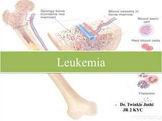
• Leukemia is a group of blood cancers that usually begin in the bone
marrow and result in high numbers of abnormal blood cells.
• Blood cells are not fully developed and are called blasts or leukemia cells.
• In 2015, leukemia was present in 2.3 million people worldwide and caused
353,500 deaths.
• Most common type of cancer in children, with three-quarters of leukemia cases
in children being the acute lymphoblastic type.
• However, over 90% of all leukemias are diagnosed in adults, with CLL and
AML being most common in adults and children respectively.
• It occurs more commonly in the developed world.
3. History
• 1st description – surgeon- Alfred Armand in 1827
• Complete description by pathologist
Rudolf Virchow in 1845
• He observed abnormally large no. of
WBCs in a sample and called it
Leukamie (German)
• Leukos- white ,Haima- blood
• 10 yrs later Pathologist Christian Neuman found that deceased
leukemic patients had greenish yellow bone marrow as
opposed to red.
4. Etiology
• Exact cause not identified.
• Genetic Predisposition
• Familial syndromes Eg Down’s syndrome
• Germline mutations in the gene AML1 (RUNX1, CBFA2) occur in the
familial platelet disorder
• Chemical, Radiation, and Other Exposures-
• Exposure to ionizing radiation, benzene, chloramphenicol, phenylbutazone,
and other drugs can uncommonly result in bone marrow failure that may
evolve into AML.
• Infections- EBV, AIDS
5. Classification
• Basis of cell types- myeloid, lymphoid
• Basis of natural history
• Acute leukemia-
1. Acute lymphoblastic leukemia –ALL – children, young adults
2. Acute myeloid leukemia- AML – all ages
• Chronic Leukemia-
1. Chronic lymphocytic leukemia- CLL - elderly
2. Chronic myeloid leukemia-CML- middle aged
6. • FAB classification
• Leukemia is acute if bone marrow consists of >30% of blasts
• OTHER TYPES
1. Burkitt’s Leukemia
• 30 % of ALL
• 3 subgroups
• African endemic- presents as jaw tumour ,associated with EBV
• Sporadic- multinucleated tumour cells
• Immunodeficient – associated with HIV
2. Aleukemia
7. 3. Prolymphocytic leukaemia-
• Variant of chronic lymphocytic leukaemia found mainly in males over the age
of 60 years.
• Typically massive splenomegaly with little lymphadenopathy and a very high
leucocyte count, often in excess of 400 × 10/L.
• Treatment is generally unsuccessful and the prognosis very poor.
4. Hairy cell leukaemia-
• Rare chronic B-cell lymphoproliferative disorder.
• Presenting symptoms - general ill health and recurrent infections.
Splenomegaly occurs in 90% cases.
• Severe neutropenia, monocytopenia and the characteristic hairy cells in the
blood and bone marrow are typical.
5. Myelodysplastic syndrome-
• Consists of a group of clonal haematopoietic disorders which represent steps in
the progression to the development of leukaemia.
8. Patho-physiology
• Due to the immature cells or some defect in the cells of the bone marrow
• Genetic damage to target WBC Proliferation abn. differenciation
• Also, they obstruct the production of other blood cells by dividing quickly
and crowding among normal cells.
AML ALL
9. • Chromosomal translocation-
In 70-90% cases with CML there is reciprocal rtranslocation of
chromosome 22 to chromosome 9, t(9;22) Philadelphia chromosome
• Organ infiltration
to spleen, liver, etc
10. Acute Vs Chronic
• Acute leukemia-
• Arise from immature cells in the bone marrow (myeloblasts or
lymphoblasts).
• Short history of days to months.
• These cells do not function like fully mature ones in fighting off infections.
• In addition, they often crowd the bone marrow, preventing the production
of other blood cells such as red blood cells, other white blood cells, and
platelets.
• Without treatment, acute leukemias often progress very rapidly.
11. • Chronic leukemia -
• Arises from mature, but abnormal white blood cells.
• These cancers grow much more slowly and may be discovered accidentally
when a blood count is done for another reason
• The disease has three phases:
• A chronic phase, in which the disease is responsive to treatment and is
easily controlled, which used to last 3–5 years.
• An accelerated phase (not always seen), in which disease control becomes
more difficult.
• Blast crisis, in which the disease transforms into an acute leukaemia, either
myeloid (70%) or lymphoblastic (30%), which is relatively refractory to
treatment. This is the cause of death in the majority of patients.
12. Clinical features
• Anemia- Pallor, easy fatiguability and dyspnoea
• Thrombocytopaenia causes bleeding, especially purpura and easy bruising on
dependent and traumatised areas.
• Granulocytopaenia results in increased incidence of infection, especially of
the skin, gums, perianal region, lungs and urinary tract.
• Infiltration of leukaemic cells into other organs leads to hepatosplenomegaly,
lymphadenopathy, mediastinal masses, sarcomas (soft tissue masses).
• CNS involvement may present with headache, vomiting, papilloedema and
convulsions. Testicular involvement presents with a mass lesion or pain.
• Overt bleeding due to disseminated intravascular coagulation (DIC).
• Gum hypertrophy and leukaemia cutis.
13.
14. Investigations
• CBC: usually shows anaemia with a normal or raised MCV. The leucocyte
count may vary from as low as 1 × 10⁹/L to as high as 500 × 109/L or more.
• Bone marrow aspiration - hypercellular, with replacement of normal elements
by leukaemic blast cells in varying degrees (but more than 20% of the cells)
• Tissue biopsy scan: A test in which a small tissue is taken from the lymph
nodes or bone marrow to identify the presence of leukaemia, its type and its
growth rate.
• Genetic abnormality test: A test to determine the Philadelphia chromosome,
genetic defect present in leukaemia cells.
• Cytochemistry: A test in which stains are used to identify the tissue structure
of the bone marrow cells.
• Other methods of diagnosing include CT scan, MRI and ultrasound.
17. • Common treatments used to fight leukemia include:
• Chemotherapy. This drug treatment uses chemicals to kill
leukemia cells.
• Targeted therapy. Targeted drug treatments focus on specific
abnormalities present within cancer cells. By blocking these
abnormalities, targeted drug treatments can cause cancer cells to die.
• Radiation therapy. Radiation therapy uses X-rays or other high-
energy beams to damage leukemia cells and stop their growth.
• Radiation therapy may be used to prepare for a bone marrow
transplant.
• Bone marrow transplant. A bone marrow transplant, also called a
stem cell transplant, helps reestablish healthy stem cells by replacing
unhealthy bone marrow with leukemia-free stem cells that will
regenerate healthy bone marrow.
18. • Immunotherapy. Immune system may not attack cancer cells
because they produce proteins that help them hide from the immune
system cells. Immunotherapy works by interfering with that process.
• Engineering immune cells to fight leukemia. A specialized
treatment called chimeric antigen receptor (CAR)-T cell therapy
takes your body's germ-fighting T cells, engineers them to fight
cancer and infuses them back into your body. CAR-T cell therapy
might be an option for certain types of leukemia.
19. Phases of therapy
• Induction therapy.
• The goal is to kill as many leukemia cells as possible in blood and bone
marrow to achieve remission.
• In remission, blood cell counts return to normal levels, no leukemia cells
are found in your blood, and all signs and symptoms of the disease
disappear. Induction therapy usually lasts four to six weeks.
• Consolidation. The goal is to kill any remaining undetected leukemia
cells. Usually lasts for over four to six months.
• Maintenance therapy. The goal is to kill any leukemia cells that may have
survived the first two treatment phases and prevent the cancer from
returning (relapse). Treatment lasts about two years.